Advertisement

Main menu

  • Home
  • Content
    • Current Issue
    • Accepted Manuscripts
    • Article Preview
    • Past Issue Archive
    • Video Articles
    • AJNR Case Collection
    • Case of the Week Archive
    • Case of the Month Archive
    • Classic Case Archive
  • Special Collections
    • AJNR Awards
    • Low-Field MRI
    • Alzheimer Disease
    • ASNR Foundation Special Collection
    • Photon-Counting CT
    • View All
  • Multimedia
    • AJNR Podcasts
    • AJNR SCANtastic
    • Trainee Corner
    • MRI Safety Corner
    • Imaging Protocols
  • For Authors
    • Submit a Manuscript
    • Submit a Video Article
    • Submit an eLetter to the Editor/Response
    • Manuscript Submission Guidelines
    • Statistical Tips
    • Fast Publishing of Accepted Manuscripts
    • Graphical Abstract Preparation
    • Imaging Protocol Submission
    • Author Policies
  • About Us
    • About AJNR
    • Editorial Board
    • Editorial Board Alumni
  • More
    • Become a Reviewer/Academy of Reviewers
    • Subscribers
    • Permissions
    • Alerts
    • Feedback
    • Advertisers
    • ASNR Home

User menu

  • Alerts
  • Log in

Search

  • Advanced search
American Journal of Neuroradiology

American Journal of Neuroradiology

ASHNR American Society of Functional Neuroradiology ASHNR American Society of Pediatric Neuroradiology ASSR
  • Alerts
  • Log in

Advanced Search

  • Home
  • Content
    • Current Issue
    • Accepted Manuscripts
    • Article Preview
    • Past Issue Archive
    • Video Articles
    • AJNR Case Collection
    • Case of the Week Archive
    • Case of the Month Archive
    • Classic Case Archive
  • Special Collections
    • AJNR Awards
    • Low-Field MRI
    • Alzheimer Disease
    • ASNR Foundation Special Collection
    • Photon-Counting CT
    • View All
  • Multimedia
    • AJNR Podcasts
    • AJNR SCANtastic
    • Trainee Corner
    • MRI Safety Corner
    • Imaging Protocols
  • For Authors
    • Submit a Manuscript
    • Submit a Video Article
    • Submit an eLetter to the Editor/Response
    • Manuscript Submission Guidelines
    • Statistical Tips
    • Fast Publishing of Accepted Manuscripts
    • Graphical Abstract Preparation
    • Imaging Protocol Submission
    • Author Policies
  • About Us
    • About AJNR
    • Editorial Board
    • Editorial Board Alumni
  • More
    • Become a Reviewer/Academy of Reviewers
    • Subscribers
    • Permissions
    • Alerts
    • Feedback
    • Advertisers
    • ASNR Home
  • Follow AJNR on Twitter
  • Visit AJNR on Facebook
  • Follow AJNR on Instagram
  • Join AJNR on LinkedIn
  • RSS Feeds

AJNR Awards, New Junior Editors, and more. Read the latest AJNR updates

Research ArticleAdult Brain
Open Access

Quantitative Susceptibility Mapping of Time-Dependent Susceptibility Changes in Multiple Sclerosis Lesions

S. Zhang, T.D. Nguyen, S.M. Hurtado Rúa, U.W. Kaunzner, S. Pandya, I. Kovanlikaya, P. Spincemaille, Y. Wang and S.A. Gauthier
American Journal of Neuroradiology June 2019, 40 (6) 987-993; DOI: https://doi.org/10.3174/ajnr.A6071
S. Zhang
aFrom the Departments of Radiology (S.Z., T.D.N., S.P., I.K., P.S., Y.W., S.A.G.)
dDepartment of Radiology (S.Z.), Tongji Hospital, Tongji Medical College, Huazhong University of Science and Technology, Wuhan, China
  • Find this author on Google Scholar
  • Find this author on PubMed
  • Search for this author on this site
  • ORCID record for S. Zhang
T.D. Nguyen
aFrom the Departments of Radiology (S.Z., T.D.N., S.P., I.K., P.S., Y.W., S.A.G.)
  • Find this author on Google Scholar
  • Find this author on PubMed
  • Search for this author on this site
  • ORCID record for T.D. Nguyen
S.M. Hurtado Rúa
eDepartment of Mathematics and Statistics (S.M.H.R.), College of Science and Health Professions, Cleveland State University, Cleveland, Ohio
  • Find this author on Google Scholar
  • Find this author on PubMed
  • Search for this author on this site
  • ORCID record for S.M. Hurtado Rúa
U.W. Kaunzner
bNeurology (U.W.K., S.A.G.)
  • Find this author on Google Scholar
  • Find this author on PubMed
  • Search for this author on this site
  • ORCID record for U.W. Kaunzner
S. Pandya
aFrom the Departments of Radiology (S.Z., T.D.N., S.P., I.K., P.S., Y.W., S.A.G.)
  • Find this author on Google Scholar
  • Find this author on PubMed
  • Search for this author on this site
  • ORCID record for S. Pandya
I. Kovanlikaya
aFrom the Departments of Radiology (S.Z., T.D.N., S.P., I.K., P.S., Y.W., S.A.G.)
  • Find this author on Google Scholar
  • Find this author on PubMed
  • Search for this author on this site
  • ORCID record for I. Kovanlikaya
P. Spincemaille
aFrom the Departments of Radiology (S.Z., T.D.N., S.P., I.K., P.S., Y.W., S.A.G.)
  • Find this author on Google Scholar
  • Find this author on PubMed
  • Search for this author on this site
  • ORCID record for P. Spincemaille
Y. Wang
aFrom the Departments of Radiology (S.Z., T.D.N., S.P., I.K., P.S., Y.W., S.A.G.)
fDepartment of Biomedical Engineering (Y.W.), Cornell University, Ithaca, New York.
  • Find this author on Google Scholar
  • Find this author on PubMed
  • Search for this author on this site
  • ORCID record for Y. Wang
S.A. Gauthier
aFrom the Departments of Radiology (S.Z., T.D.N., S.P., I.K., P.S., Y.W., S.A.G.)
bNeurology (U.W.K., S.A.G.)
cFeil Family Brain and Mind Research Institute (S.A.G.), Weill Cornell Medicine, New York, New York
  • Find this author on Google Scholar
  • Find this author on PubMed
  • Search for this author on this site
  • ORCID record for S.A. Gauthier
  • Article
  • Figures & Data
  • Info & Metrics
  • Responses
  • References
  • PDF
Loading

Article Figures & Data

Figures

  • Tables
  • Fig 1.
    • Download figure
    • Open in new tab
    • Download powerpoint
    Fig 1.

    Longitudinal QSM and T2-weighted FLAIR images of a new Gd-enhancing MS lesion without a QSM rim appearance (rim−). At baseline, the enhancing lesion was isointense on QSM (mean lesion susceptibility = −3.31 ppb), became most hyperintense at 1 year (22.50 ppb), and gradually disappeared in subsequent years (−1.18 ppb at year 5).

  • Fig 2.
    • Download figure
    • Open in new tab
    • Download powerpoint
    Fig 2.

    Longitudinal QSM and T2-weighted FLAIR images of a new Gd-enhancing MS lesion with a QSM rim appearance (rim+). This lesion was slightly hyperintense on QSM at the time of Gd-enhancement (mean lesion susceptibility = 12.74 ppb), became most hyperintense at 3 years (34.28 ppb), and remained hyperintense at 6 years (25.15 ppb).
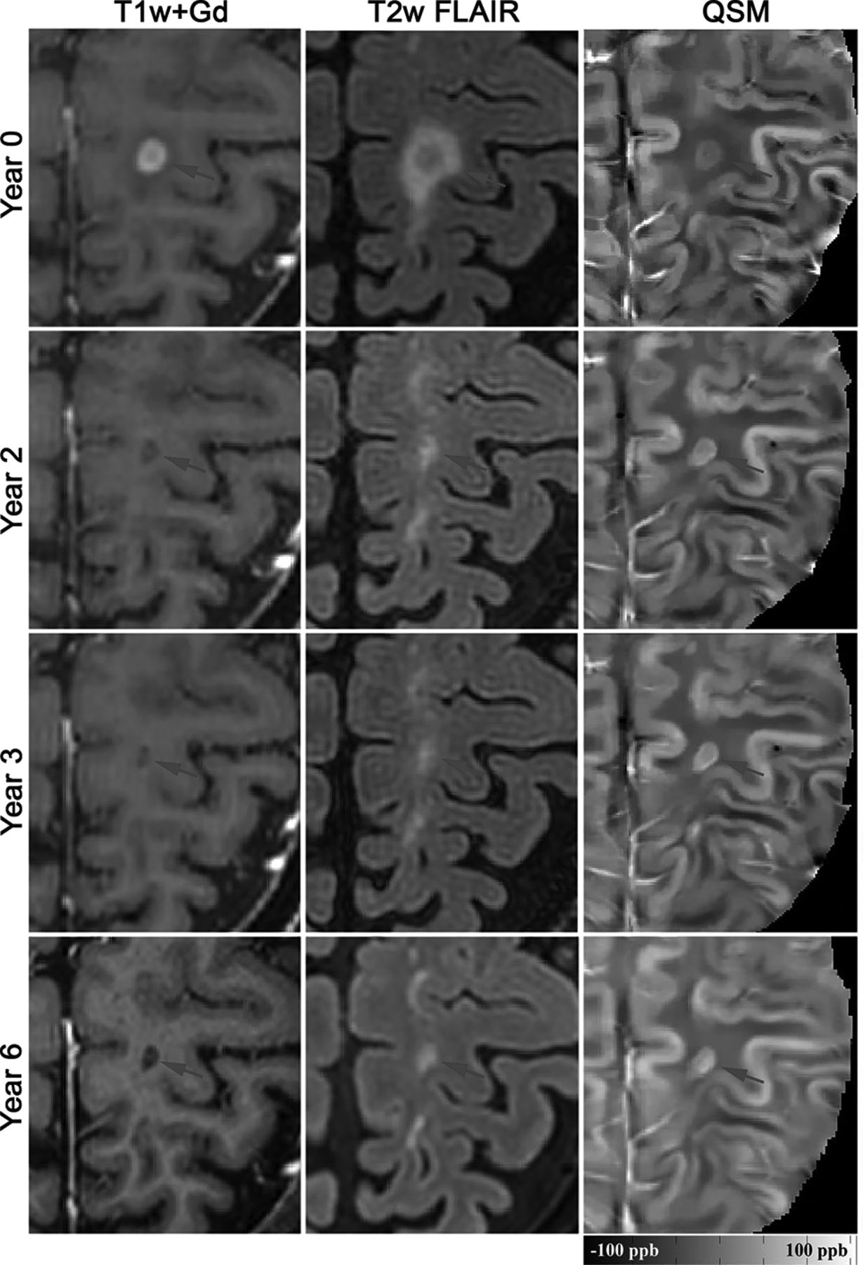
  • Fig 3.
    • Download figure
    • Open in new tab
    • Download powerpoint
    Fig 3.

    Longitudinal QSM and T2-weighted FLAIR images of a new Gd-enhancing MS lesion with a QSM rim appearance (rim+). This lesion was isointense on QSM at baseline and demonstrated an increase in susceptibility and rim appearance on subsequent QSM scans. The lesion susceptibility increased continually from −0.34 ppb at baseline to 26.35 ppb at 28 months. Gd-enhancement remained in this lesion until month 2.

  • Fig 4.
    • Download figure
    • Open in new tab
    • Download powerpoint
    Fig 4.

    Longitudinal lesion-volume evolution changes among QSM rim+ and rim− lesions. Rim+ lesions were statistically larger at Gd-enhancement (time = 0), 0.5, 2, and 4 years (all P < .0001).
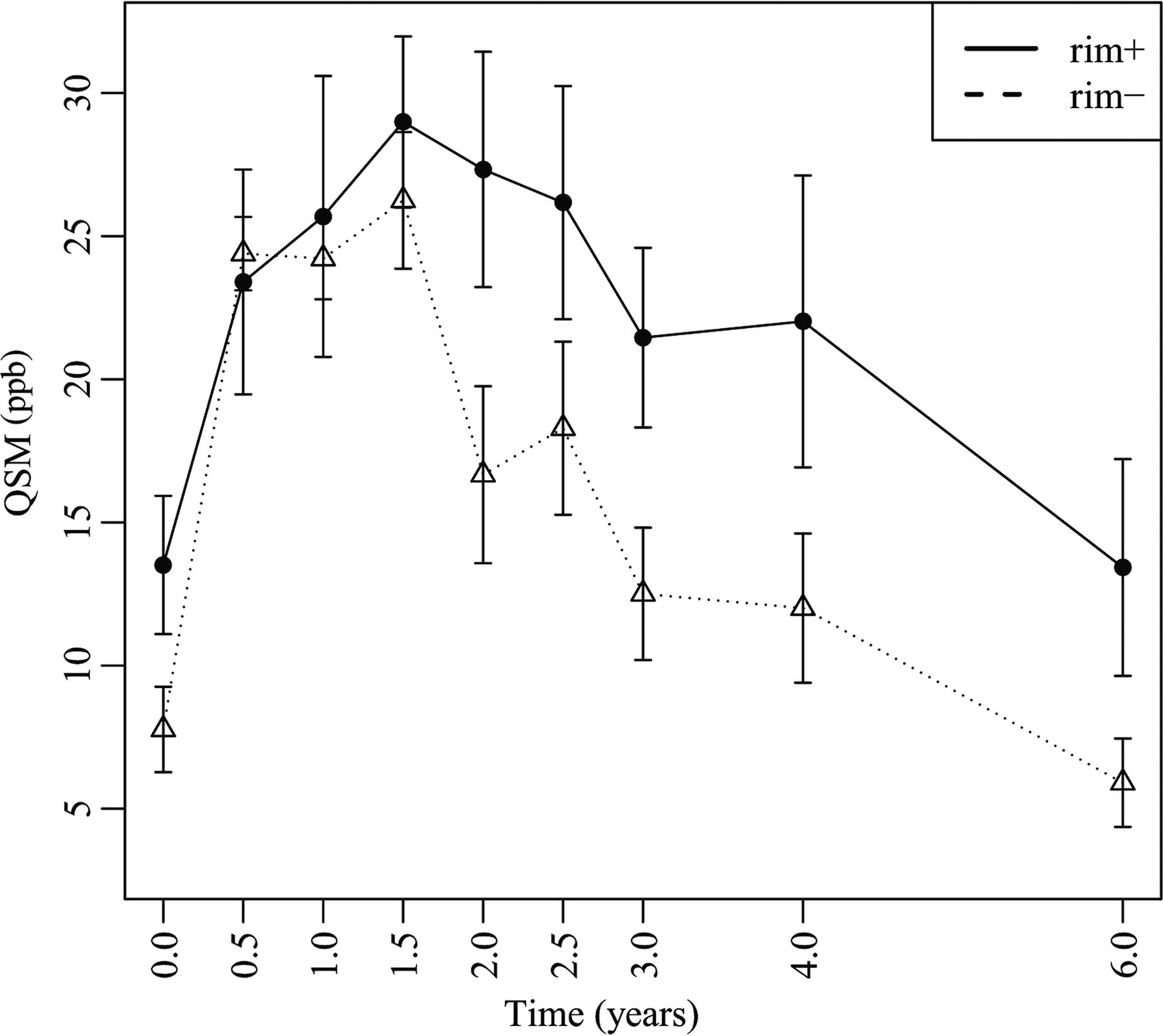
  • Fig 5.
    • Download figure
    • Open in new tab
    • Download powerpoint
    Fig 5.

    Longitudinal lesion age-dependent susceptibility time course of QSM rim+ and rim− MS lesions. Rim+ lesions demonstrate a higher peak QSM value and significantly slower decay rate compared with rim− (see text).

Tables

  • Figures
  • The mean susceptibility of rim+ and rim− lesions derived from the regression model

    Time (yr)Rim+Rim−
    No. of LesionsMean (ppb)95% CINo. of LesionsMean (ppb)95% CI
    01613.5111.09–15.93167.776.27–9.26
    0.51023.4019.47–27.33924.3923.11–25.67
    1725.6920.78–30.601124.2222.79–25.64
    1.5528.9926.00–31.98526.2523.86–28.63
    2627.3323.22–31.44716.6613.57–19.76
    2.5826.1722.10–30.251018.2915.27–21.31
    3721.4518.31–24.59912.5010.19–14.81
    4822.0216.92–27.121012.009.40–14.61
    6513.429.64–17.2165.904.36–7.45
PreviousNext
Back to top

In this issue

American Journal of Neuroradiology: 40 (6)
American Journal of Neuroradiology
Vol. 40, Issue 6
1 Jun 2019
  • Table of Contents
  • Index by author
  • Complete Issue (PDF)
Advertisement
Print
Download PDF
Email Article

Thank you for your interest in spreading the word on American Journal of Neuroradiology.

NOTE: We only request your email address so that the person you are recommending the page to knows that you wanted them to see it, and that it is not junk mail. We do not capture any email address.

Enter multiple addresses on separate lines or separate them with commas.
Quantitative Susceptibility Mapping of Time-Dependent Susceptibility Changes in Multiple Sclerosis Lesions
(Your Name) has sent you a message from American Journal of Neuroradiology
(Your Name) thought you would like to see the American Journal of Neuroradiology web site.
CAPTCHA
This question is for testing whether or not you are a human visitor and to prevent automated spam submissions.
Cite this article
S. Zhang, T.D. Nguyen, S.M. Hurtado Rúa, U.W. Kaunzner, S. Pandya, I. Kovanlikaya, P. Spincemaille, Y. Wang, S.A. Gauthier
Quantitative Susceptibility Mapping of Time-Dependent Susceptibility Changes in Multiple Sclerosis Lesions
American Journal of Neuroradiology Jun 2019, 40 (6) 987-993; DOI: 10.3174/ajnr.A6071

Citation Manager Formats

  • BibTeX
  • Bookends
  • EasyBib
  • EndNote (tagged)
  • EndNote 8 (xml)
  • Medlars
  • Mendeley
  • Papers
  • RefWorks Tagged
  • Ref Manager
  • RIS
  • Zotero
0 Responses
Respond to this article
Share
Bookmark this article
Quantitative Susceptibility Mapping of Time-Dependent Susceptibility Changes in Multiple Sclerosis Lesions
S. Zhang, T.D. Nguyen, S.M. Hurtado Rúa, U.W. Kaunzner, S. Pandya, I. Kovanlikaya, P. Spincemaille, Y. Wang, S.A. Gauthier
American Journal of Neuroradiology Jun 2019, 40 (6) 987-993; DOI: 10.3174/ajnr.A6071
del.icio.us logo Twitter logo Facebook logo Mendeley logo
  • Tweet Widget
  • Facebook Like
  • Google Plus One
Purchase

Jump to section

  • Article
    • Abstract
    • ABBREVIATIONS:
    • Materials and Methods
    • Results
    • Discussion
    • Conclusions
    • Footnotes
    • REFERENCES
  • Figures & Data
  • Info & Metrics
  • Responses
  • References
  • PDF

Related Articles

  • No related articles found.
  • PubMed
  • Google Scholar

Cited By...

  • Quantifying the Impact of Ocrelizumab on Paramagnetic Rim Lesions in Multiple Sclerosis
  • The Presence of a Paramagnetic Phase Rim is Linked to Lesion Age in Multiple Sclerosis
  • The sequence of regional structural disconnectivity due to multiple sclerosis lesions
  • SWI as an Alternative to Contrast-Enhanced Imaging to Detect Acute MS Lesions
  • Dimethyl Fumarate Reduces Inflammation in Chronic Active Multiple Sclerosis Lesions
  • Quantitative susceptibility mapping captures chronic multiple sclerosis rim lesions with greater myelin damage: Comparison with high-pass filtered phase MRI
  • Increased Risk for Cerebral Small Vessel Disease is Associated with Quantitative Susceptibility Mapping in HIV Infected and Uninfected Individuals
  • Structural disconnectivity from quantitative susceptibility mapping rim+ lesions is related to disability in people with multiple sclerosis
  • Crossref
  • Google Scholar

This article has not yet been cited by articles in journals that are participating in Crossref Cited-by Linking.

More in this TOC Section

  • Diagnostic Neuroradiology of Monoclonal Antibodies
  • Clinical Outcomes After Chiari I Decompression
  • Segmentation of Brain Metastases with BLAST
Show more ADULT BRAIN

Similar Articles

Advertisement

Indexed Content

  • Current Issue
  • Accepted Manuscripts
  • Article Preview
  • Past Issues
  • Editorials
  • Editor's Choice
  • Fellows' Journal Club
  • Letters to the Editor
  • Video Articles

Cases

  • Case Collection
  • Archive - Case of the Week
  • Archive - Case of the Month
  • Archive - Classic Case

More from AJNR

  • Trainee Corner
  • Imaging Protocols
  • MRI Safety Corner
  • Book Reviews

Multimedia

  • AJNR Podcasts
  • AJNR Scantastics

Resources

  • Turnaround Time
  • Submit a Manuscript
  • Submit a Video Article
  • Submit an eLetter to the Editor/Response
  • Manuscript Submission Guidelines
  • Statistical Tips
  • Fast Publishing of Accepted Manuscripts
  • Graphical Abstract Preparation
  • Imaging Protocol Submission
  • Evidence-Based Medicine Level Guide
  • Publishing Checklists
  • Author Policies
  • Become a Reviewer/Academy of Reviewers
  • News and Updates

About Us

  • About AJNR
  • Editorial Board
  • Editorial Board Alumni
  • Alerts
  • Permissions
  • Not an AJNR Subscriber? Join Now
  • Advertise with Us
  • Librarian Resources
  • Feedback
  • Terms and Conditions
  • AJNR Editorial Board Alumni

American Society of Neuroradiology

  • Not an ASNR Member? Join Now

© 2025 by the American Society of Neuroradiology All rights, including for text and data mining, AI training, and similar technologies, are reserved.
Print ISSN: 0195-6108 Online ISSN: 1936-959X

Powered by HighWire