
Article Figures & Data
Figures
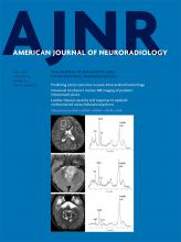
- Fig 1.
Proportion of white matter lesions demonstrating the central vein sign per case and the 40% rule. Eighteen patients with RIS (90%) met the 40% rule, whereas 2 did not.
- Fig 2.
White matter lesions with evident central vein signs in 2 different radiologically isolated syndrome cases, as seen on axial reconstructed 3D-T2*-weighted segmented echo-planar imaging sequences and 3D-T2-weighted FLAIR sequences of the brain, and sagittal T1-weighted phase-sensitive inversion recovery of the cervical spinal cord. A 50-year-old woman in whom most (90%) of the white matter lesions demonstrate the central vein sign (A, arrows). This individual also has evident infratentorial (B, arrows) and cervical spinal cord lesions (C, arrow). A 53-year-old woman with a small proportion (29%) of white matter lesions demonstrating the central vein sign (D, arrow). This individual did not have any infratentorial (E) or cervical spinal cord lesions (F).
- Fig 3.
White matter lesions demonstrating the central vein sign in different brain regions, using 3D-T2*-weighted echo-planar imaging. A, Infratentorial. B, Deep white matter. C, Periventricular. D, juxtacortical.


Tables
Clinical Characteristics Participants (No.) 20 Age (mean) (SD) (yr) 46 (11) Female (No.) (%) 15 (75%) No. of cases with positive oligoclonal band (No.) (%)a 5 (83%) MRI characteristics Brain lesions Total brain lesion count (No.) 997 No. of brain lesions per case (median) (range) 33 (9–165) Total brain lesion volume (median) (range) (cm3)b 3.9 (0.3–17.9) No. of cases with T1 black hole lesions (No.) (%) 15 (75%) No. of T1 black hole lesion count 206 (21%) No. of T1 black hole lesions per case (median) (range) 3.5 (0–43) No. of brain lesions included in the analysis (No.) (%) 391 (39%) No. of brain lesions excluded from analysis (No.) (%) 606 (61%) Brain volume (median) (range) (cm3)b 1208 (1066–1468) Cerebral volume fraction (median) (range)b 0.90 (0.88–0.91) No. of assessed brain lesion/total brain lesion by region (%) Cortical/juxtacortical 90/193 (47%) Subcortical/deep 228/562 (40%) Periventricular 60/203 (30%) Infratentorial 13/39 (33%) Cervical spinal cord lesions No. of cases with spinal cord lesions (No.) (%) 13 (65%) Total spinal cord lesion count 30 No. of spinal cord lesions per case (median) (range) 1 (0–4) Reasons (No.) (%) Headache 9 (45%) Work-up for pituitary adenoma 2 (10%) Transient paraphasic symptoms atypical for demyelinating disease 2 (10%) Intermittent subjective cognitive symptoms 1 (5%) Intermittent nocturnal tremor 1 (5%) Pars planitis 1 (5%) Sinusitis 1 (5%) Back pain 1 (5%) Dental pain 1 (5%) Tinnitus 1 (5%) MS Diagnostic Criteria Using the CVS No. of RIS Participants Positive for CVS Negative for CVS 40% rule 18 (90%) 2 (10%) Rule of 6 19 (95%) 1 (5%) - Table 4:
Relationships between the proportion of white matter lesions demonstrating the central vein sign and demographic and MRI variables in cases of RIS
Variables Univariable Regression (P Value) Multivariable Regression (P Value) Age .27 .01 Sex .30 .12 Total No. of brain lesions .33 .29 No. of infratentorial lesions .21 .06 No. of cervical spinal cord lesions .04 .002


