
Article Figures & Data
Figures
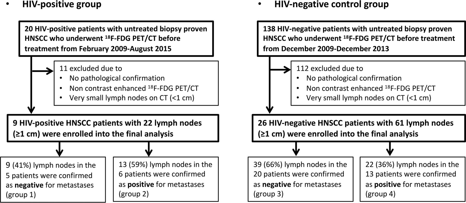
- Fig 1.
Flowchart shows patient selection for the study.
- Fig 2.
Representative case of false-positive findings on FDG-PET for benign nodes in a 74-year-old woman with unknown primary squamous cell carcinoma who had both benign and malignant nodes (HIV-positive; absolute CD4 = 473). Axial contrast-enhanced CT images (A and D), [18F] FDG-PET/CT fusion images (B and E), and corresponding axial section ROI mask-segmented lymph nodes (C and F) for pathologically benign (A–C) and malignant (D–F) lymph nodes. [18F] FDG-PET/CT shows abnormal uptake (cutoff = >5.5) for both lymph nodes with SUVmax = 8.1 (benign, B) and 9.5 (malignant, E). However, texture features with SRE (cutoff = <0.164) of both lymph nodes were correctly diagnosed as follows: 0.188 (benign, C) and 0.151 (malignant, F).


Tables
- Table 1:
Patient demographics and tumor characteristics of patients with head and neck cancera
Characteristic HIV-Positive Group (n = 9) HIV-Negative Group (n = 26) P Value Age (yr) .034b Median (range) 48 (29–62) 59 (30–86) Sex .490 Male 7 (78) 17 (65) Female 2 (22) 9 (35) HPV status (protein 16) .283 Positive 3 12 Negative 1 14 Unknown 5 0 Primary site .169 Oropharynx 3 (33) 10 (38) Hypopharynx 0 (0) 1 (4) Larynx 1 (11) 3 (12) Oral cavity 3 (33) 12 (46) Primary unknown 1 (11) 0 (0) Maxilla 1 (11) 0 (0) T-Stage .444 T0 1 (11) 0 (0) T1 1 (11) 7 (27) T2 3 (33) 9 (35) T3 1 (11) 2 (8) T4 3 (33) 8 (30) - Table 2:
[18F] FDG-PET/CT characteristics and selected texture parameters differentiating lymph node characterization in patients with HIV infection (group 1 vs 2)
Texture Parameter Benign Nodes (n = 9) Malignant Nodes (n = 13) P Valuea Cutoff AUCb (GLIMMROC) Mean SD Mean SD Node characteristics Size (cm) 1.400 0.300 1.892 0.690 .024 <2.0 0.752 Volume (cm3) 0.547 0.363 2.535 2.218 .007 >0.76 0.889 SUVmax 5.111 1.610 8.562 3.886 .042 >5.5 0.803 Histogram Mean 624.4 74.6 735.3 75.2 .017 >683.3 0.872 Median 774.0 217.9 1013.1 71.2 .018 >877.8 0.872 Second SD 93.76 28.70 59.85 19.19 .017 <76.3 0.872 Range 273.7 82.9 175.4 54.5 .017 <232.7 0.863 Geometric mean 199.6 44.7 272.8 63.8 .032 >237.3 0.829 SD 5 81.9 29.4 16.8 19.2 .018 <62.89 0.872 SD 9 99.7 49.3 19.5 17.3 .017 <63.16 0.889 GLCM Contrast 113.1 20.2 84.1 16.9 .009 <97.7 0.889 Energy 0.036 0.019 0.081 0.047 .025 >0.047 0.812 Homogeneity 0.451 0.065 0.542 0.069 .020 >0.498 0.821 GLRL SRE 0.177 0.007 0.157 0.009 <.001 <0.164 0.966c LRE 0.197 0.010 0.173 0.012 .001 <0.186 0.957 GLN 0.183 0.009 0.160 0.011 .001 <0.172 0.949 RLN 0.196 0.010 0.173 0.012 .002 <0.184 0.932 LRHGE 224.6 51.8 300.0 68.7 .035 >261.8 0.863 Note:—GLIMMROC indicates generalized linear mixed model receiver operating characteristic.
↵a Indicates a significant difference by the mixed linear regression model (Proc MIXED) to adjust the variance-covariance matrix among multiple values recorded for each patient (P < .05).
↵b Using the generalized linear mixed model (GLIMMROC).
↵c The highest AUC among 41 texture features.
- Table 3:
[18F] FDG-PET/CT characteristics and selected texture parameters differentiating lymph node characterization in patients with head and neck squamous cell carcinoma without HIV infection (group 3 vs 4)
Texture Parameter Benign Nodes (n = 39) Malignant Nodes (n = 22) P Valuea Cutoff AUCb (GLIMMROC) Mean SD Mean SD Node characteristics Size (cm) 1.439 0.048 1.687 0.831 .079 DNC 0.643 Volume (cm3) 1.122 0.147 3.468 4.718 .001c >2.599 0.702 SUVmax 3.228 0.315 6.438 4.811 .007c >3.980 0.731 Histogram Mean 670.6 68.4 751.8 137.3 .022c >738.1 0.705 Geometric mean 225.9 50.6 297.6 124.5 .020c >257.5 0.667 Harmonic mean 24.12 4.21 29.57 9.42 .020c >25.5 0.678 IQR 1064.3 63.9 865.0 360.2 .022c <1017.0 0.723 Fourth moment 8.71E+10 5.74E+09 9.41E+10 1.00E+10 .018c >9.38E+10 0.698 GLCM Contrast 102.5 18.2 81.8 28.3 .015c <75.7 0.754 Energy 0.041 0.022 0.072 0.055 .034c >0.058 0.712 Homogeneity 0.475 0.056 0.541 0.104 .029c >0.528 0.735 GLRL SRE 0.164 0.011 0.154 0.019 .036c <0.153 0.695 RLN 0.183 0.014 0.170 0.023 .034c <0.172 0.698 RHGE 254.2 77.2 367.0 193.6 .016c >305.4 0.739 LRLGE 322.6 91.4 469.8 231.9 .006c >397.1 0.759 LRHGE 246.2 60.5 352.7 168.8 .003c >340.9 0.760d Laws features L1 1,130,272.9 280,670.9 876,676.6 413,942.2 .024c <911,080.5 0.722 Note:—DNC indicates did not converge; IQR, interquartile range; GLIMMROC, generalized linear mixed model receiver operating characteristic.
↵a Indicates a significant difference by the mixed linear regression model (Proc MIXED) to adjust the variance-covariance matrix among multiple values recorded for each patient (P < .05).
↵b Using the generalized linear mixed model (GLIMMROC).
↵c Significant.
↵d The highest AUC among 41 texture features.


Jump to section
Related Articles
Cited By...
- No citing articles found.