Advertisement

Main menu

  • Home
  • Content
    • Current Issue
    • Accepted Manuscripts
    • Article Preview
    • Past Issue Archive
    • Video Articles
    • AJNR Case Collection
    • Case of the Week Archive
    • Case of the Month Archive
    • Classic Case Archive
  • Special Collections
    • Advancing NeuroMRI with High-Relaxivity Contrast Agents
    • AJNR Awards
    • Low-Field MRI
    • Alzheimer Disease
    • ASNR Foundation Special Collection
    • Photon-Counting CT
    • View All
  • Multimedia
    • AJNR Podcasts
    • AJNR SCANtastic
    • Trainee Corner
    • MRI Safety Corner
    • Imaging Protocols
  • For Authors
    • Submit a Manuscript
    • Submit a Video Article
    • Submit an eLetter to the Editor/Response
    • Manuscript Submission Guidelines
    • Statistical Tips
    • Fast Publishing of Accepted Manuscripts
    • Graphical Abstract Preparation
    • Imaging Protocol Submission
    • Author Policies
  • About Us
    • About AJNR
    • Editorial Board
    • Editorial Board Alumni
  • More
    • Become a Reviewer/Academy of Reviewers
    • Subscribers
    • Permissions
    • Alerts
    • Feedback
    • Advertisers
    • ASNR Home

User menu

  • Alerts
  • Log in

Search

  • Advanced search
American Journal of Neuroradiology

American Journal of Neuroradiology

ASHNR American Society of Functional Neuroradiology ASHNR American Society of Pediatric Neuroradiology ASSR
  • Alerts
  • Log in

Advanced Search

  • Home
  • Content
    • Current Issue
    • Accepted Manuscripts
    • Article Preview
    • Past Issue Archive
    • Video Articles
    • AJNR Case Collection
    • Case of the Week Archive
    • Case of the Month Archive
    • Classic Case Archive
  • Special Collections
    • Advancing NeuroMRI with High-Relaxivity Contrast Agents
    • AJNR Awards
    • Low-Field MRI
    • Alzheimer Disease
    • ASNR Foundation Special Collection
    • Photon-Counting CT
    • View All
  • Multimedia
    • AJNR Podcasts
    • AJNR SCANtastic
    • Trainee Corner
    • MRI Safety Corner
    • Imaging Protocols
  • For Authors
    • Submit a Manuscript
    • Submit a Video Article
    • Submit an eLetter to the Editor/Response
    • Manuscript Submission Guidelines
    • Statistical Tips
    • Fast Publishing of Accepted Manuscripts
    • Graphical Abstract Preparation
    • Imaging Protocol Submission
    • Author Policies
  • About Us
    • About AJNR
    • Editorial Board
    • Editorial Board Alumni
  • More
    • Become a Reviewer/Academy of Reviewers
    • Subscribers
    • Permissions
    • Alerts
    • Feedback
    • Advertisers
    • ASNR Home
  • Follow AJNR on Twitter
  • Visit AJNR on Facebook
  • Follow AJNR on Instagram
  • Join AJNR on LinkedIn
  • RSS Feeds

AJNR Awards, New Junior Editors, and more. Read the latest AJNR updates


Improved Turnaround Times | Median time to first decision: 12 days

Research ArticleAdult Brain
Open Access

Complementary Roles of Dynamic Contrast-Enhanced MR Imaging and Postcontrast Vessel Wall Imaging in Detecting High-Risk Intracranial Aneurysms

H. Qi, X. Liu, P. Liu, W. Yuan, A. Liu, Y. Jiang, Y. Li, J. Sun and H. Chen
American Journal of Neuroradiology March 2019, 40 (3) 490-496; DOI: https://doi.org/10.3174/ajnr.A5983
H. Qi
aFrom the Department of Biomedical Engineering (H.Q., X.L., H.C.), Tsinghua University, Beijing, China
  • Find this author on Google Scholar
  • Find this author on PubMed
  • Search for this author on this site
  • ORCID record for H. Qi
X. Liu
aFrom the Department of Biomedical Engineering (H.Q., X.L., H.C.), Tsinghua University, Beijing, China
  • Find this author on Google Scholar
  • Find this author on PubMed
  • Search for this author on this site
  • ORCID record for X. Liu
P. Liu
bDepartment of Interventional Neuroradiology (P.L., A.L.,Y.J., Y.L.), Beijing Neurosurgical Institute and Beijing Tiantan Hospital, Capital Medical University, Beijing, China
  • Find this author on Google Scholar
  • Find this author on PubMed
  • Search for this author on this site
  • ORCID record for P. Liu
W. Yuan
cDepartment of Radiology (W.Y.), Navy Qingdao No. 1 Sanatorium of People's Liberation Army, Qingdao, China
  • Find this author on Google Scholar
  • Find this author on PubMed
  • Search for this author on this site
  • ORCID record for W. Yuan
A. Liu
bDepartment of Interventional Neuroradiology (P.L., A.L.,Y.J., Y.L.), Beijing Neurosurgical Institute and Beijing Tiantan Hospital, Capital Medical University, Beijing, China
  • Find this author on Google Scholar
  • Find this author on PubMed
  • Search for this author on this site
  • ORCID record for A. Liu
Y. Jiang
bDepartment of Interventional Neuroradiology (P.L., A.L.,Y.J., Y.L.), Beijing Neurosurgical Institute and Beijing Tiantan Hospital, Capital Medical University, Beijing, China
  • Find this author on Google Scholar
  • Find this author on PubMed
  • Search for this author on this site
  • ORCID record for Y. Jiang
Y. Li
bDepartment of Interventional Neuroradiology (P.L., A.L.,Y.J., Y.L.), Beijing Neurosurgical Institute and Beijing Tiantan Hospital, Capital Medical University, Beijing, China
  • Find this author on Google Scholar
  • Find this author on PubMed
  • Search for this author on this site
  • ORCID record for Y. Li
J. Sun
dDepartment of Radiology (J.S.), University of Washington, Seattle, Washington.
  • Find this author on Google Scholar
  • Find this author on PubMed
  • Search for this author on this site
  • ORCID record for J. Sun
H. Chen
aFrom the Department of Biomedical Engineering (H.Q., X.L., H.C.), Tsinghua University, Beijing, China
  • Find this author on Google Scholar
  • Find this author on PubMed
  • Search for this author on this site
  • ORCID record for H. Chen
  • Article
  • Figures & Data
  • Info & Metrics
  • Responses
  • References
  • PDF
Loading

Article Figures & Data

Figures

  • Tables
  • Fig 1.
    • Download figure
    • Open in new tab
    • Download powerpoint
    Fig 1.

    Measuring aneurysm wall permeability by DCE-MR imaging. Signal intensity in the lumen was measured on DCE imaging series (red contour in A) to obtain contrast concentration in plasma at different time points, which gave the arterial input function Cp(t) (red curve in C). The concentration-time curve of tissue C(t) was fitted voxel by voxel using the extended Kety/Tofts model, which generated parametric maps that were overlaid onto DCE images (B). ROIs were placed on the slice with highest Ktrans to measure Ktrans in the region adjacent to intracranial aneurysms (IA-ROI, white contour on Ktrans map in B) and near a normal artery. The concentration-time curves of points 1 and 2 (red dots in B) are shown in D. ref-ROI indicates reference ROI.

  • Fig 2.
    • Download figure
    • Open in new tab
    • Download powerpoint
    Fig 2.

    Enhanced aneurysm wall with and without increased permeability. Case 1: A 51-year-old woman who presented with a 19.5-mm aneurysm in the internal carotid artery. Pre- and postcontrast vessel wall images show conspicuous wall enhancement. DCE-MR imaging shows increased wall permeability (Ktrans = 0.0346 min−1). Case 2: A 61-year-old woman who presented with a 9-mm aneurysm in the anterior communicating artery. Pre- and postcontrast vessel wall imaging shows conspicuous wall enhancement, but DCE-MR imaging did not show increased wall permeability (Ktrans = 0.0096 min−1).
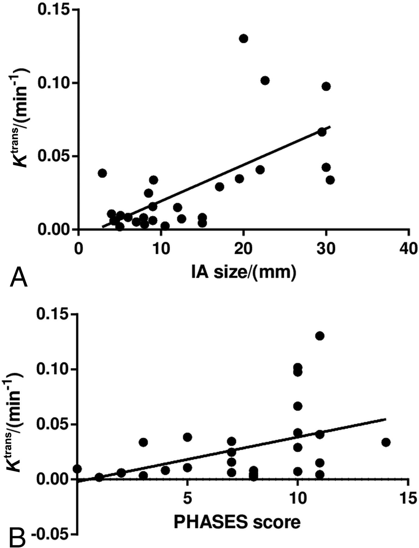
  • Fig 3.
    • Download figure
    • Open in new tab
    • Download powerpoint
    Fig 3.

    Associations between aneurysm wall permeability and size (A) and the PHASES score (B). Spearman correlation analysis shows that Ktrans is positively correlated with aneurysm size (ρ = 0.54, P = .002) and the PHASES score (ρ = 0.40, P = .030).
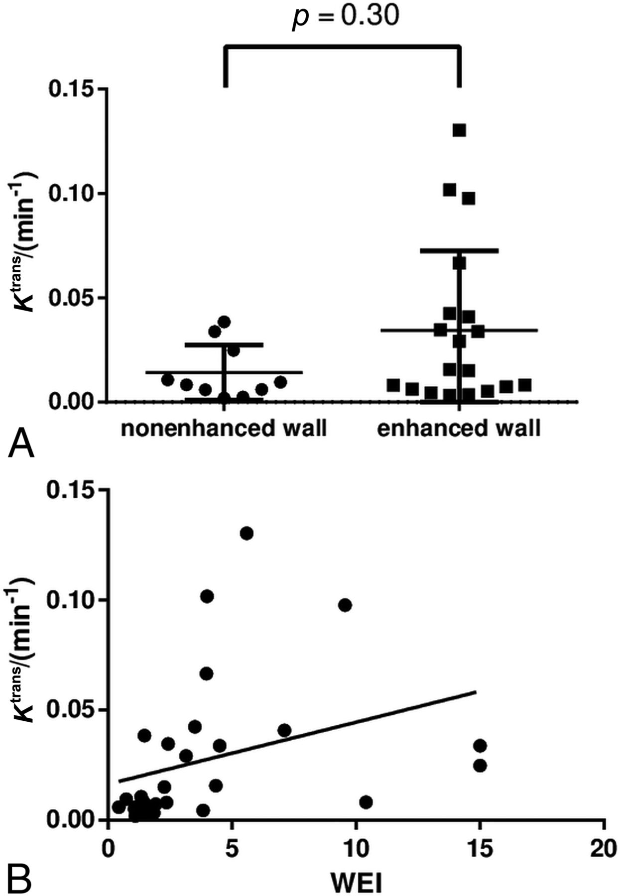
  • Fig 4.
    • Download figure
    • Open in new tab
    • Download powerpoint
    Fig 4.

    Association between Ktrans and aneurysm wall enhancement. Ktrans shows no statistically significant association with the presence of aneurysm wall enhancement (A) and a poor concordance with the wall enhancement index (B).

  • Fig 5.
    • Download figure
    • Open in new tab
    • Download powerpoint
    Fig 5.

    Baseline MR images of the 2 aneurysms that ruptured during follow-up. Case 1: A 36-year-old man has a 17.1-mm aneurysm in the basilar artery. The patient was concerned about surgical risk and received conservative treatment. Subarachnoid hemorrhage occurred 5 months after the baseline scan (no aneurysm wall enhancement; Ktrans = 0.0449 min−1). Case 2: A 68-year-old man had an 8.5-mm aneurysm in the basilar artery. The patient was concerned about surgical risk and received conservative treatment. Subarachnoid hemorrhage occurred 1 month after baseline scan (no aneurysm wall enhancement; Ktrans = 0.0524 min−1).

Tables

  • Figures
    • View popup
    Table 1:

    Components of the PHASES score14

    PHASES Aneurysm Risk ScorePoints
    (P) Population
        North American, European (other than Finnish)0
        Japanese3
        Finnish5
    (H) Hypertension
        No0
        Yes1
    (A) Age
        Younger than 70 yr0
        70 yr or older1
    (S) Size of aneurysm
        <7.0 mm0
        7.0–9.9 mm3
        10.0–19.9 mm6
        ≥20.0 mm10
    (E) Earlier SAH from another aneurysm
        No0
        Yes1
    (S) Site of aneurysm
        ICA0
        MCA2
        ACA/PcomA/posterior circulation4
    • Note:—ACA indicates anterior cerebral arteries (including the anterior cerebral artery, anterior communicating artery, and pericallosal artery); ICA, internal carotid artery; MCA, middle cerebral artery; PcomA, posterior communicating artery; SAH, subarachnoid hemorrhage.

    • View popup
    Table 2:

    The demography and aneurysm characteristics of the study sample (N = 29)a

    No. (%), Mean, or Median (IQR)
    Age (yr)53.9 ± 13.5
    Male sex7 (24%)
    Current smoking5 (17%)
    Hypertension11 (38%)
    Diabetes2 (7%)
    Location
        Anterior circulation18 (62%)
        Posterior circulation11 (38%)
    Size
        <7.0 mm7 (24%)
        7.0–9.9 mm8 (28%)
        10.0–19.9 mm7 (24%)
        ≥20.0 mm7 (24%)
    Blebs3 (10%)
    PHASES score8 (4.75–10)
    Ktrans (min−1)0.0107 (0.0060–0.0390)
    Aneurysm wall enhancement19 (66%)
    • ↵a See Table 1 for details of the PHASES score.

PreviousNext
Back to top

In this issue

American Journal of Neuroradiology: 40 (3)
American Journal of Neuroradiology
Vol. 40, Issue 3
1 Mar 2019
  • Table of Contents
  • Index by author
  • Complete Issue (PDF)
Advertisement
Print
Download PDF
Email Article

Thank you for your interest in spreading the word on American Journal of Neuroradiology.

NOTE: We only request your email address so that the person you are recommending the page to knows that you wanted them to see it, and that it is not junk mail. We do not capture any email address.

Enter multiple addresses on separate lines or separate them with commas.
Complementary Roles of Dynamic Contrast-Enhanced MR Imaging and Postcontrast Vessel Wall Imaging in Detecting High-Risk Intracranial Aneurysms
(Your Name) has sent you a message from American Journal of Neuroradiology
(Your Name) thought you would like to see the American Journal of Neuroradiology web site.
CAPTCHA
This question is for testing whether or not you are a human visitor and to prevent automated spam submissions.
Cite this article
H. Qi, X. Liu, P. Liu, W. Yuan, A. Liu, Y. Jiang, Y. Li, J. Sun, H. Chen
Complementary Roles of Dynamic Contrast-Enhanced MR Imaging and Postcontrast Vessel Wall Imaging in Detecting High-Risk Intracranial Aneurysms
American Journal of Neuroradiology Mar 2019, 40 (3) 490-496; DOI: 10.3174/ajnr.A5983

Citation Manager Formats

  • BibTeX
  • Bookends
  • EasyBib
  • EndNote (tagged)
  • EndNote 8 (xml)
  • Medlars
  • Mendeley
  • Papers
  • RefWorks Tagged
  • Ref Manager
  • RIS
  • Zotero
0 Responses
Respond to this article
Share
Bookmark this article
Complementary Roles of Dynamic Contrast-Enhanced MR Imaging and Postcontrast Vessel Wall Imaging in Detecting High-Risk Intracranial Aneurysms
H. Qi, X. Liu, P. Liu, W. Yuan, A. Liu, Y. Jiang, Y. Li, J. Sun, H. Chen
American Journal of Neuroradiology Mar 2019, 40 (3) 490-496; DOI: 10.3174/ajnr.A5983
del.icio.us logo Twitter logo Facebook logo Mendeley logo
  • Tweet Widget
  • Facebook Like
  • Google Plus One
Purchase

Jump to section

  • Article
    • Abstract
    • ABBREVIATIONS:
    • Materials and Methods
    • Results
    • Discussion
    • Conclusions
    • Footnotes
    • References
  • Figures & Data
  • Info & Metrics
  • Responses
  • References
  • PDF

Related Articles

  • No related articles found.
  • PubMed
  • Google Scholar

Cited By...

  • A Review of Intracranial Aneurysm Imaging Modalities, from CT to State-of-the-Art MR
  • 3D aneurysm wall enhancement is associated with symptomatic presentation
  • Quantitative analysis of unruptured intracranial aneurysm wall thickness and enhancement using 7T high resolution, black blood magnetic resonance imaging
  • Atorvastatin for unruptured intracranial vertebrobasilar dissecting aneurysm (ATREAT-VBD): protocol for a randomised, double-blind, blank-controlled trial
  • In situ decellularization of a large animal saccular aneurysm model: sustained inflammation and active aneurysm wall remodeling
  • Crossref
  • Google Scholar

This article has not yet been cited by articles in journals that are participating in Crossref Cited-by Linking.

More in this TOC Section

Adult Brain

  • Diagnostic Neuroradiology of Monoclonal Antibodies
  • Segmentation of Brain Metastases with BLAST
  • Cerebral ADC Changes in Fabry Disease
Show more Adult Brain

Neurointervention

  • Prophylactic Endovascular Treatments for CBS
  • Smoking & Aneurysm Outcomes: A Meta-Analysis
  • “Buddy-wire anchoring” technique for TVE
Show more Neurointervention

Similar Articles

Advertisement

Indexed Content

  • Current Issue
  • Accepted Manuscripts
  • Article Preview
  • Past Issues
  • Editorials
  • Editor's Choice
  • Fellows' Journal Club
  • Letters to the Editor
  • Video Articles

Cases

  • Case Collection
  • Archive - Case of the Week
  • Archive - Case of the Month
  • Archive - Classic Case

More from AJNR

  • Trainee Corner
  • Imaging Protocols
  • MRI Safety Corner
  • Book Reviews

Multimedia

  • AJNR Podcasts
  • AJNR Scantastics

Resources

  • Turnaround Time
  • Submit a Manuscript
  • Submit a Video Article
  • Submit an eLetter to the Editor/Response
  • Manuscript Submission Guidelines
  • Statistical Tips
  • Fast Publishing of Accepted Manuscripts
  • Graphical Abstract Preparation
  • Imaging Protocol Submission
  • Evidence-Based Medicine Level Guide
  • Publishing Checklists
  • Author Policies
  • Become a Reviewer/Academy of Reviewers
  • News and Updates

About Us

  • About AJNR
  • Editorial Board
  • Editorial Board Alumni
  • Alerts
  • Permissions
  • Not an AJNR Subscriber? Join Now
  • Advertise with Us
  • Librarian Resources
  • Feedback
  • Terms and Conditions
  • AJNR Editorial Board Alumni

American Society of Neuroradiology

  • Not an ASNR Member? Join Now

© 2025 by the American Society of Neuroradiology All rights, including for text and data mining, AI training, and similar technologies, are reserved.
Print ISSN: 0195-6108 Online ISSN: 1936-959X

Powered by HighWire