
Article Figures & Data
Figures
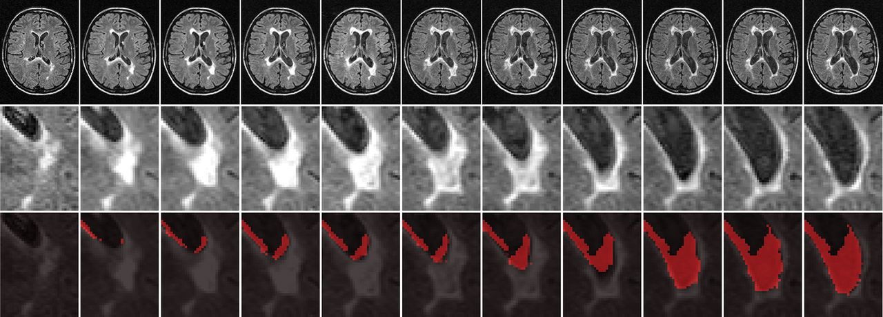
- FIGURE.
Representative example of a serially enlarging and then atrophying lesion. The upper row shows raw T2-FLAIR images from baseline to 10-year follow-up in 1-year increments, and the middle row provides an enlarged view of the relevant lesion. In the lower row, cumulative atrophied lesion volume is shown for the same area, in which red voxels indicate regions that were lesions at any prior time point and are CSF at the current time point.

Tables
- Table 1:
Demographic and clinical characteristics at baseline and during the follow-up in 176 patients with MS, according to the confirmed disability progression status at the 10-year follow-up
Total Study Cohort (N = 176) Stable Group (n = 76) CDP Group (n = 100) P Valuea Female sex (No.) (%) 137 (77.8) 57 (75) 80 (80) .429 Age at baseline (mean) (SD) (yr) 30.7 (7.9) 28.7 (7.1) 31.8 (7.9) .008b Disease duration at baseline (mean) (SD) (yr) 4.9 (5.2) 4.0 (3.2) 5.7 (6.2) .02 EDSS at baseline (median) (IQR) 2.0 (1.0–2.5) 2.0 (1.0–2.0) 2.0 (1.5–2.0) .093 EDSS at follow-up (median) (IQR) 3.0 (2.0–4.0) 2.0 (1.5–2.5) 4.0 (3.0–5.0) <.001b EDSS absolute change during follow-up (median) (IQR) 1.3 (0–2) 1.0 0.13 (0–0.5) 0 2.2 (1.5–3) 2.0 <.001b No. of relapses between baseline and follow-up (mean) (SD) 5.2 (3.8) 5 4.6 (3.9) 5.8 (3.7) .048 Annual relapse rate during the follow-up (mean) (SD) 0.5 (0.4) 0.5 (0.4) 0.6 (0.4) .048 Relapse-free from baseline to follow-up (No.) (%) 7 (4) 5 (6.6) 2 (2) .124 Treatment status at follow-up (No.) (%) Remained on IM interferon β-1a 74 (42) 45 (59.2) 29 (29) <.001b Switched to other DMTs 79 (44.9) 23 (30.3) 56 (56) Discontinued DMT 23 (13.1) 8 (10.5) 15 (15) Time on interferon β-1a IM (mean) (SD) (mo) 87.3 (99.5) 91.3 (16.6) 84.1 (24.4) .022 - Table 2:
Time course of cumulative atrophied T2 lesion volume on serial MRI in patients with MS, according to the confirmed disability progression status at the 10-year follow-upa
Months from Baseline No. in Stable Group Atrophied T2-LV Stable Group (Mean) (SD) No. of Patients with CDP Atrophied T2-LV CDP Group (Mean) (SD) % Difference Cohen d P Valueb 6 mo 74 0.05 (0.09) 94 0.12 (0.16) 140 0.54 .004 12 mo 76 0.09 (0.14) 95 0.21 (0.30) 133 0.51 <.001 24 mo 68 0.14 (0.21) 85 0.32 (0.41) 129 0.52 <.001 36 mo 67 0.21 (0.36) 89 0.40 (0.50) 90.5 0.44 <.001 48 mo 68 0.30 (0.50) 87 0.52 (0.70) 73.3 0.36 .007 60 mo 67 0.36 (0.57) 91 0.62 (0.83) 72.2 0.37 .004 72 mo 66 0.43 (0.70) 87 0.83 (1.10) 93 0.43 <.001 84 mo 68 0.44 (0.54) 85 1.10 (1.40) 150 0.62 <.001 96 mo 67 0.52 (0.66) 87 1.36 (1.72) 162 0.64 .004 108 mo 68 0.60 (0.76) 84 1.39 (1.68) 132 0.61 <.001 120 mo 68 0.68 (0.85) 85 1.54 (1.90) 126.5 0.58 <.001 ↵a The volumes are presented in milliliters.
↵b P values, percentage difference, and Cohen d effect size represent the CDP-vs-stable group comparisons. The follow-up changes in P values were calculated using analysis of covariance corrected for age, sex, and treatment change at each time point. The Benjamini-Hochberg correction was used to minimize the false discovery rate, and all P values < .05 were considered significant.
- Table 3:
Time course of whole-brain atrophy on serial MRI in patients with MS, according to the confirmed disability progression status at the 10-year follow-up
Months from Baseline No. in Stable Group PBVC Stable Group (Mean) (SD) No. of Patients with CDP PBVC CDP Group (Mean) (SD) % Difference Cohen d P Valuea 6 mo 74 −0.24 (0.80) 94 −0.34 (1.00) 47.8 0.11 .396 12 mo 76 −0.50 (0.88) 95 −0.77 (1.33) 54.0 0.27 .098 24 mo 68 −0.99 (1.13) 85 −1.49 (1.66) 50.5 0.36 .022b 36 mo 67 −1.70 (1.66) 89 −2.51 (2.42) 46.8 0.38 .010b 48 mo 68 −2.24 (1.76) 87 −3.50 (3.11) 55.8 0.51 <.001b 60 mo 67 −2.63 (2.10) 91 −4.46 (3.80) 69.6 0.62 <.001b 72 mo 66 −3.18 (2.30) 87 −5.03 (3.16) 58.2 0.68 <.001b 84 mo 68 −3.92 (2.60) 85 −6.00 (3.58) 53.3 0.67 <.001b 96 mo 67 −4.49 (2.81) 87 −6.30 (3.40) 40.3 0.58 <.001b 108 mo 68 −4.58 (2.91) 84 −6.86 (3.64) 49.8 0.70 <.001b 120 mo 68 −5.23 (3.00) 85 −7.52 (3.84) 43.8 0.55 <.001b ↵a P values, percentage difference, and Cohen d effect size represent the CDP-vs-stable group comparisons. The follow-up changes in P values were calculated using analysis of covariance corrected for age, sex, and treatment change at each time point. The Benjamini-Hochberg correction was used to minimize the false discovery rate, and P values < .05 were considered significant.
↵b Significant P values < .05.
- Table 4:
Early MRI predictors (0–6 and 0–12 mo) of time to confirmed disability progression status using Cox regression analysis and Kaplan-Meier survival analysisa
Cox Regression Analysis AUC Analysis Kaplan-Meier Survival Analysis at 80% Specificity HR P Value AUC P Value Cutoff Sensitivity P Value New/enlarging T2 lesions 0–6 mo 1.01 .726 0.56 .293 3.5 0.25 .582 New/enlarging T2 lesions 0–12 mo 1.01 .435 0.59 .072 5.3 0.33 .171 T2-LV absolute change 0–6 mo 1.02 .711 0.54 .501 1.16 0.27 .415 T2-LV absolute change 0–12 mo 1.03 .567 0.57 .190 1.41 0.32 .188 Atrophied T2-LV 0–6 mo 4.23 .04b 0.61 .022b 0.09 0.40 .017b Atrophied T2-LV 0–12 mo 2.41 .022b 0.61 .022b 0.18 0.36 .076 PBVC 0–6 mo 0.89 .431 0.54 .366 −0.83 0.29 .128 PBVC 0–12 mo 0.85 .105 0.55 .379 −1.38 0.21 .704 PVVC 0–6 mo 1.01 .419 0.52 .707 13.46 0.22 .482 PVVC 0–12 mo 1.07 .375 0.53 .451 16.29 0.26 .487 PCVC 0–6 mo 0.92 .123 0.55 .272 −2.28 0.31 .152 PCVC 0–12 mo 0.92 .115 0.62 .011b −2.47 0.39 .091 Note:—HR indicates hazard ratio; AUC, area under the curve.
↵a Cox regression and Kaplan-Meier analyses were used to analyze the association of early MRI outcome changes (0–6 and 0–12 mo) and time to development of CDP. The Benjamini-Hochberg correction was used to minimize the false discovery rate, and P values < .05 were considered significant.
↵b Significant P value <.05.


