Advertisement

Main menu

  • Home
  • Content
    • Current Issue
    • Accepted Manuscripts
    • Article Preview
    • Past Issue Archive
    • Video Articles
    • AJNR Case Collection
    • Case of the Week Archive
    • Case of the Month Archive
    • Classic Case Archive
  • Special Collections
    • AJNR Awards
    • Low-Field MRI
    • Alzheimer Disease
    • ASNR Foundation Special Collection
    • Photon-Counting CT
    • View All
  • Multimedia
    • AJNR Podcasts
    • AJNR SCANtastic
    • Trainee Corner
    • MRI Safety Corner
    • Imaging Protocols
  • For Authors
    • Submit a Manuscript
    • Submit a Video Article
    • Submit an eLetter to the Editor/Response
    • Manuscript Submission Guidelines
    • Statistical Tips
    • Fast Publishing of Accepted Manuscripts
    • Graphical Abstract Preparation
    • Imaging Protocol Submission
    • Author Policies
  • About Us
    • About AJNR
    • Editorial Board
    • Editorial Board Alumni
  • More
    • Become a Reviewer/Academy of Reviewers
    • Subscribers
    • Permissions
    • Alerts
    • Feedback
    • Advertisers
    • ASNR Home

User menu

  • Alerts
  • Log in

Search

  • Advanced search
American Journal of Neuroradiology

American Journal of Neuroradiology

ASHNR American Society of Functional Neuroradiology ASHNR American Society of Pediatric Neuroradiology ASSR
  • Alerts
  • Log in

Advanced Search

  • Home
  • Content
    • Current Issue
    • Accepted Manuscripts
    • Article Preview
    • Past Issue Archive
    • Video Articles
    • AJNR Case Collection
    • Case of the Week Archive
    • Case of the Month Archive
    • Classic Case Archive
  • Special Collections
    • AJNR Awards
    • Low-Field MRI
    • Alzheimer Disease
    • ASNR Foundation Special Collection
    • Photon-Counting CT
    • View All
  • Multimedia
    • AJNR Podcasts
    • AJNR SCANtastic
    • Trainee Corner
    • MRI Safety Corner
    • Imaging Protocols
  • For Authors
    • Submit a Manuscript
    • Submit a Video Article
    • Submit an eLetter to the Editor/Response
    • Manuscript Submission Guidelines
    • Statistical Tips
    • Fast Publishing of Accepted Manuscripts
    • Graphical Abstract Preparation
    • Imaging Protocol Submission
    • Author Policies
  • About Us
    • About AJNR
    • Editorial Board
    • Editorial Board Alumni
  • More
    • Become a Reviewer/Academy of Reviewers
    • Subscribers
    • Permissions
    • Alerts
    • Feedback
    • Advertisers
    • ASNR Home
  • Follow AJNR on Twitter
  • Visit AJNR on Facebook
  • Follow AJNR on Instagram
  • Join AJNR on LinkedIn
  • RSS Feeds

AJNR Awards, New Junior Editors, and more. Read the latest AJNR updates

Research ArticleAdult Brain
Open Access

Preoperative MR Imaging to Differentiate Chordoid Meningiomas from Other Meningioma Histologic Subtypes

J.D. Baal, W.C. Chen, D.A. Solomon, J.S. Pai, C.-H. Lucas, J.H. Hara, N.A. Oberheim Bush, M.W. McDermott, D.R. Raleigh and J.E. Villanueva-Meyer
American Journal of Neuroradiology March 2019, 40 (3) 433-439; DOI: https://doi.org/10.3174/ajnr.A5996
J.D. Baal
aFrom the Departments of Radiology and Biomedical Imaging (J.D.B., J.E.V.-M.)
  • Find this author on Google Scholar
  • Find this author on PubMed
  • Search for this author on this site
  • ORCID record for J.D. Baal
W.C. Chen
bRadiation Oncology (W.C.C., J.S.P., J.H.H., D.R.R.)
  • Find this author on Google Scholar
  • Find this author on PubMed
  • Search for this author on this site
  • ORCID record for W.C. Chen
D.A. Solomon
cPathology (D.A.S., C.-H.L.)
  • Find this author on Google Scholar
  • Find this author on PubMed
  • Search for this author on this site
  • ORCID record for D.A. Solomon
J.S. Pai
bRadiation Oncology (W.C.C., J.S.P., J.H.H., D.R.R.)
  • Find this author on Google Scholar
  • Find this author on PubMed
  • Search for this author on this site
  • ORCID record for J.S. Pai
C.-H. Lucas
cPathology (D.A.S., C.-H.L.)
  • Find this author on Google Scholar
  • Find this author on PubMed
  • Search for this author on this site
  • ORCID record for C.-H. Lucas
J.H. Hara
bRadiation Oncology (W.C.C., J.S.P., J.H.H., D.R.R.)
  • Find this author on Google Scholar
  • Find this author on PubMed
  • Search for this author on this site
  • ORCID record for J.H. Hara
N.A. Oberheim Bush
dNeurological Surgery (N.A.O.B., M.W.M.), University of California San Francisco, San Francisco, California.
  • Find this author on Google Scholar
  • Find this author on PubMed
  • Search for this author on this site
  • ORCID record for N.A. Oberheim Bush
M.W. McDermott
dNeurological Surgery (N.A.O.B., M.W.M.), University of California San Francisco, San Francisco, California.
  • Find this author on Google Scholar
  • Find this author on PubMed
  • Search for this author on this site
  • ORCID record for M.W. McDermott
D.R. Raleigh
bRadiation Oncology (W.C.C., J.S.P., J.H.H., D.R.R.)
  • Find this author on Google Scholar
  • Find this author on PubMed
  • Search for this author on this site
  • ORCID record for D.R. Raleigh
J.E. Villanueva-Meyer
aFrom the Departments of Radiology and Biomedical Imaging (J.D.B., J.E.V.-M.)
  • Find this author on Google Scholar
  • Find this author on PubMed
  • Search for this author on this site
  • ORCID record for J.E. Villanueva-Meyer
  • Article
  • Figures & Data
  • Info & Metrics
  • Responses
  • References
  • PDF
Loading

Article Figures & Data

Figures

  • Tables
  • Fig 1.
    • Download figure
    • Open in new tab
    • Download powerpoint
    Fig 1.

    Meningiomas with chordoid histology. A–D, Chordoid meningioma. Axial T2-weighted image (A) demonstrates a T2 hyperintense falcotentorial meningioma with facilitated diffusion on the ADC map (B, white arrow). Hematoxylin-eosin (H&E) stained sections at 20× (C) and 40x (D) magnification demonstrate chains and clusters of epithelioid cells in a basophilic myxoid stroma characteristic of chordoid meningioma. E–H, Meningiomas with focal chordoid features. Axial T2-weighted image (E) demonstrates a T2-hyperintense left posterior parasagittal meningioma. The corresponding ADC map (F) demonstrates a dominant area of signal isointensity (black arrow) with focal facilitated diffusion (white arrow). H&E-stained sections at 40× magnification demonstrate regions of chordoid (G) and conventional meningothelial (H) histology. I–L, Anaplastic meningioma with focal chordoid features. Axial T2-weighted image (I) demonstrates a heterogeneous right sphenoid wing meningioma. The corresponding ADC map (J) demonstrates regions of reduced diffusion (black arrow), suggesting increased tumoral cellularity, with a small focus of facilitated diffusion (white arrow). H&E-stained sections at 40× magnification demonstrate focal regions of chordoid histology (K), with a predominant component of anaplastic meningioma lacking chordoid features (L).

  • Fig 2.
    • Download figure
    • Open in new tab
    • Download powerpoint
    Fig 2.

    Left frontal chordoid meningioma. A, Postcontrast T1-weighted image of a 5.5 × 3.6 cm chordoid meningioma shows clear enhancement with areas of nonenhancement. B, T2-weighted image shows moderate-to-hyperintense signal. C, DWI shows hypointense signal. D, A corresponding ADC map demonstrates marked hyperintense signals from the tumor with resulting ADC and nADC values of 1.84 × 10⁻³mm2/s and 2.36, respectively.

  • Fig 3.
    • Download figure
    • Open in new tab
    • Download powerpoint
    Fig 3.

    Left cerebellopontine angle nonchordoid meningioma, WHO grade I. A, Postcontrast T1-weighted image shows homogeneous enhancement in the 3.0 × 2.8 cm nonchordoid meningioma. B, T2-weighted image shows moderately intense signals. C, DWI shows isointense signals. D, A corresponding ADC map demonstrates isointense signal with resulting ADC and nADC values of 0.94 × 10⁻³mm2/s and 1.26, respectively.

  • Fig 4.
    • Download figure
    • Open in new tab
    • Download powerpoint
    Fig 4.

    Distribution of ADC and nADC values between chordoid and nonchordoid meningiomas. Boxplots of ADC (×10−6mm2/s) (A) and nADC (B) values of nonchordoid (gray) and chordoid meningiomas (red).
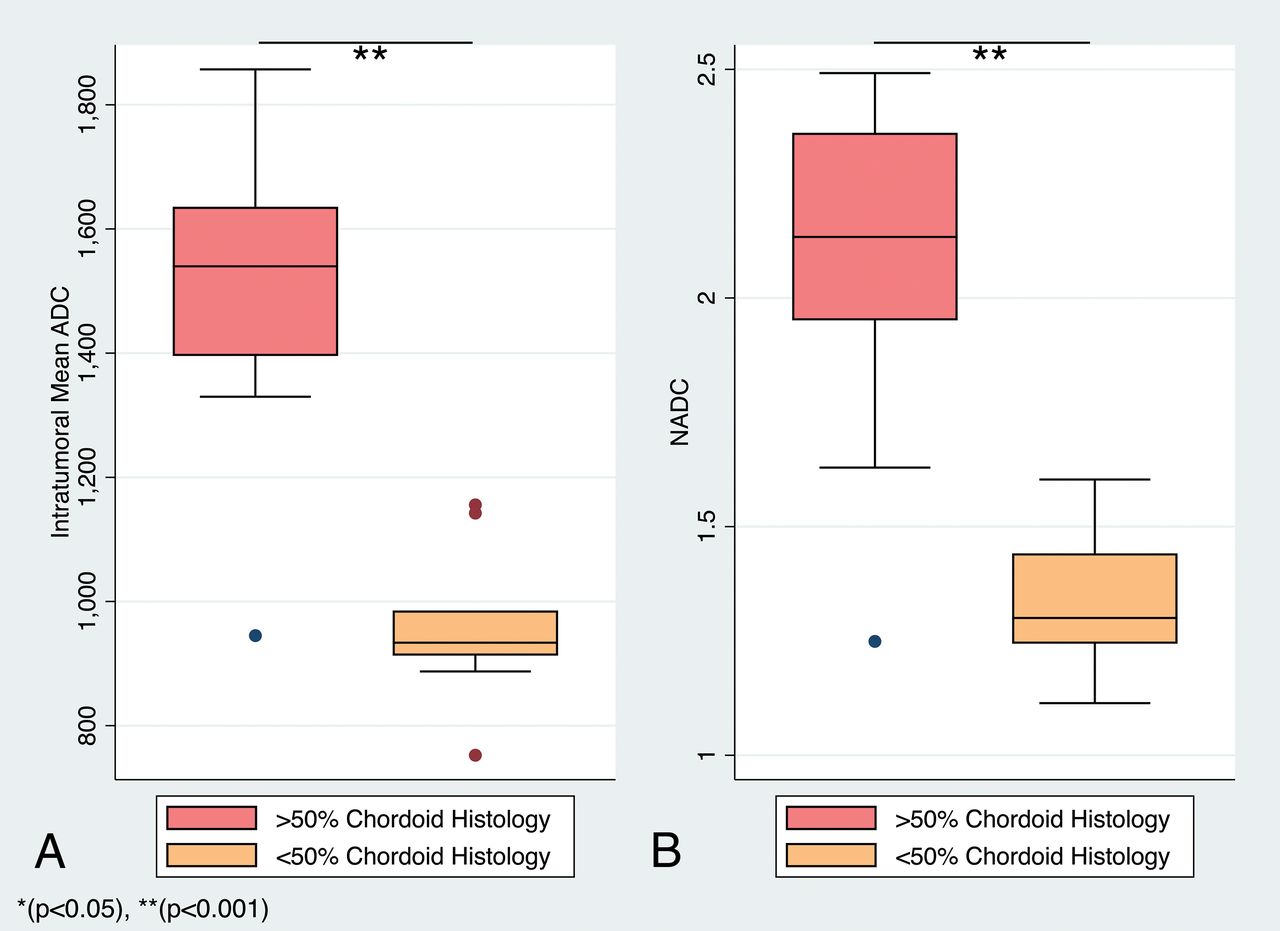
  • Fig 5.
    • Download figure
    • Open in new tab
    • Download powerpoint
    Fig 5.

    Distribution of ADC and nADC values by an intratumoral proportion of chordoid histology. Boxplots of ADC (×10−6mm2/s) (A) and nADC (B) values of chordoid meningiomas (red) and meningiomas with focal chordoid features (orange).

  • Fig 6.
    • Download figure
    • Open in new tab
    • Download powerpoint
    Fig 6.

    ADC and nADC values among chordoid meningioma, meningiomas with focal chordoid histology, and nonchordoid meningiomas. Boxplots of ADC (×10−6mm2/s) (A) and nADC (B) values of chordoid meningiomas (red), meningiomas with focal chordoid features (orange), and nonchordoid meningiomas (green).

Tables

  • Figures
  • Univariate analysis of qualitative MR imaging features for chordoid meningioma

    Predictor VariablesChordoid Meningioma (N = 21)
    OR (95% CI)P Value
    Supratentorial location1.43 (0.43–4.75).555
    Multifocality4.06 (0.87–19.04).075
    T1 hyperintensity1.34 (0.46–3.91).590
    T1+ marked CEa6.49 (0.93 to +inf).061
    T2 hyperintensity2.91 (0.90–9.40).074
    ADC hyperintensity4.29 (1.31–13.98).016b
    Presence of dural taila0.50 (0–19.50).667
    Bony involvement3.10 (0.86–8.06).090
    Cystic/necrotic change2.31 (0.59–9.11).231
    Sunburst vessels1.00 (0.29–3.42)1.000
    Venous involvementa1.57 (0.21–10.40).859
    Arterial narrowing0.48 (0.05–4.54).518
    CSF cleft0.44 (0.14–1.40).165
    Parenchymal edema1.10 (0.38–3.24).856
    Irregular marginsa6.05 (0.88–69.7).072
    • Note:—CE indicates contrast enhancement; inf, infinity.

    • ↵a Exact logistic regression.

    • ↵b Significant.

PreviousNext
Back to top

In this issue

American Journal of Neuroradiology: 40 (3)
American Journal of Neuroradiology
Vol. 40, Issue 3
1 Mar 2019
  • Table of Contents
  • Index by author
  • Complete Issue (PDF)
Advertisement
Print
Download PDF
Email Article

Thank you for your interest in spreading the word on American Journal of Neuroradiology.

NOTE: We only request your email address so that the person you are recommending the page to knows that you wanted them to see it, and that it is not junk mail. We do not capture any email address.

Enter multiple addresses on separate lines or separate them with commas.
Preoperative MR Imaging to Differentiate Chordoid Meningiomas from Other Meningioma Histologic Subtypes
(Your Name) has sent you a message from American Journal of Neuroradiology
(Your Name) thought you would like to see the American Journal of Neuroradiology web site.
CAPTCHA
This question is for testing whether or not you are a human visitor and to prevent automated spam submissions.
Cite this article
J.D. Baal, W.C. Chen, D.A. Solomon, J.S. Pai, C.-H. Lucas, J.H. Hara, N.A. Oberheim Bush, M.W. McDermott, D.R. Raleigh, J.E. Villanueva-Meyer
Preoperative MR Imaging to Differentiate Chordoid Meningiomas from Other Meningioma Histologic Subtypes
American Journal of Neuroradiology Mar 2019, 40 (3) 433-439; DOI: 10.3174/ajnr.A5996

Citation Manager Formats

  • BibTeX
  • Bookends
  • EasyBib
  • EndNote (tagged)
  • EndNote 8 (xml)
  • Medlars
  • Mendeley
  • Papers
  • RefWorks Tagged
  • Ref Manager
  • RIS
  • Zotero
0 Responses
Respond to this article
Share
Bookmark this article
Preoperative MR Imaging to Differentiate Chordoid Meningiomas from Other Meningioma Histologic Subtypes
J.D. Baal, W.C. Chen, D.A. Solomon, J.S. Pai, C.-H. Lucas, J.H. Hara, N.A. Oberheim Bush, M.W. McDermott, D.R. Raleigh, J.E. Villanueva-Meyer
American Journal of Neuroradiology Mar 2019, 40 (3) 433-439; DOI: 10.3174/ajnr.A5996
del.icio.us logo Twitter logo Facebook logo Mendeley logo
  • Tweet Widget
  • Facebook Like
  • Google Plus One
Purchase

Jump to section

  • Article
    • Abstract
    • ABBREVIATIONS:
    • Materials and Methods
    • Results
    • Discussion
    • Conclusions
    • Footnotes
    • References
  • Figures & Data
  • Info & Metrics
  • Responses
  • References
  • PDF

Related Articles

  • No related articles found.
  • PubMed
  • Google Scholar

Cited By...

  • No citing articles found.
  • Crossref (6)
  • Google Scholar

This article has been cited by the following articles in journals that are participating in Crossref Cited-by Linking.

  • Predicting the grade of meningiomas by clinical–radiological features: A comparison of precontrast and postcontrast MRI
    Yuan Yao, Yifan Xu, Shihe Liu, Feng Xue, Bao Wang, Shanshan Qin, Xiubin Sun, Jingzhen He
    Frontiers in Oncology 2022 12
  • A Case of Tuberculum Sellae Chordoid Meningioma Treated via Extended Endoscopic Endonasal Trans-sphenoidal Surgery
    Chihiro Yagi, Shun Yamamuro, Yoshinari Ozawa, Sodai Yoshimura, Koichiro Sumi, Atsuo Yoshino
    NMC Case Report Journal 2020 7 2
  • Utility of multiparametric pre-operative magnetic resonance imaging in differentiation of chordoid meningioma from the other histopathological subtypes of meningioma—a retrospective study
    Sameer Peer, Jitender Saini, Chandrajit Prasad, Karthik Kulanthaivelu, Nishanth Sadashiva, Bevinahalli N Nandeesh, Alok Mohan Uppar, Shilpa Rao
    Neuroradiology 2022 64 2
  • Chordoid Meningioma: The Rarest Subtype of Grade II Meningioma
    Jose Valerio, Noe Santiago Rea, Jorge Zumaeta, Luis Rey Martinez
    Cureus 2024
  • Evaluation of Magnetic Resonance Imaging for Microsurgical Efficacy and Relapse of Rolandic Meningioma
    Peng Cao, Nianhua Wang, Arpit Bhardwaj
    Computational Intelligence and Neuroscience 2022 2022
  • Radiopaedia.org
    Manar osman
    2023

More in this TOC Section

  • Diagnostic Neuroradiology of Monoclonal Antibodies
  • Cerebral ADC Changes in Fabry Disease
  • ML for Glioma Molecular Subtype Prediction
Show more ADULT BRAIN

Similar Articles

Advertisement

Indexed Content

  • Current Issue
  • Accepted Manuscripts
  • Article Preview
  • Past Issues
  • Editorials
  • Editor's Choice
  • Fellows' Journal Club
  • Letters to the Editor
  • Video Articles

Cases

  • Case Collection
  • Archive - Case of the Week
  • Archive - Case of the Month
  • Archive - Classic Case

More from AJNR

  • Trainee Corner
  • Imaging Protocols
  • MRI Safety Corner
  • Book Reviews

Multimedia

  • AJNR Podcasts
  • AJNR Scantastics

Resources

  • Turnaround Time
  • Submit a Manuscript
  • Submit a Video Article
  • Submit an eLetter to the Editor/Response
  • Manuscript Submission Guidelines
  • Statistical Tips
  • Fast Publishing of Accepted Manuscripts
  • Graphical Abstract Preparation
  • Imaging Protocol Submission
  • Evidence-Based Medicine Level Guide
  • Publishing Checklists
  • Author Policies
  • Become a Reviewer/Academy of Reviewers
  • News and Updates

About Us

  • About AJNR
  • Editorial Board
  • Editorial Board Alumni
  • Alerts
  • Permissions
  • Not an AJNR Subscriber? Join Now
  • Advertise with Us
  • Librarian Resources
  • Feedback
  • Terms and Conditions
  • AJNR Editorial Board Alumni

American Society of Neuroradiology

  • Not an ASNR Member? Join Now

© 2025 by the American Society of Neuroradiology All rights, including for text and data mining, AI training, and similar technologies, are reserved.
Print ISSN: 0195-6108 Online ISSN: 1936-959X

Powered by HighWire