Advertisement

Main menu

  • Home
  • Content
    • Current Issue
    • Accepted Manuscripts
    • Article Preview
    • Past Issue Archive
    • Video Articles
    • AJNR Case Collection
    • Case of the Week Archive
    • Case of the Month Archive
    • Classic Case Archive
  • Special Collections
    • AJNR Awards
    • Low-Field MRI
    • Alzheimer Disease
    • ASNR Foundation Special Collection
    • Photon-Counting CT
    • View All
  • Multimedia
    • AJNR Podcasts
    • AJNR SCANtastic
    • Trainee Corner
    • MRI Safety Corner
    • Imaging Protocols
  • For Authors
    • Submit a Manuscript
    • Submit a Video Article
    • Submit an eLetter to the Editor/Response
    • Manuscript Submission Guidelines
    • Statistical Tips
    • Fast Publishing of Accepted Manuscripts
    • Graphical Abstract Preparation
    • Imaging Protocol Submission
    • Author Policies
  • About Us
    • About AJNR
    • Editorial Board
    • Editorial Board Alumni
  • More
    • Become a Reviewer/Academy of Reviewers
    • Subscribers
    • Permissions
    • Alerts
    • Feedback
    • Advertisers
    • ASNR Home

User menu

  • Alerts
  • Log in

Search

  • Advanced search
American Journal of Neuroradiology

American Journal of Neuroradiology

ASHNR American Society of Functional Neuroradiology ASHNR American Society of Pediatric Neuroradiology ASSR
  • Alerts
  • Log in

Advanced Search

  • Home
  • Content
    • Current Issue
    • Accepted Manuscripts
    • Article Preview
    • Past Issue Archive
    • Video Articles
    • AJNR Case Collection
    • Case of the Week Archive
    • Case of the Month Archive
    • Classic Case Archive
  • Special Collections
    • AJNR Awards
    • Low-Field MRI
    • Alzheimer Disease
    • ASNR Foundation Special Collection
    • Photon-Counting CT
    • View All
  • Multimedia
    • AJNR Podcasts
    • AJNR SCANtastic
    • Trainee Corner
    • MRI Safety Corner
    • Imaging Protocols
  • For Authors
    • Submit a Manuscript
    • Submit a Video Article
    • Submit an eLetter to the Editor/Response
    • Manuscript Submission Guidelines
    • Statistical Tips
    • Fast Publishing of Accepted Manuscripts
    • Graphical Abstract Preparation
    • Imaging Protocol Submission
    • Author Policies
  • About Us
    • About AJNR
    • Editorial Board
    • Editorial Board Alumni
  • More
    • Become a Reviewer/Academy of Reviewers
    • Subscribers
    • Permissions
    • Alerts
    • Feedback
    • Advertisers
    • ASNR Home
  • Follow AJNR on Twitter
  • Visit AJNR on Facebook
  • Follow AJNR on Instagram
  • Join AJNR on LinkedIn
  • RSS Feeds

AJNR Awards, New Junior Editors, and more. Read the latest AJNR updates

Research ArticleAdult Brain
Open Access

Utility of Dynamic Susceptibility Contrast Perfusion-Weighted MR Imaging and 11C-Methionine PET/CT for Differentiation of Tumor Recurrence from Radiation Injury in Patients with High-Grade Gliomas

Z. Qiao, X. Zhao, K. Wang, Y. Zhang, D. Fan, T. Yu, H. Shen, Q. Chen and L. Ai
American Journal of Neuroradiology February 2019, 40 (2) 253-259; DOI: https://doi.org/10.3174/ajnr.A5952
Z. Qiao
aFrom the Departments of Nuclear Medicine (Z.Q., X.Z., K.W., Y.Z., D.F., Q.C., L.A.)
  • Find this author on Google Scholar
  • Find this author on PubMed
  • Search for this author on this site
  • ORCID record for Z. Qiao
X. Zhao
aFrom the Departments of Nuclear Medicine (Z.Q., X.Z., K.W., Y.Z., D.F., Q.C., L.A.)
  • Find this author on Google Scholar
  • Find this author on PubMed
  • Search for this author on this site
  • ORCID record for X. Zhao
K. Wang
aFrom the Departments of Nuclear Medicine (Z.Q., X.Z., K.W., Y.Z., D.F., Q.C., L.A.)
  • Find this author on Google Scholar
  • Find this author on PubMed
  • Search for this author on this site
  • ORCID record for K. Wang
Y. Zhang
aFrom the Departments of Nuclear Medicine (Z.Q., X.Z., K.W., Y.Z., D.F., Q.C., L.A.)
  • Find this author on Google Scholar
  • Find this author on PubMed
  • Search for this author on this site
  • ORCID record for Y. Zhang
D. Fan
aFrom the Departments of Nuclear Medicine (Z.Q., X.Z., K.W., Y.Z., D.F., Q.C., L.A.)
  • Find this author on Google Scholar
  • Find this author on PubMed
  • Search for this author on this site
  • ORCID record for D. Fan
T. Yu
cDepartment of Medical Imaging (T.Y.), Cancer Hospital of China Medical University, Shenyang, China
dDepartment of Medical Imaging (T.Y.), Liaoning Cancer Hospital and Institute, Shenyang, China.
  • Find this author on Google Scholar
  • Find this author on PubMed
  • Search for this author on this site
  • ORCID record for T. Yu
H. Shen
bRadiology (H.S.), Beijing Tian Tan Hospital, Capital Medical University, Beijing, China
  • Find this author on Google Scholar
  • Find this author on PubMed
  • Search for this author on this site
  • ORCID record for H. Shen
Q. Chen
aFrom the Departments of Nuclear Medicine (Z.Q., X.Z., K.W., Y.Z., D.F., Q.C., L.A.)
  • Find this author on Google Scholar
  • Find this author on PubMed
  • Search for this author on this site
  • ORCID record for Q. Chen
L. Ai
aFrom the Departments of Nuclear Medicine (Z.Q., X.Z., K.W., Y.Z., D.F., Q.C., L.A.)
  • Find this author on Google Scholar
  • Find this author on PubMed
  • Search for this author on this site
  • ORCID record for L. Ai
  • Article
  • Figures & Data
  • Supplemental
  • Info & Metrics
  • Responses
  • References
  • PDF
Loading

Article Figures & Data

Figures

  • Tables
  • Fig 1.
    • Download figure
    • Open in new tab
    • Download powerpoint
    Fig 1.

    Receiver operating curves for perfusion-weighted MR imaging and MET PET/CT parameters for the differentiation of recurrence and radiation injury in patients with high-grade gliomas.
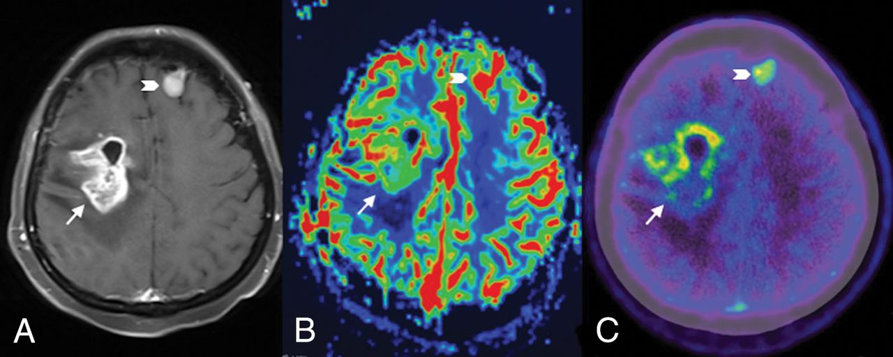
  • Fig 2.
    • Download figure
    • Open in new tab
    • Download powerpoint
    Fig 2.

    Contrast-enhanced T1-weighted MR image (A) and a relative CBV map (B) and an MET PET/CT image (C) for a 41-year-old woman with recurrent glioblastoma. The lesion in the right frontal lobe (arrow) shows enhancement (A) and positive findings on both the rCBV map (B) and MET PET/CT image (C). The lesion localized in the left frontal lobe (chevron) might be a meningioma, which has been stable for several years.
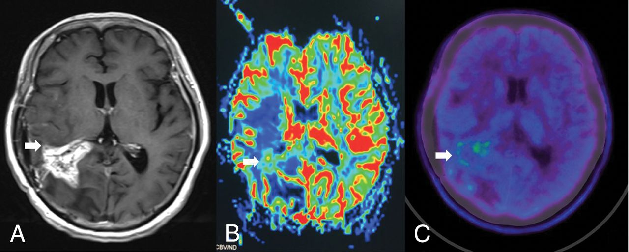
  • Fig 3.
    • Download figure
    • Open in new tab
    • Download powerpoint
    Fig 3.

    Contrast-enhanced T1-weighted MR image (A) and relative CBV map (B) and MET PET/CT image (C) for a 44-year-old man with necrosis. The lesion in the right parietal lobe (arrow) shows enhancement (A) and negative findings on both the rCBV map (B) and the MET PET/CT image (C).

Tables

  • Figures
    • View popup
    Table 1:

    Results of visual assessments of MET PET/CT for patients with HGGs

    Final DiagnosisVisual AssessmentTotal
    PositiveEquivocalNegative
    Recurrence303033
    Radiation injury4329
    Total346242
    • View popup
    Table 2:

    Results of visual assessments of PWI for patients with HGGs

    Final DiagnosisVisual AssessmentTotal
    PositiveEquivocalNegative
    Recurrence228333
    Radiation injury2169
    Total249942
    • View popup
    Table 3:

    Results of semiquantitative analysis of MET PET/CT and PWI parameters for the differentiation of recurrence from radiation injury in patients with HGGs

    Final DiagnosisP Valuea
    RecurrenceRadiation Injury
    SUVmax5.10 ± 2.412.41 ± 1.67.003
    SUVmean2.83 ± 1.271.39 ± 0.9.003
    TBRSUVmax3.48 ± 1.171.96 ± 0.96.003
    rCBVmean2.68 ± 1.141.33 ± 0.77.004
    • ↵a All P values for discrimination between recurrence and radiation injury are significant.

    • View popup
    Table 4:

    Comparison of patients with recurrence with patients with radiation injury, according to cutoff values for TBRSUVmax and rCBVmean

    PETRecurrence PWIRadiation Injury PWI
    +−Total+−Total
    +25732033
    −101156
    Total26733189
    • Note:—+ indicates TBRSUVmax ≥ 1.85 or rCBVmean ≥ 1.83; −, TBRSUVmax < 1.85 or rCBVmean < 1.83.

PreviousNext
Back to top

In this issue

American Journal of Neuroradiology: 40 (2)
American Journal of Neuroradiology
Vol. 40, Issue 2
1 Feb 2019
  • Table of Contents
  • Index by author
  • Complete Issue (PDF)
Advertisement
Print
Download PDF
Email Article

Thank you for your interest in spreading the word on American Journal of Neuroradiology.

NOTE: We only request your email address so that the person you are recommending the page to knows that you wanted them to see it, and that it is not junk mail. We do not capture any email address.

Enter multiple addresses on separate lines or separate them with commas.
Utility of Dynamic Susceptibility Contrast Perfusion-Weighted MR Imaging and 11C-Methionine PET/CT for Differentiation of Tumor Recurrence from Radiation Injury in Patients with High-Grade Gliomas
(Your Name) has sent you a message from American Journal of Neuroradiology
(Your Name) thought you would like to see the American Journal of Neuroradiology web site.
CAPTCHA
This question is for testing whether or not you are a human visitor and to prevent automated spam submissions.
Cite this article
Z. Qiao, X. Zhao, K. Wang, Y. Zhang, D. Fan, T. Yu, H. Shen, Q. Chen, L. Ai
Utility of Dynamic Susceptibility Contrast Perfusion-Weighted MR Imaging and 11C-Methionine PET/CT for Differentiation of Tumor Recurrence from Radiation Injury in Patients with High-Grade Gliomas
American Journal of Neuroradiology Feb 2019, 40 (2) 253-259; DOI: 10.3174/ajnr.A5952

Citation Manager Formats

  • BibTeX
  • Bookends
  • EasyBib
  • EndNote (tagged)
  • EndNote 8 (xml)
  • Medlars
  • Mendeley
  • Papers
  • RefWorks Tagged
  • Ref Manager
  • RIS
  • Zotero
0 Responses
Respond to this article
Share
Bookmark this article
Utility of Dynamic Susceptibility Contrast Perfusion-Weighted MR Imaging and 11C-Methionine PET/CT for Differentiation of Tumor Recurrence from Radiation Injury in Patients with High-Grade Gliomas
Z. Qiao, X. Zhao, K. Wang, Y. Zhang, D. Fan, T. Yu, H. Shen, Q. Chen, L. Ai
American Journal of Neuroradiology Feb 2019, 40 (2) 253-259; DOI: 10.3174/ajnr.A5952
del.icio.us logo Twitter logo Facebook logo Mendeley logo
  • Tweet Widget
  • Facebook Like
  • Google Plus One
Purchase

Jump to section

  • Article
    • Abstract
    • ABBREVIATIONS:
    • Materials and Methods
    • Results
    • Discussion
    • Conclusions
    • Footnotes
    • References
  • Figures & Data
  • Supplemental
  • Info & Metrics
  • Responses
  • References
  • PDF

Related Articles

  • PubMed
  • Google Scholar

Cited By...

  • Diagnostic Value of PET Tracers in Differentiating Glioma Tumor Recurrence from Treatment-Related Changes: A Systematic Review and Meta-Analysis
  • Diagnostic value of PET with different radiotracers and MRI for recurrent glioma: a Bayesian network meta-analysis
  • Positron emission tomography and perfusion weighted imaging in the detection of brain tumors recurrence: A meta-analysis
  • Diagnostic Performance of PET and Perfusion-Weighted Imaging in Differentiating Tumor Recurrence or Progression from Radiation Necrosis in Posttreatment Gliomas: A Review of Literature
  • Crossref (34)
  • Google Scholar

This article has been cited by the following articles in journals that are participating in Crossref Cited-by Linking.

  • Advanced Imaging Techniques for Radiotherapy Planning of Gliomas
    Antonella Castellano, Michele Bailo, Francesco Cicone, Luciano Carideo, Natale Quartuccio, Pietro Mortini, Andrea Falini, Giuseppe Lucio Cascini, Giuseppe Minniti
    Cancers 2021 13 5
  • Brain tumor segmentation using 3D Mask R-CNN for dynamic susceptibility contrast enhanced perfusion imaging
    Jiwoong Jeong, Yang Lei, Shannon Kahn, Tian Liu, Walter J Curran, Hui-Kuo Shu, Hui Mao, Xiaofeng Yang
    Physics in Medicine & Biology 2020 65 18
  • Diagnostic Accuracy of PET for Differentiating True Glioma Progression From Post Treatment-Related Changes: A Systematic Review and Meta-Analysis
    Meng Cui, Rocío Isabel Zorrilla-Veloz, Jian Hu, Bing Guan, Xiaodong Ma
    Frontiers in Neurology 2021 12
  • Evaluation of perfusion MRI value for tumor progression assessment after glioma radiotherapy
    Longlong Wang, Lizhou Wei, Jingjian Wang, Na Li, Yanzhong Gao, Hongge Ma, Xinran Qu, Ming Zhang
    Medicine 2020 99 52
  • Clinical Imaging for Diagnostic Challenges in the Management of Gliomas: A Review
    Alipi V. Bonm, Reed Ritterbusch, Patrick Throckmorton, Jerome J. Graber
    Journal of Neuroimaging 2020 30 2
  • The Molecular Effects of Ionizing Radiations on Brain Cells: Radiation Necrosis vs. Tumor Recurrence
    Vincenzo Cuccurullo, Giuseppe Danilo Di Stasio, Giuseppe Lucio Cascini, Gianluca Gatta, Cataldo Bianco
    Diagnostics 2019 9 4
  • The Utility of Conventional Amino Acid PET Radiotracers in the Evaluation of Glioma Recurrence also in Comparison with MRI
    Giulia Santo, Riccardo Laudicella, Flavia Linguanti, Anna Giulia Nappi, Elisabetta Abenavoli, Vittoria Vergura, Giuseppe Rubini, Roberto Sciagrà, Gaspare Arnone, Orazio Schillaci, Fabio Minutoli, Sergio Baldari, Natale Quartuccio, Sotirios Bisdas
    Diagnostics 2022 12 4
  • Comparison of Amino Acid PET to Advanced and Emerging MRI Techniques for Neurooncology Imaging: A Systematic Review of the Recent Studies
    Brittany M. Stopa, Csaba Juhász, Sandeep Mittal
    Molecular Imaging 2021 2021
  • Research progress on mechanism and imaging of temporal lobe injury induced by radiotherapy for head and neck cancer
    Zhuangzhuang Zheng, Bin Wang, Qin Zhao, Yuyu Zhang, Jinlong Wei, Lingbin Meng, Ying Xin, Xin Jiang
    European Radiology 2022 32 1
  • Combined amino acid PET-MRI for identifying recurrence in post-treatment gliomas: together we grow
    Shumyla Jabeen, Arpana Arbind, Dinesh Kumar, Pardeep Kumar Singh, Jitender Saini, Nishanth Sadashiva, Uday Krishna, Arivazhagan Arimappamagan, Vani Santosh, Chandana Nagaraj
    European Journal of Hybrid Imaging 2021 5 1

More in this TOC Section

Adult Brain

  • Diagnostic Neuroradiology of Monoclonal Antibodies
  • Clinical Outcomes After Chiari I Decompression
  • Segmentation of Brain Metastases with BLAST
Show more Adult Brain

Functional

  • Kurtosis and Epileptogenic Tubers: A Pilot Study
  • Glutaric Aciduria Type 1: DK vs. Conventional MRI
  • Multiparametric MRI in PEDS Pontine Glioma
Show more Functional

Similar Articles

Advertisement

Indexed Content

  • Current Issue
  • Accepted Manuscripts
  • Article Preview
  • Past Issues
  • Editorials
  • Editor's Choice
  • Fellows' Journal Club
  • Letters to the Editor
  • Video Articles

Cases

  • Case Collection
  • Archive - Case of the Week
  • Archive - Case of the Month
  • Archive - Classic Case

More from AJNR

  • Trainee Corner
  • Imaging Protocols
  • MRI Safety Corner
  • Book Reviews

Multimedia

  • AJNR Podcasts
  • AJNR Scantastics

Resources

  • Turnaround Time
  • Submit a Manuscript
  • Submit a Video Article
  • Submit an eLetter to the Editor/Response
  • Manuscript Submission Guidelines
  • Statistical Tips
  • Fast Publishing of Accepted Manuscripts
  • Graphical Abstract Preparation
  • Imaging Protocol Submission
  • Evidence-Based Medicine Level Guide
  • Publishing Checklists
  • Author Policies
  • Become a Reviewer/Academy of Reviewers
  • News and Updates

About Us

  • About AJNR
  • Editorial Board
  • Editorial Board Alumni
  • Alerts
  • Permissions
  • Not an AJNR Subscriber? Join Now
  • Advertise with Us
  • Librarian Resources
  • Feedback
  • Terms and Conditions
  • AJNR Editorial Board Alumni

American Society of Neuroradiology

  • Not an ASNR Member? Join Now

© 2025 by the American Society of Neuroradiology All rights, including for text and data mining, AI training, and similar technologies, are reserved.
Print ISSN: 0195-6108 Online ISSN: 1936-959X

Powered by HighWire