Advertisement

Main menu

  • Home
  • Content
    • Current Issue
    • Accepted Manuscripts
    • Article Preview
    • Past Issue Archive
    • Video Articles
    • AJNR Case Collection
    • Case of the Week Archive
    • Case of the Month Archive
    • Classic Case Archive
  • Special Collections
    • Advancing NeuroMRI with High-Relaxivity Contrast Agents
    • AJNR Awards
    • Low-Field MRI
    • Alzheimer Disease
    • ASNR Foundation Special Collection
    • Photon-Counting CT
    • View All
  • Multimedia
    • AJNR Podcasts
    • AJNR SCANtastic
    • Trainee Corner
    • MRI Safety Corner
    • Imaging Protocols
  • For Authors
    • Submit a Manuscript
    • Submit a Video Article
    • Submit an eLetter to the Editor/Response
    • Manuscript Submission Guidelines
    • Statistical Tips
    • Fast Publishing of Accepted Manuscripts
    • Graphical Abstract Preparation
    • Imaging Protocol Submission
    • Author Policies
  • About Us
    • About AJNR
    • Editorial Board
    • Editorial Board Alumni
  • More
    • Become a Reviewer/Academy of Reviewers
    • Subscribers
    • Permissions
    • Alerts
    • Feedback
    • Advertisers
    • ASNR Home

User menu

  • Alerts
  • Log in

Search

  • Advanced search
American Journal of Neuroradiology

American Journal of Neuroradiology

ASHNR American Society of Functional Neuroradiology ASHNR American Society of Pediatric Neuroradiology ASSR
  • Alerts
  • Log in

Advanced Search

  • Home
  • Content
    • Current Issue
    • Accepted Manuscripts
    • Article Preview
    • Past Issue Archive
    • Video Articles
    • AJNR Case Collection
    • Case of the Week Archive
    • Case of the Month Archive
    • Classic Case Archive
  • Special Collections
    • Advancing NeuroMRI with High-Relaxivity Contrast Agents
    • AJNR Awards
    • Low-Field MRI
    • Alzheimer Disease
    • ASNR Foundation Special Collection
    • Photon-Counting CT
    • View All
  • Multimedia
    • AJNR Podcasts
    • AJNR SCANtastic
    • Trainee Corner
    • MRI Safety Corner
    • Imaging Protocols
  • For Authors
    • Submit a Manuscript
    • Submit a Video Article
    • Submit an eLetter to the Editor/Response
    • Manuscript Submission Guidelines
    • Statistical Tips
    • Fast Publishing of Accepted Manuscripts
    • Graphical Abstract Preparation
    • Imaging Protocol Submission
    • Author Policies
  • About Us
    • About AJNR
    • Editorial Board
    • Editorial Board Alumni
  • More
    • Become a Reviewer/Academy of Reviewers
    • Subscribers
    • Permissions
    • Alerts
    • Feedback
    • Advertisers
    • ASNR Home
  • Follow AJNR on Twitter
  • Visit AJNR on Facebook
  • Follow AJNR on Instagram
  • Join AJNR on LinkedIn
  • RSS Feeds

AJNR Awards, New Junior Editors, and more. Read the latest AJNR updates


Improved Turnaround Times | Median time to first decision: 12 days

Research ArticleHead and Neck Imaging
Open Access

MR Imaging of the Extracranial Facial Nerve with the CISS Sequence

J.P. Guenette, N. Ben-Shlomo, J. Jayender, R.T. Seethamraju, V. Kimbrell, N.-A. Tran, R.Y. Huang, C.J. Kim, J.I. Kass, C.E. Corrales and T.C. Lee
American Journal of Neuroradiology November 2019, 40 (11) 1954-1959; DOI: https://doi.org/10.3174/ajnr.A6261
J.P. Guenette
aFrom the Divisions of Neuroradiology (J.P.G., J.J., V.K., N.-A.T., R.Y.H., C.J.K., T.C.L.)
  • Find this author on Google Scholar
  • Find this author on PubMed
  • Search for this author on this site
  • ORCID record for J.P. Guenette
N. Ben-Shlomo
bOtolaryngology—Head and Neck Surgery (N.B.-S., J.I.K., C.E.C), Brigham & Women's Hospital, Harvard Medical School, Boston, Massachusetts
  • Find this author on Google Scholar
  • Find this author on PubMed
  • Search for this author on this site
  • ORCID record for N. Ben-Shlomo
J. Jayender
aFrom the Divisions of Neuroradiology (J.P.G., J.J., V.K., N.-A.T., R.Y.H., C.J.K., T.C.L.)
  • Find this author on Google Scholar
  • Find this author on PubMed
  • Search for this author on this site
  • ORCID record for J. Jayender
R.T. Seethamraju
cSiemens Medical Solutions USA (R.T.S.), Boston, Massachusetts
  • Find this author on Google Scholar
  • Find this author on PubMed
  • Search for this author on this site
  • ORCID record for R.T. Seethamraju
V. Kimbrell
aFrom the Divisions of Neuroradiology (J.P.G., J.J., V.K., N.-A.T., R.Y.H., C.J.K., T.C.L.)
  • Find this author on Google Scholar
  • Find this author on PubMed
  • Search for this author on this site
  • ORCID record for V. Kimbrell
N.-A. Tran
aFrom the Divisions of Neuroradiology (J.P.G., J.J., V.K., N.-A.T., R.Y.H., C.J.K., T.C.L.)
  • Find this author on Google Scholar
  • Find this author on PubMed
  • Search for this author on this site
  • ORCID record for N.-A. Tran
R.Y. Huang
aFrom the Divisions of Neuroradiology (J.P.G., J.J., V.K., N.-A.T., R.Y.H., C.J.K., T.C.L.)
  • Find this author on Google Scholar
  • Find this author on PubMed
  • Search for this author on this site
  • ORCID record for R.Y. Huang
C.J. Kim
aFrom the Divisions of Neuroradiology (J.P.G., J.J., V.K., N.-A.T., R.Y.H., C.J.K., T.C.L.)
  • Find this author on Google Scholar
  • Find this author on PubMed
  • Search for this author on this site
  • ORCID record for C.J. Kim
J.I. Kass
bOtolaryngology—Head and Neck Surgery (N.B.-S., J.I.K., C.E.C), Brigham & Women's Hospital, Harvard Medical School, Boston, Massachusetts
  • Find this author on Google Scholar
  • Find this author on PubMed
  • Search for this author on this site
  • ORCID record for J.I. Kass
C.E. Corrales
bOtolaryngology—Head and Neck Surgery (N.B.-S., J.I.K., C.E.C), Brigham & Women's Hospital, Harvard Medical School, Boston, Massachusetts
  • Find this author on Google Scholar
  • Find this author on PubMed
  • Search for this author on this site
  • ORCID record for C.E. Corrales
T.C. Lee
aFrom the Divisions of Neuroradiology (J.P.G., J.J., V.K., N.-A.T., R.Y.H., C.J.K., T.C.L.)
  • Find this author on Google Scholar
  • Find this author on PubMed
  • Search for this author on this site
  • ORCID record for T.C. Lee
  • Article
  • Figures & Data
  • Supplemental
  • Info & Metrics
  • Responses
  • References
  • PDF
Loading

Article Figures & Data

Figures

  • Tables
  • Fig 1.
    • Download figure
    • Open in new tab
    • Download powerpoint
    Fig 1.

    Sagittal midline localizer MR image. Horizontal white lines through the basisphenoid and cranial aspect of the extrinsic tongue muscles serve as an accurate approximation for the CISS craniocaudal slab selection when imaging the facial nerve.
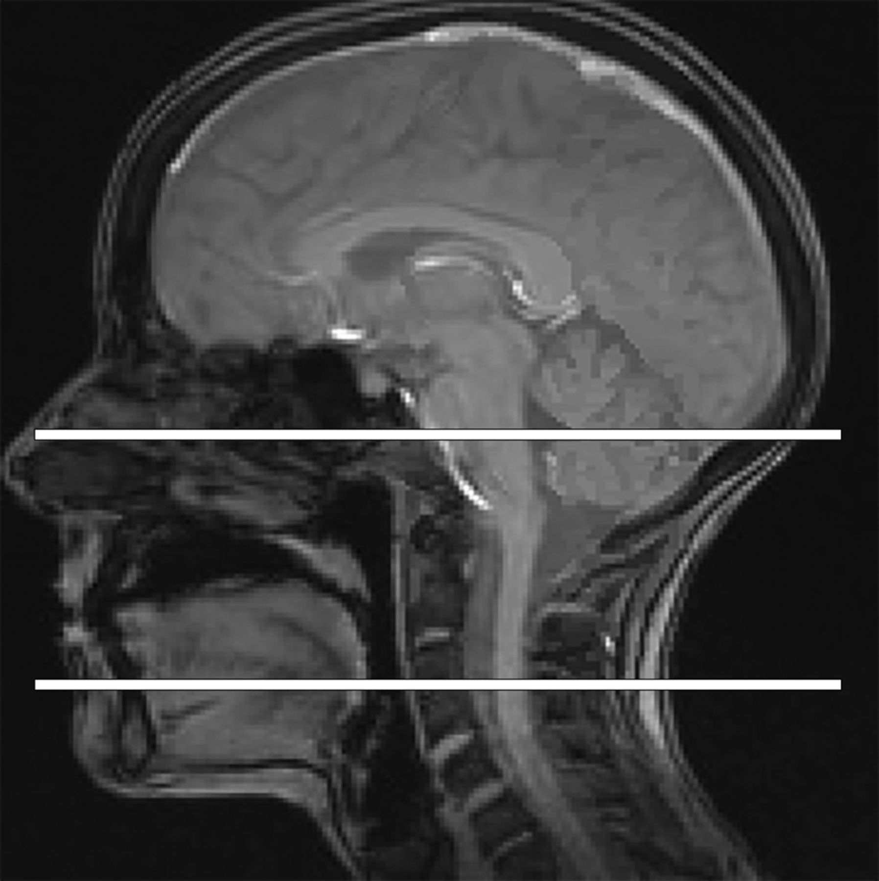
  • Fig 2.
    • Download figure
    • Open in new tab
    • Download powerpoint
    Fig 2.

    Representative sagittal-oblique CISS minimum intensity projection MR image shows the visible course of the facial nerve trunk (curved arrow) from the stylomastoid foramen to the distal aspects of the temporofacial (arrowhead) and cervicofacial (arrow) trunks.
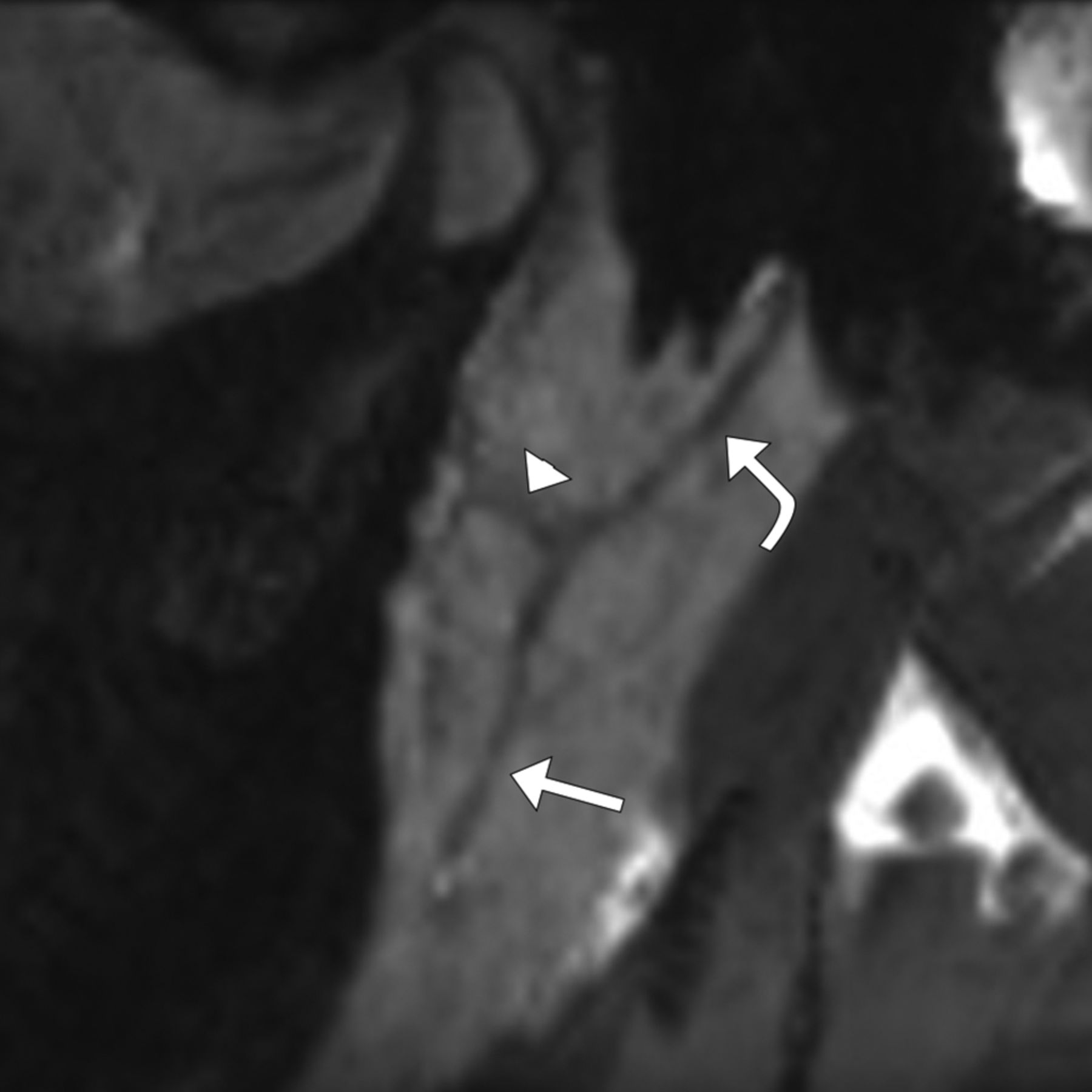
  • Fig 3.
    • Download figure
    • Open in new tab
    • Download powerpoint
    Fig 3.

    Representative segmentations of the left facial nerve of subject 20 with an average Hausdorff distance of 0.64, Dice coefficient of 0.60, length difference of <1%, and segmentation volume difference of 8%. A, Segmentations of observer 1 (green outline) and observer 4 (red outline) superimposed on the CISS image show similar agreement along the main nerve root, bifurcation, and proximal aspects of the temporofacial and cervicofacial roots, with more distal variability along the cervicofacial root, even despite the similar length segmented. B, 3D rendering of the observer 1 segmentation. C, 3D rendering of the observer 4 segmentation. D, Dice overlap map shows the spectrum of agreement, with blue being good agreement and red being poor agreement.
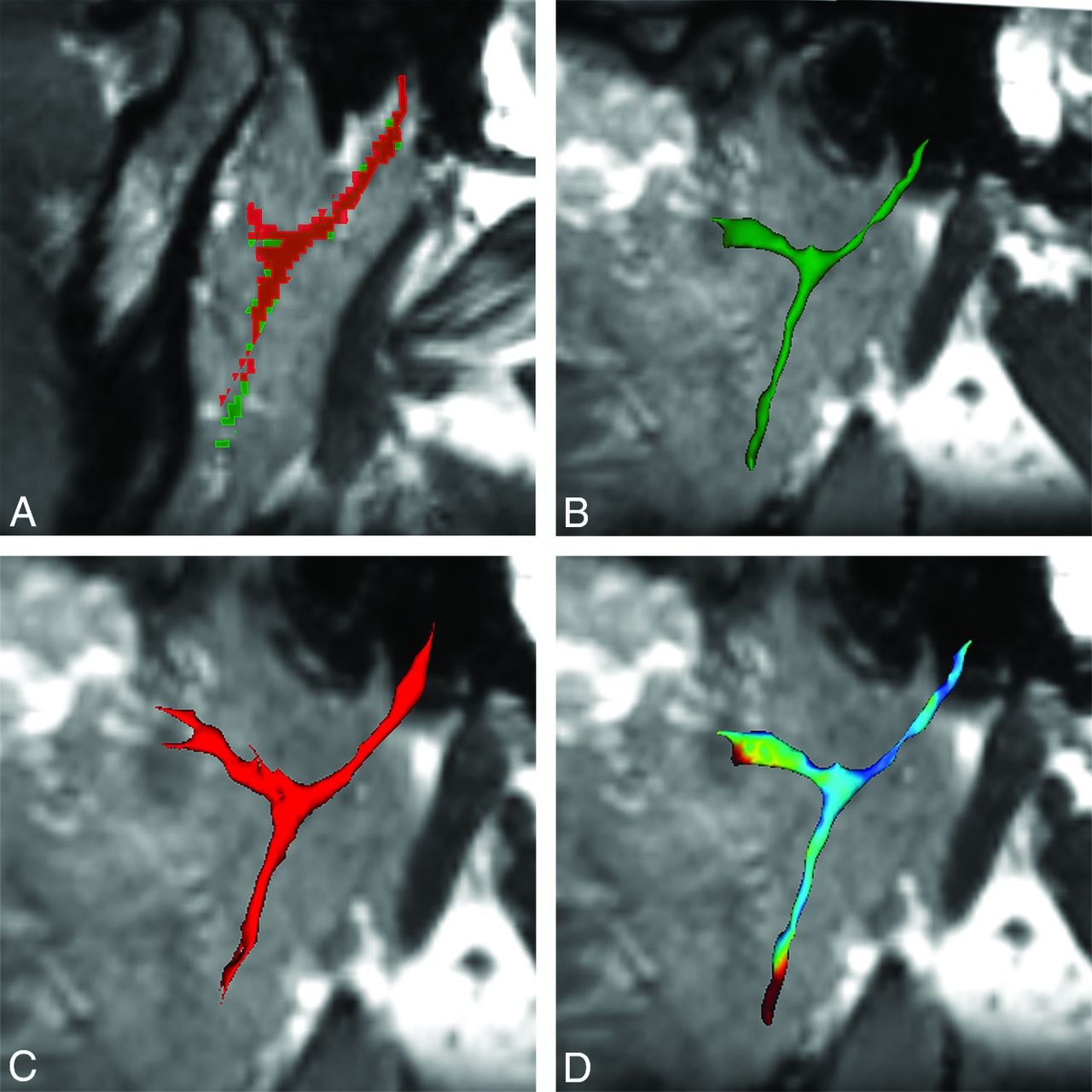
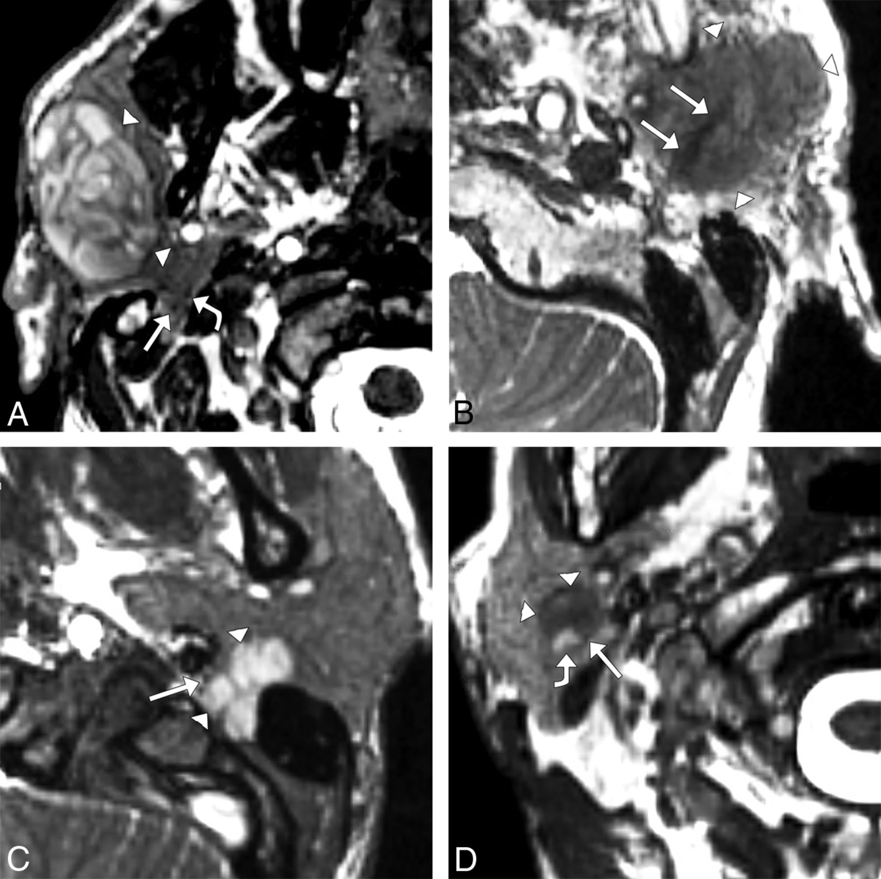
  • Fig 4.
    • Download figure
    • Open in new tab
    • Download powerpoint
    Fig 4.

    Axial CISS images in patients with parotid gland tumors. A, A 55-year-old man with a right parotid gland, 35 × 22 × 44 mm (anteroposterior × transverse × craniocaudal) Warthin tumor (arrowheads) and temporofacial (straight arrow) and superiorly displaced cervicofacial (curved arrow) trunks of the facial nerve (straight arrow), apparently deep to the tumor and just posterior to the retromandibular vein. In the operating room, the distal branches of the facial nerve were confirmed to be in the plane of the tumor with distal divisions displaced above and below the tumor and with superior displacement of the parotid plexus. B, An 83-year-old woman with left parotid gland, 23 × 21 × 29 mm poorly differentiated carcinoma with sarcomatoid features (arrowheads) and an apparently expanded facial nerve with irregular margins (arrows) coursing through the tumor, suggestive of perineural tumor invasion. A radical parotidectomy was performed, and pathology analysis confirmed extensive perineural invasion. C, A 32-year-old woman with a left parotid gland 18 × 19 × 24 mm pleomorphic adenoma (arrowheads) extending into the stylomastoid foramen and anteromedially displacing the facial nerve (arrow). Due to these imaging findings, a postauricular infratemporal fossa surgical approach was used, confirming the location of the nerve and confirming impingement of the nerve as it entered the stylomastoid foramen. D, A 75-year-old man with a right parotid gland, 12 × 11 × 19 mm Warthin tumor (arrowheads) just superficial to and between the distal, small-caliber, low-signal cervicofacial trunk of the facial nerve (straight arrow) and high-signal retromandibular vein (curved arrow). This patient has elected observation, and there is thus no surgical confirmation of facial nerve location.

Tables

  • Figures
  • Parameters for CISS sequence

    Parameter
    TR4.97 ms
    TE2.19 ms
    Averages1
    Section thickness0.8 mm
    FOV240 × 240
    Matrix320 × 320
    Voxel size0.8 × 0.8 × 0.8 mm
    Bandwidth521 Hz/pixel
    Flip angle33°
    Elliptic scanningOn
    Acquisition time4:44 minutes
PreviousNext
Back to top

In this issue

American Journal of Neuroradiology: 40 (11)
American Journal of Neuroradiology
Vol. 40, Issue 11
1 Nov 2019
  • Table of Contents
  • Index by author
  • Complete Issue (PDF)
Advertisement
Print
Download PDF
Email Article

Thank you for your interest in spreading the word on American Journal of Neuroradiology.

NOTE: We only request your email address so that the person you are recommending the page to knows that you wanted them to see it, and that it is not junk mail. We do not capture any email address.

Enter multiple addresses on separate lines or separate them with commas.
MR Imaging of the Extracranial Facial Nerve with the CISS Sequence
(Your Name) has sent you a message from American Journal of Neuroradiology
(Your Name) thought you would like to see the American Journal of Neuroradiology web site.
CAPTCHA
This question is for testing whether or not you are a human visitor and to prevent automated spam submissions.
Cite this article
J.P. Guenette, N. Ben-Shlomo, J. Jayender, R.T. Seethamraju, V. Kimbrell, N.-A. Tran, R.Y. Huang, C.J. Kim, J.I. Kass, C.E. Corrales, T.C. Lee
MR Imaging of the Extracranial Facial Nerve with the CISS Sequence
American Journal of Neuroradiology Nov 2019, 40 (11) 1954-1959; DOI: 10.3174/ajnr.A6261

Citation Manager Formats

  • BibTeX
  • Bookends
  • EasyBib
  • EndNote (tagged)
  • EndNote 8 (xml)
  • Medlars
  • Mendeley
  • Papers
  • RefWorks Tagged
  • Ref Manager
  • RIS
  • Zotero
0 Responses
Respond to this article
Share
Bookmark this article
MR Imaging of the Extracranial Facial Nerve with the CISS Sequence
J.P. Guenette, N. Ben-Shlomo, J. Jayender, R.T. Seethamraju, V. Kimbrell, N.-A. Tran, R.Y. Huang, C.J. Kim, J.I. Kass, C.E. Corrales, T.C. Lee
American Journal of Neuroradiology Nov 2019, 40 (11) 1954-1959; DOI: 10.3174/ajnr.A6261
del.icio.us logo Twitter logo Facebook logo Mendeley logo
  • Tweet Widget
  • Facebook Like
  • Google Plus One
Purchase

Jump to section

  • Article
    • Abstract
    • MATERIALS AND METHODS
    • RESULTS
    • DISCUSSION
    • CONCLUSIONS
    • Footnotes
    • References
  • Figures & Data
  • Supplemental
  • Info & Metrics
  • Responses
  • References
  • PDF

Related Articles

  • PubMed
  • Google Scholar

Cited By...

  • Comparing the Double-Echo Steady-State with Water Excitation and Constructive Interference in Steady-State Sequence Techniques for Identifying Extracranial Facial Nerve and Tumor Positions in Patients with Parotid Tumors
  • Visualization of the Extracranial Branches of the Trigeminal Nerve Using Improved Motion-Sensitized Driven Equilibrium-Prepared 3D Inversion Recovery TSE Sequence
  • Time-Saving 3D MR Imaging Protocols with Millimeter and Submillimeter Isotropic Spatial Resolution for Face and Neck Imaging as Implemented at a Single-Site Major Referral Center
  • Crossref
  • Google Scholar

This article has not yet been cited by articles in journals that are participating in Crossref Cited-by Linking.

More in this TOC Section

  • Diagnostic Performance of US in Neck Node NIRADS 2
  • Hydrops Herniation into the Semicircular Canals
  • DIVr of NPC Predicted Outcome
Show more HEAD AND NECK IMAGING

Similar Articles

Advertisement

Indexed Content

  • Current Issue
  • Accepted Manuscripts
  • Article Preview
  • Past Issues
  • Editorials
  • Editor's Choice
  • Fellows' Journal Club
  • Letters to the Editor
  • Video Articles

Cases

  • Case Collection
  • Archive - Case of the Week
  • Archive - Case of the Month
  • Archive - Classic Case

More from AJNR

  • Trainee Corner
  • Imaging Protocols
  • MRI Safety Corner
  • Book Reviews

Multimedia

  • AJNR Podcasts
  • AJNR Scantastics

Resources

  • Turnaround Time
  • Submit a Manuscript
  • Submit a Video Article
  • Submit an eLetter to the Editor/Response
  • Manuscript Submission Guidelines
  • Statistical Tips
  • Fast Publishing of Accepted Manuscripts
  • Graphical Abstract Preparation
  • Imaging Protocol Submission
  • Evidence-Based Medicine Level Guide
  • Publishing Checklists
  • Author Policies
  • Become a Reviewer/Academy of Reviewers
  • News and Updates

About Us

  • About AJNR
  • Editorial Board
  • Editorial Board Alumni
  • Alerts
  • Permissions
  • Not an AJNR Subscriber? Join Now
  • Advertise with Us
  • Librarian Resources
  • Feedback
  • Terms and Conditions
  • AJNR Editorial Board Alumni

American Society of Neuroradiology

  • Not an ASNR Member? Join Now

© 2025 by the American Society of Neuroradiology All rights, including for text and data mining, AI training, and similar technologies, are reserved.
Print ISSN: 0195-6108 Online ISSN: 1936-959X

Powered by HighWire