
Article Figures & Data
Figures
- Fig 1.
Study schema.
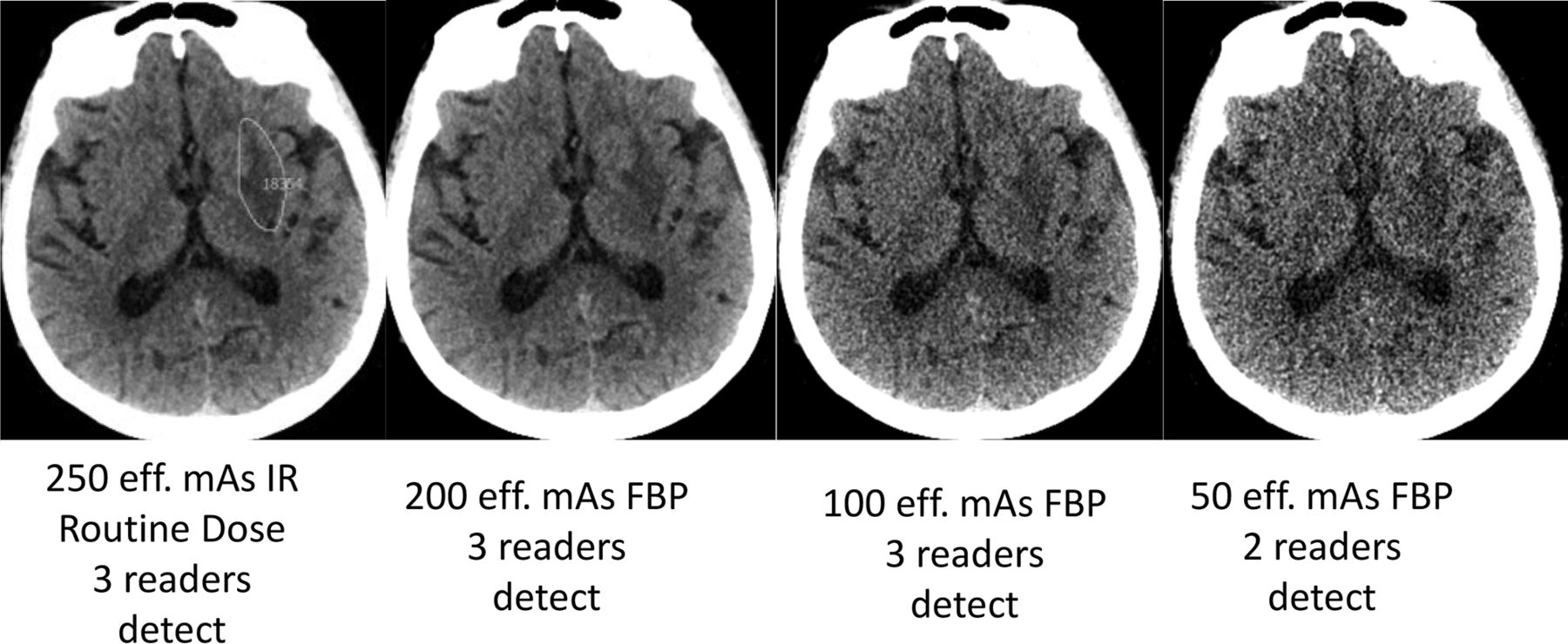
- Fig 2.
Small right thalamic hemorrhage (white arrow) shown on routine-dose CT image (250-eff. mAs IR) along with lower-dose configurations. The small left inset shows reference neuroradiologist markings of the target lesion (green circle). This CT examination was performed after trauma, with hemorrhage confirmed surgically, and the final diagnosis was recorded as right thalamic hemorrhage consistent with shear injury.
- Fig 3.
Acute left lentiform nucleus infarct (green circle indicates reference neuroradiologist markings at routine dose) with corresponding lower-dose FBP CT images along with reader results. The imaging finding on this CT examination evolved with time, with corresponding clinical confirmation of corresponding neurologic deficit by a staff neurologist, and the final diagnosis was recorded as acute left striatal infarct.
- Fig 4.
Noninferiority analysis showing the difference between JAFROC FOM at a routine dose and the lower-dose configurations for CT findings causing acute neurologic deficit. The limit of noninferiority was established a priori to be −0.10, meaning that if the lower limit of the 95% confidence interval is greater than −0.10, then noninferiority was shown.
- Fig 5.
Image-quality metrics for routine and lower-dose configurations in this study. Optimal ratings were 5 for image quality and 1 for individual image metrics (i.e. image sharpness, noise, and texture).


Tables
- Table 1:
Dose levels and reconstruction kernels for unenhanced CT examinations used in this study
Tube Current Setting CTDIvol (mGy) Reconstruction Kernel (Type, Strength) 250 eff. mAs 38.1 J40 (IR 2) 200 eff. mAs 30.5 H40 (FBP) 100 eff. mAs 15.2 J40 (IR 2) 100 eff. mAs 15.2 H40 (FBP) 50 eff. mAs 7.6 J40 (IR 2) 50 eff. mAs 7.6 H40 (FBP) 25 eff. mAs 3.8 J40 (IR 2) - Table 2:
Reference documentation and conspicuity of proved lesions in positive CT examinations with imaging findings corresponding to cause of acute neurologic deficit (n = 42)
Target Diagnosis No. of Imaging Findings with Target Diagnosis No. with Reference Criterion (Nonexclusive List) Ranking of Conspicuity Scoresa (Mean) (SD) Infarct 29 Clinical confirmation of corresponding deficit = 29 2.10 (0.76) Progression/confirmation on another imaging study = 23 Confirmation at surgery = 0 Mass 25 Clinical confirmation of corresponding deficit = 22
Progression/confirmation on another imaging study = 21
Confirmation at surgery = 22.80 (0.70) Extra-axial hemorrhage 10 Clinical confirmation of corresponding deficit = 10
Progression/confirmation on another imaging study = 3
Confirmation at surgery = 02.60 (0.66) Intra-axial hemorrhage 6 Clinical confirmation of corresponding deficit = 6
Progression/confirmation on another imaging study = 2
Confirmation at surgery = 02.67 (0.47) ↵a Please see Materials and Methods. In brief, conspicuity scores: 1, minimally evident; 2, subtle; 3, distinct focal abnormality; 4, obvious.
- Table 3:
Reader agreement of lower-dose reconstruction configurations compared with routine-dose unenhanced head CT examinations, along with JAFROC FOMsa
Lower-Dose–Reconstruction Configuration % of the 47 Essential Lesionsb Detected by Readers at Lower-Dose Configurations No. of Successful Interpretations per Lower- Dose–Reconstruction Configuration JAFROC FOM (95% CI) 2 of 3 3 of 3 Cases with at Least 1 Essential Lesion (n = 34) Cases without Any Essential Lesions (n = 49) No. Successful Interpretations (≥71 Required per Design) 200 eff. mAs FBP 44 (94%) 39 (83%) 30 48 78 0.846 (0.78–0.912) 100 eff. mAs IR 43 (92%) 37 (79%) 29 46 75 0.831 (0.764–0.898) 100 eff. mAs FBP 42 (89%) 36 (77%) 28 45 73 0.805 (0.732–0.878) 50 eff. mAs IR 41 (87%) 32 (68%) 26 47 73 0.795 (0.727–0.864) 50 eff. mAs FBP 38 (81%) 31 (66%) 25 47 72 0.789 (0.717–0.861) 25 eff. mAs IR 34 (72%) 25 (53%) 22 45 67c 0.754 (0.681–0.827) ↵a The JAFROC FOM for routine unenhanced head CT (250 eff. mAs with IR) was 0.867 (0.805–0.929).
↵b Essential lesions are described in the Materials and Methods. Briefly, they represent lesions correctly localized and classified at the routine dose (250 eff. mAs with IR) by majority of readers.
↵c Dose-reconstruction configuration did not meet preset criteria for agreement with routine-dose interpretation, which was defined as agreement in 71 of the 83 examinations.
- Table 4:
Per-patient and per-lesion sensitivity and specificity using GEEs for target neurologic findings accounting for acute neurologic deficits
Dose-Kernel Configuration Per-Patient Sensitivity for CT Findings Accounting for Acute Neurologic Deficits (GEE) (%) (95% CI) (Range) (%) Per-Patient Specificity for CT Findings Accounting for Acute Neurologic Deficits (GEE) (%) (95% CI) (Range) (%) Target Lesion Sensitivity for CT Findings Accounting for Acute Neurologic Deficits (GEE) (%) (95% CI) (Range) (%) 250-eff. mAs IR 81.7 (71.1–92.3) (78.6–83.3) 93.5 (88.9–98.1) (85.4–100.0) 200-eff. mAs FBP 79.4 (68.2–90.6) (76.2–83.3) 91.9 (87.5–96.3) (80.5–100.0) 68.6 (62.3–74.9) (61.4–72.9) 100-eff. mAs IR 77.0 (65.5–88.5) (73.8–81.0) 88.6 (82.8–94.4) (73.2–95.1) 68.1 (61.8–74.4) (64.3–71.4) 100-eff. mAs FBP 74.6 (62.4–86.8) (69.0–78.6) 87.0 (81.1–92.9) (75.6–95.1) 62.9 (56.3–69.4) (57.1–65.7) 50-eff. mAs IR 73.8 (62.3–85.4) (66.7–78.6) 88.6 (82.8–94.4) (75.6–97.6) 60.5 (53.9–67.1) (52.9–65.7) 50-eff. mAs FBP 72.2 (60.6–83.8) (71.4–73.8) 83.7 (77.7–89.8) (69.8–86.0) 61.0 (54.4–67.6) (60.0–62.9) 25-eff. mAs IR 65.9 (53.3–78.4) (61.9–71.4) 88.6 (82.4–94.8) (85.4–92.7) 53.3 (46.6–60.1 (45.7–62.9)a ↵a The 95% confidence interval does not overlap the routine dose, so the dose-reconstruction configuration is significantly worse.


Jump to section
Related Articles
Cited By...
- No citing articles found.