
Article Figures & Data
Figures
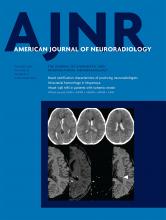
- Fig 1.
A 47-year-old female patient who presented with right-sided disabling pulsatile tinnitus disappearing on compression of the right jugular vein. MR imaging with an axial CISS sequence (A) shows enlargement and CSF infiltration of the sella turcica. Sagittal T1-weighted MR imaging with gadolinium shows flattening of the pituitary gland (B). Ratios of pituitary gland height/sellar height were calculated on sagittal reconstruction of a 3D gadolinium-enhanced T1 MRI sequence using the cut showing the thickest section of pituitary gland (B). Sellar volumes were estimated on the basis of dimensions measured on a preoperative CTA. The laterolateral dimension of the sella was measured on the coronal MPR of the preoperative CTA as shown in C using the longest measurement between the medial walls of the cavernous segments of the internal carotid arteries (white arrow). The sellar height and anterior-posterior diameter were measured on the 3D-MPR in the midsagittal plane as shown in D. Height was the longest measurement intersecting a line joining the tuberculum and dorsum sellae and the lowest point in the sella. The anterior-posterior measurement was defined as the longest measure on the midsagittal plane (white arrows).
- Fig 2.
Study flow chart.
- Fig 3.
Estimated sellar volumes (cubic millimeters) based on preoperative CTA measurements for patients with isolated venous pulsatile tinnitus and idiopathic intracranial hypertension.
- Fig 4.
Trans-stenotic gradients (millimeters of mercury) based on local anesthesia venous manometry measurements for patients with isolated venous pulsatile tinnitus and IIH.


Tables
Patients' baseline characteristics
IPT IIH Control P Value No. 34 54 39 Age (median in years) 39 ± 15 35.5 ± 113 35 ± 14 .65 Women (No. and %) 32 (94.1) 51 (94.4) 36 (92.3) .91 BMI (mean) 27.7 ± 5.3 31.9 ± 6.7 N/A .06 Pulsatile tinnitus (%) 100 53.7 N/A Lumbar puncture opening pressure (cm H2O, mean and range) N/A 31.8 ± 10.6 (21–53) N/A Note:—N/A indicates not available.


