Advertisement
  • Main menu
  • User menu
  • Search
  • English ▼
    • English
    • Afrikaans
    • Albanian
    • Amharic
    • Arabic
    • Armenian
    • Azerbaijani
    • Basque
    • Belarusian
    • Bengali
    • Bosnian
    • Bulgarian
    • Catalan
    • Cebuano
    • Chichewa
    • Chinese (Simplified)
    • Chinese (Traditional)
    • Corsican
    • Croatian
    • Czech
    • Danish
    • Dutch
    • Esperanto
    • Estonian
    • Filipino
    • Finnish
    • French
    • Frisian
    • Galician
    • Georgian
    • German
    • Greek
    • Gujarati
    • Haitian Creole
    • Hausa
    • Hawaiian
    • Hebrew
    • Hindi
    • Hmong
    • Hungarian
    • Icelandic
    • Igbo
    • Indonesian
    • Irish
    • Italian
    • Japanese
    • Javanese
    • Kannada
    • Kazakh
    • Khmer
    • Korean
    • Kurdish (Kurmanji)
    • Kyrgyz
    • Lao
    • Latin
    • Latvian
    • Lithuanian
    • Luxembourgish
    • Macedonian
    • Malagasy
    • Malay
    • Malayalam
    • Maltese
    • Maori
    • Marathi
    • Mongolian
    • Myanmar (Burmese)
    • Nepali
    • Norwegian
    • Pashto
    • Persian
    • Polish
    • Portuguese
    • Punjabi
    • Romanian
    • Russian
    • Samoan
    • Scottish Gaelic
    • Serbian
    • Sesotho
    • Shona
    • Sindhi
    • Sinhala
    • Slovak
    • Slovenian
    • Somali
    • Spanish
    • Sudanese
    • Swahili
    • Swedish
    • Tajik
    • Tamil
    • Telugu
    • Thai
    • Turkish
    • Ukrainian
    • Urdu
    • Uzbek
    • Vietnamese
    • Welsh
    • Xhosa
    • Yiddish
    • Yoruba
    • Zulu

Main menu

  • Home
  • Content
    • Current Issue
    • Accepted Manuscripts
    • Article Preview
    • Past Issue Archive
    • Video Articles
    • AJNR Case Collection
    • Case of the Week Archive
    • Case of the Month Archive
    • Classic Case Archive
  • Special Collections
    • Advancing NeuroMRI with High-Relaxivity Contrast Agents
    • AJNR Awards
    • Low-Field MRI
    • Alzheimer Disease
    • ASNR Foundation Special Collection
    • Photon-Counting CT
    • View All
  • Multimedia
    • AJNR Podcasts
    • AJNR SCANtastic
    • Trainee Corner
    • MRI Safety Corner
    • Imaging Protocols
  • For Authors
    • Submit a Manuscript
    • Submit a Video Article
    • Submit an eLetter to the Editor/Response
    • Manuscript Submission Guidelines
    • Statistical Tips
    • Fast Publishing of Accepted Manuscripts
    • Graphical Abstract Preparation
    • Imaging Protocol Submission
    • Author Policies
  • About Us
    • About AJNR
    • Editorial Board
    • Editorial Board Alumni
  • More
    • Become a Reviewer/Academy of Reviewers
    • Subscribers
    • Permissions
    • Alerts
    • Feedback
    • Advertisers
    • ASNR Home

User menu

  • Alerts
  • Log in

Search

  • Advanced search
American Journal of Neuroradiology

American Journal of Neuroradiology

ASHNR American Society of Functional Neuroradiology ASHNR American Society of Pediatric Neuroradiology ASSR
  • Alerts
  • Log in

Advanced Search

English ▼
  • English
  • Afrikaans
  • Albanian
  • Amharic
  • Arabic
  • Armenian
  • Azerbaijani
  • Basque
  • Belarusian
  • Bengali
  • Bosnian
  • Bulgarian
  • Catalan
  • Cebuano
  • Chichewa
  • Chinese (Simplified)
  • Chinese (Traditional)
  • Corsican
  • Croatian
  • Czech
  • Danish
  • Dutch
  • Esperanto
  • Estonian
  • Filipino
  • Finnish
  • French
  • Frisian
  • Galician
  • Georgian
  • German
  • Greek
  • Gujarati
  • Haitian Creole
  • Hausa
  • Hawaiian
  • Hebrew
  • Hindi
  • Hmong
  • Hungarian
  • Icelandic
  • Igbo
  • Indonesian
  • Irish
  • Italian
  • Japanese
  • Javanese
  • Kannada
  • Kazakh
  • Khmer
  • Korean
  • Kurdish (Kurmanji)
  • Kyrgyz
  • Lao
  • Latin
  • Latvian
  • Lithuanian
  • Luxembourgish
  • Macedonian
  • Malagasy
  • Malay
  • Malayalam
  • Maltese
  • Maori
  • Marathi
  • Mongolian
  • Myanmar (Burmese)
  • Nepali
  • Norwegian
  • Pashto
  • Persian
  • Polish
  • Portuguese
  • Punjabi
  • Romanian
  • Russian
  • Samoan
  • Scottish Gaelic
  • Serbian
  • Sesotho
  • Shona
  • Sindhi
  • Sinhala
  • Slovak
  • Slovenian
  • Somali
  • Spanish
  • Sudanese
  • Swahili
  • Swedish
  • Tajik
  • Tamil
  • Telugu
  • Thai
  • Turkish
  • Ukrainian
  • Urdu
  • Uzbek
  • Vietnamese
  • Welsh
  • Xhosa
  • Yiddish
  • Yoruba
  • Zulu
  • Home
  • Content
    • Current Issue
    • Accepted Manuscripts
    • Article Preview
    • Past Issue Archive
    • Video Articles
    • AJNR Case Collection
    • Case of the Week Archive
    • Case of the Month Archive
    • Classic Case Archive
  • Special Collections
    • Advancing NeuroMRI with High-Relaxivity Contrast Agents
    • AJNR Awards
    • Low-Field MRI
    • Alzheimer Disease
    • ASNR Foundation Special Collection
    • Photon-Counting CT
    • View All
  • Multimedia
    • AJNR Podcasts
    • AJNR SCANtastic
    • Trainee Corner
    • MRI Safety Corner
    • Imaging Protocols
  • For Authors
    • Submit a Manuscript
    • Submit a Video Article
    • Submit an eLetter to the Editor/Response
    • Manuscript Submission Guidelines
    • Statistical Tips
    • Fast Publishing of Accepted Manuscripts
    • Graphical Abstract Preparation
    • Imaging Protocol Submission
    • Author Policies
  • About Us
    • About AJNR
    • Editorial Board
    • Editorial Board Alumni
  • More
    • Become a Reviewer/Academy of Reviewers
    • Subscribers
    • Permissions
    • Alerts
    • Feedback
    • Advertisers
    • ASNR Home
  • Follow AJNR on Twitter
  • Visit AJNR on Facebook
  • Follow AJNR on Instagram
  • Join AJNR on LinkedIn
  • RSS Feeds

AJNR Awards, New Junior Editors, and more. Read the latest AJNR updates


Improved Turnaround Times | Median time to first decision: 12 days

Research ArticleAdult Brain
Open Access

Cerebral Damage after Carbon Monoxide Poisoning: A Longitudinal Diffusional Kurtosis Imaging Study

Y. Zhang, T. Wang, J. Lei, S. Guo, S. Wang, Y. Gu, S. Wang, Y. Dou and X. Zhuang
American Journal of Neuroradiology October 2019, 40 (10) 1630-1637; DOI: https://doi.org/10.3174/ajnr.A6201
Y. Zhang
aFrom the Department of Radiology (Y.Z., J.L., S.G., Shuaiwen Wang, Y.D., X.Z.), The First Hospital of Lan Zhou University, Intelligent Imaging Medical Engineering Research Center of Gansu Province, Accurate Image Collaborative Innovation International Science and Technology Cooperation Base of Gansu Province, Lanzhou, China
  • Find this author on Google Scholar
  • Find this author on PubMed
  • Search for this author on this site
  • ORCID record for Y. Zhang
T. Wang
bDepartment of Neurology (T.W., Y.G.), The First Hospital of Lan Zhou University, Lan Zhou, China
  • Find this author on Google Scholar
  • Find this author on PubMed
  • Search for this author on this site
  • ORCID record for T. Wang
J. Lei
aFrom the Department of Radiology (Y.Z., J.L., S.G., Shuaiwen Wang, Y.D., X.Z.), The First Hospital of Lan Zhou University, Intelligent Imaging Medical Engineering Research Center of Gansu Province, Accurate Image Collaborative Innovation International Science and Technology Cooperation Base of Gansu Province, Lanzhou, China
  • Find this author on Google Scholar
  • Find this author on PubMed
  • Search for this author on this site
  • ORCID record for J. Lei
S. Guo
aFrom the Department of Radiology (Y.Z., J.L., S.G., Shuaiwen Wang, Y.D., X.Z.), The First Hospital of Lan Zhou University, Intelligent Imaging Medical Engineering Research Center of Gansu Province, Accurate Image Collaborative Innovation International Science and Technology Cooperation Base of Gansu Province, Lanzhou, China
  • Find this author on Google Scholar
  • Find this author on PubMed
  • Search for this author on this site
  • ORCID record for S. Guo
S. Wang
cMR Scientific Marketing (Shaoyu Wang), Siemens Healthineers, Xi'an, China.
  • Find this author on Google Scholar
  • Find this author on PubMed
  • Search for this author on this site
  • ORCID record for S. Wang
Y. Gu
bDepartment of Neurology (T.W., Y.G.), The First Hospital of Lan Zhou University, Lan Zhou, China
  • Find this author on Google Scholar
  • Find this author on PubMed
  • Search for this author on this site
  • ORCID record for Y. Gu
S. Wang
cMR Scientific Marketing (Shaoyu Wang), Siemens Healthineers, Xi'an, China.
  • Find this author on Google Scholar
  • Find this author on PubMed
  • Search for this author on this site
  • ORCID record for S. Wang
Y. Dou
aFrom the Department of Radiology (Y.Z., J.L., S.G., Shuaiwen Wang, Y.D., X.Z.), The First Hospital of Lan Zhou University, Intelligent Imaging Medical Engineering Research Center of Gansu Province, Accurate Image Collaborative Innovation International Science and Technology Cooperation Base of Gansu Province, Lanzhou, China
  • Find this author on Google Scholar
  • Find this author on PubMed
  • Search for this author on this site
  • ORCID record for Y. Dou
X. Zhuang
aFrom the Department of Radiology (Y.Z., J.L., S.G., Shuaiwen Wang, Y.D., X.Z.), The First Hospital of Lan Zhou University, Intelligent Imaging Medical Engineering Research Center of Gansu Province, Accurate Image Collaborative Innovation International Science and Technology Cooperation Base of Gansu Province, Lanzhou, China
  • Find this author on Google Scholar
  • Find this author on PubMed
  • Search for this author on this site
  • ORCID record for X. Zhuang
  • Article
  • Figures & Data
  • Supplemental
  • Info & Metrics
  • Responses
  • References
  • PDF
Loading

Article Figures & Data

Figures

  • Tables
  • Fig 1.
    • Download figure
    • Open in new tab
    • Download powerpoint
    Fig 1.

    Location of the ROIs. A, Centrum semiovale. B, Genu, body, and splenium of the corpus callosum. C, Frontal and parietal lobes. D, Globus pallidus, caudate nucleus, thalamus, and occipital lobe. E, Temporal lobe.

  • Fig 2.
    • Download figure
    • Open in new tab
    • Download powerpoint
    Fig 2.

    Boxplots of diffusional kurtosis imaging in ROIs in the acute phase (red bar), DNS phase (green bar), and chronic phase (blue bar) with CO intoxication. The hashtag indicates P < .05 (acute versus DNS); ampersand, P < .05 (acute versus chronic); and asterisk, P < .05 (DNS versus chronic).
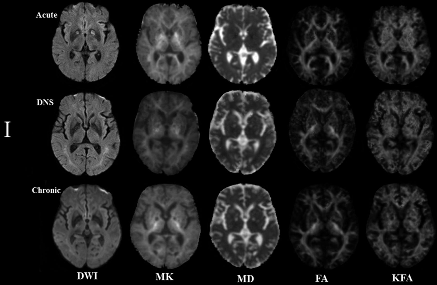
  • Fig 3.
    • Download figure
    • Open in new tab
    • Download powerpoint
    Fig 3.

    Patient 1: A 54-year-old man. The lesion evolution in the globus pallidus in the acute (5 days), delayed neuropsychiatric (39 days), and chronic (192 days) phases.
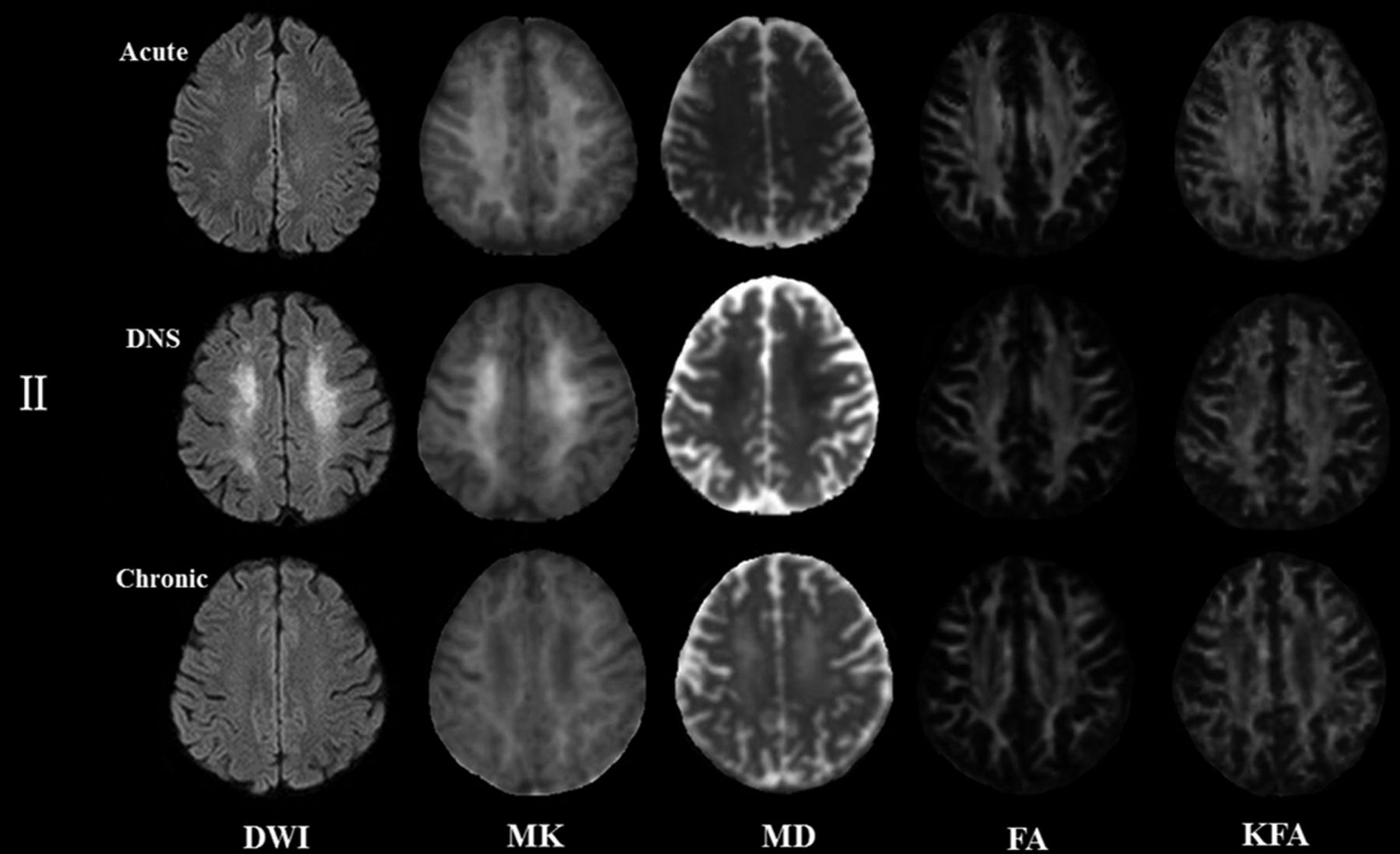
  • Fig 4.
    • Download figure
    • Open in new tab
    • Download powerpoint
    Fig 4.

    Patient 2: A 42-year-old woman. The lesion evolution in the centrum semiovale in the acute (4 days), delayed neuropsychiatric (25 days), and chronic (223 days) phases.

Tables

  • Figures
    • View popup
    Table 1:

    Clinical variables of patients in different stages of CO exposurea

    CO-Exposure Group (n = 17)Controls (n = 30)
    Acute StageDNS StageChronic Stage
    Average days to evaluation5.53 ± 1.0730.76 ± 5.90196.41 ± 9.21
    Sex (male/female) (No.)8:98:98:915:15
    Age (yr)47.41 ± 11.5047.41 ± 11.5047.41 ± 11.5048.40 ± 9.84
    Education (yr)14.00 (9.50 ± 14.00)14.00 (9.50 ± 14.00)14.00 (9.50 ± 14.00)14.00 (11.00 ± 14.00)
    MMSE26.00 ± 2.50b,c18.59 ± 4.21c,d24.59 ± 2.81c29.00 (28.00–29.00)
    Barthel Index90.00 (80.00–100.00)b,c25.00 (16.00–45.00)c,d98.00 (92.50–100.00)c100.00 (100.00-100.00)
    Digit span test
        Forward6.00 ± 1.46b,c2.00 (1.50–3.00)c,d5.06 ± 1.98c7.00 (7.00–8.00)
        Backward3.76 ± 1.25b,c1.00 (1.00–1.00)c,d2.41 ± 0.94c5.00 (4.00–5.00)
    Verbal fluency test
        Animals11.18 ± 3.36b,c5.47 ± 2.90c,d9.53 ± 3.15c15.00 (14.75.00–16.00)
        Vegetables11.59 ± 2.29b,c5.00 (3.50–6.00)c,d11.00 (9.50–12.00)c13.87 ± 1.50
    • Note:—MMSE indicates Mini-Mental State Examination.

    • ↵a Data are expressed as mean ± SD and median (25th to 75th quartiles).

    • ↵b P < .05 (acute versus DNS).

    • ↵c P < .05 (versus controls).

    • ↵d P < .05 (DNS versus chronic).

    • View popup
    Table 2:

    Diagnostic performance of DKI parameters in differentiating patients with CO exposure from controls

    Brain Regions/ROIsArea under the ROC Curve
    Acute StageDNS StageChronic Stage
    MKMDFAKFAMKMDFAKFAMKMDFAKFA
    White matter
        Centrum semiovale0.588a0.5490.5550.6670.774a0.770a0.6670.812a0.5160.5210.732a0.802a
        Corpus callosum
            Genu0.1260.5220.4990.701a0.5990.6420.700a0.753a0.5740.663a0.681a0.766a
            Body0.6180.6010.5700.6020.759a0.714a0.699a0.780a0.6490.5860.789a0.810
            Splenium0.5780.719a0.6000.749a0.6740.7680.707a0.770a0.720a0.4160.729a0.811a
        Frontal0.6510.5640.6020.6710.726a0.6400.736a0.741a0.5030.6120.757a0.771a
        Parietal0.5320.5830.5570.6460.6370.6390.6560.697a0.6050.4290.697a0.762a
        Temporal0.5670.6220.5750.731a0.6710.745a0.749a0.806a0.6040.5090.6990.775a
        Occipital0.6450.5150.6710.6930.701a0.6090.733a0.775a0.287a0.5030.761a0.791a
    Gray matter
        Globus pallidus0.800a0.76a0.5410.6330.6490.4480.5530.5410.724a0.6400.6600.602
        Caudate nucleus0.5620.5160.5200.5010.5390.5140.5510.5140.659a0.5350.6030.613
        Thalamus0.5900.5850.5560.4770.5500.3790.5090.5200.700a0.4000.5340.537
    • Note:—ROC indicates receiver operating characteristic.

    • ↵a <.05 indicates significance.

    • View popup
    Table 3:

    Correlation study between KFA and cognitive tests in patients with CO intoxication

    Brain Regions/ROIsMMSEBarthel IndexDigit SpanVerbal Fluency
    ForwardBackwardAnimalsVegetables
    White matter
        Centrum semiovaler = 0.399ar = 0.125r = 0.212r = 0.476ar = 0.164r = 0.221a
    P = .000P = .268P = .057P = .000P = .145P = .048
        Corpus callosum
            Genur = 0.283ar = 0.141r = 0.218r = 0.374ar = 0.167r = 0.188
    P = .010P = .209P = .051P = .001P = .136P = .092
            Bodyr = 0.206r = 0.211r = 0.232ar = 0.364ar = 0.219ar = 0.176
    P = .065P = .059P = .037P = .001P = .049P = .117
            Spleniumr = 0.346ar = 0.252ar = 0.201r = 0.270ar = 0.200r = 0.282a
    P = .002P = .023P = .073P = .015P = .073P = .011
        Frontalr = 0.324ar = 0.155r = 0.173r = 0.391ar = 0.203r = 0.170
    P = .003P = .167P = .123P = .000P = .069P = .129
        Parietalr = 0.222ar = 0.122r = 0.158r = 0.413ar = 0.141r = 0.152
    P = .047P = .279P = .159P = .000P = .211P = .174
        Temporalr = 0.174r = 0.171r = 0.246ar = 0.172r = 0.091r = −0.007
    P = .119P = .127P = .027P = .124P = .417P = .948
        Occipitalr = 0.151r = 0.115r = 0.187r = 0.141r = 0.142r = 0.087
    P = .178P = .305P = .095P = .210P = .208P = .442
    Gray matter
        Globus pallidusr = 0.282ar = 0.206r = 0.280ar = 0.264ar = 0.217r = 0.175
    P = .011P = .065P = .011P = .017P = .051P = .119
        Caudate nucleusr = −0.052r = −0.148r = −0.185r = 0.108r = −0.115r = −0.118
    P = .645P = .188P = .099P = .338P = .308P = .294
        Thalamusr = 0.068r = 0.039r = −0.018r = 0.054r = 0.111r = 0.040
    P = .544P = .726P = .871P = .630P = .323P = .720
    • Note:—MMSE indicates Mini-Mental State Examination.

    • ↵a Significant indicated by<.05.

PreviousNext
Back to top

In this issue

American Journal of Neuroradiology: 40 (10)
American Journal of Neuroradiology
Vol. 40, Issue 10
1 Oct 2019
  • Table of Contents
  • Index by author
  • Complete Issue (PDF)
Advertisement
Print
Download PDF
Email Article
Cite this article
0 Responses
Respond to this article
Share
Bookmark this article
Cerebral Damage after Carbon Monoxide Poisoning: A Longitudinal Diffusional Kurtosis Imaging Study
Y. Zhang, T. Wang, J. Lei, S. Guo, S. Wang, Y. Gu, S. Wang, Y. Dou, X. Zhuang
American Journal of Neuroradiology Oct 2019, 40 (10) 1630-1637; DOI: 10.3174/ajnr.A6201
del.icio.us logo Twitter logo Facebook logo Mendeley logo
  • Tweet Widget
Purchase

Jump to section

  • Article
    • Abstract
    • ABBREVIATIONS:
    • MATERIALS AND METHODS
    • RESULTS
    • DISCUSSION
    • CONCLUSIONS
    • Footnotes
    • References
  • Figures & Data
  • Supplemental
  • Info & Metrics
  • Responses
  • References
  • PDF

Related Articles

  • PubMed
  • Google Scholar

Cited By...

  • An Introduction to Kurtosis Fractional Anisotropy
  • Crossref
  • Google Scholar

This article has not yet been cited by articles in journals that are participating in Crossref Cited-by Linking.

More in this TOC Section

Adult Brain

  • Diagnostic Neuroradiology of Monoclonal Antibodies
  • ML for Glioma Molecular Subtype Prediction
  • NCCT vs. MRI for Brain Atrophy in Acute Stroke
Show more Adult Brain

Functional

  • Kurtosis and Epileptogenic Tubers: A Pilot Study
  • Glutaric Aciduria Type 1: DK vs. Conventional MRI
  • WM Neuroaxonal Loss in Type 1 Diabetes
Show more Functional

Similar Articles

Advertisement

Indexed Content

  • Current Issue
  • Accepted Manuscripts
  • Article Preview
  • Past Issues
  • Editorials
  • Editor's Choice
  • Fellows' Journal Club
  • Letters to the Editor
  • Video Articles

Cases

  • Case Collection
  • Archive - Case of the Week
  • Archive - Case of the Month
  • Archive - Classic Case

More from AJNR

  • Trainee Corner
  • Imaging Protocols
  • MRI Safety Corner
  • Book Reviews

Multimedia

  • AJNR Podcasts
  • AJNR Scantastics

Resources

  • Turnaround Time
  • Submit a Manuscript
  • Submit a Video Article
  • Submit an eLetter to the Editor/Response
  • Manuscript Submission Guidelines
  • Statistical Tips
  • Fast Publishing of Accepted Manuscripts
  • Graphical Abstract Preparation
  • Imaging Protocol Submission
  • Evidence-Based Medicine Level Guide
  • Publishing Checklists
  • Author Policies
  • Become a Reviewer/Academy of Reviewers
  • News and Updates

About Us

  • About AJNR
  • Editorial Board
  • Editorial Board Alumni
  • Alerts
  • Permissions
  • Not an AJNR Subscriber? Join Now
  • Advertise with Us
  • Librarian Resources
  • Feedback
  • Terms and Conditions
  • AJNR Editorial Board Alumni

American Society of Neuroradiology

  • Not an ASNR Member? Join Now

© 2025 by the American Society of Neuroradiology All rights, including for text and data mining, AI training, and similar technologies, are reserved.
Print ISSN: 0195-6108 Online ISSN: 1936-959X

Powered by HighWire
Email this Article

Thank you for your interest in spreading the word on American Journal of Neuroradiology.

NOTE: We only request your email address so that the person you are recommending the page to knows that you wanted them to see it, and that it is not junk mail. We do not capture any email address.

Enter multiple addresses on separate lines or separate them with commas.
Cerebral Damage after Carbon Monoxide Poisoning: A Longitudinal Diffusional Kurtosis Imaging Study
(Your Name) has sent you a message from American Journal of Neuroradiology
(Your Name) thought you would like to see the American Journal of Neuroradiology web site.
CAPTCHA
This question is for testing whether or not you are a human visitor and to prevent automated spam submissions.
Cite this article
Y. Zhang, T. Wang, J. Lei, S. Guo, S. Wang, Y. Gu, S. Wang, Y. Dou, X. Zhuang
Cerebral Damage after Carbon Monoxide Poisoning: A Longitudinal Diffusional Kurtosis Imaging Study
American Journal of Neuroradiology Oct 2019, 40 (10) 1630-1637; DOI: 10.3174/ajnr.A6201

Citation Manager Formats

  • BibTeX
  • Bookends
  • EasyBib
  • EndNote (tagged)
  • EndNote 8 (xml)
  • Medlars
  • Mendeley
  • Papers
  • RefWorks Tagged
  • Ref Manager
  • RIS
  • Zotero

We use cookies on this site to enhance your user experience. By clicking any link on this page you are giving your consent for us to set cookies.