Advertisement

Main menu

  • Home
  • Content
    • Current Issue
    • Accepted Manuscripts
    • Article Preview
    • Past Issue Archive
    • Video Articles
    • AJNR Case Collection
    • Case of the Week Archive
    • Case of the Month Archive
    • Classic Case Archive
  • Special Collections
    • AJNR Awards
    • Low-Field MRI
    • Alzheimer Disease
    • ASNR Foundation Special Collection
    • Photon-Counting CT
    • View All
  • Multimedia
    • AJNR Podcasts
    • AJNR SCANtastic
    • Trainee Corner
    • MRI Safety Corner
    • Imaging Protocols
  • For Authors
    • Submit a Manuscript
    • Submit a Video Article
    • Submit an eLetter to the Editor/Response
    • Manuscript Submission Guidelines
    • Statistical Tips
    • Fast Publishing of Accepted Manuscripts
    • Graphical Abstract Preparation
    • Imaging Protocol Submission
    • Author Policies
  • About Us
    • About AJNR
    • Editorial Board
    • Editorial Board Alumni
  • More
    • Become a Reviewer/Academy of Reviewers
    • Subscribers
    • Permissions
    • Alerts
    • Feedback
    • Advertisers
    • ASNR Home

User menu

  • Alerts
  • Log in

Search

  • Advanced search
American Journal of Neuroradiology

American Journal of Neuroradiology

ASHNR American Society of Functional Neuroradiology ASHNR American Society of Pediatric Neuroradiology ASSR
  • Alerts
  • Log in

Advanced Search

  • Home
  • Content
    • Current Issue
    • Accepted Manuscripts
    • Article Preview
    • Past Issue Archive
    • Video Articles
    • AJNR Case Collection
    • Case of the Week Archive
    • Case of the Month Archive
    • Classic Case Archive
  • Special Collections
    • AJNR Awards
    • Low-Field MRI
    • Alzheimer Disease
    • ASNR Foundation Special Collection
    • Photon-Counting CT
    • View All
  • Multimedia
    • AJNR Podcasts
    • AJNR SCANtastic
    • Trainee Corner
    • MRI Safety Corner
    • Imaging Protocols
  • For Authors
    • Submit a Manuscript
    • Submit a Video Article
    • Submit an eLetter to the Editor/Response
    • Manuscript Submission Guidelines
    • Statistical Tips
    • Fast Publishing of Accepted Manuscripts
    • Graphical Abstract Preparation
    • Imaging Protocol Submission
    • Author Policies
  • About Us
    • About AJNR
    • Editorial Board
    • Editorial Board Alumni
  • More
    • Become a Reviewer/Academy of Reviewers
    • Subscribers
    • Permissions
    • Alerts
    • Feedback
    • Advertisers
    • ASNR Home
  • Follow AJNR on Twitter
  • Visit AJNR on Facebook
  • Follow AJNR on Instagram
  • Join AJNR on LinkedIn
  • RSS Feeds

AJNR Awards, New Junior Editors, and more. Read the latest AJNR updates

Article CommentaryAdult Brain

An Introduction to Kurtosis Fractional Anisotropy

B. Hansen
American Journal of Neuroradiology October 2019, 40 (10) 1638-1641; DOI: https://doi.org/10.3174/ajnr.A6235
B. Hansen
aCenter of Functionally Integrative Neuroscience Aarhus University Aarhus, Denmark
  • Find this author on Google Scholar
  • Find this author on PubMed
  • Search for this author on this site
  • ORCID record for B. Hansen
  • Article
  • Figures & Data
  • Info & Metrics
  • Responses
  • References
  • PDF
Loading

Neuroradiology and imaging-based diagnostics in general have a dire need for scan techniques with improved microstructural sensitivity for detection of subtle tissue alterations in early disease phases or for diagnostics of diseases that are currently simply radiologically invisible. Diseases in which this need is most outspoken are often neurodegenerative diseases such as Alzheimer's disease, multiple sclerosis, and Parkinson's disease but examples also include (mild, repetitive) trauma, addiction, and stroke. Less frequently reported but important nevertheless is the need for sensitive methods for diagnosis and monitoring of patients exposed to poisonous substances. In the industrialized parts of the world, the leading cause of poisoning is carbon monoxide (CO).1 In the study by Zhang et al2 in the present issue of the American Journal of Neuroradiology (AJNR), brain microstructure in CO-poisoned patients is assessed at 3 time points using diffusional kurtosis imaging (DKI) and is correlated to patient cognitive performance. It concluded that DKI metrics provide important information about the damage to the brain due to CO, and supplement cognitive scores. As a first, the study found kurtosis fractional anisotropy (KFA) to have the best diagnostic efficiency based on a standard area under the curve measure. Because this parameter is likely to be somewhat new to many working in the field of neuroradiology, this commentary aims to recapitulate the basis of KFA and what it might indicate about tissue microstructure.

Diffusion MR Imaging and Diffusional Kurtosis Imaging

Water is abundant in the brain, and the water molecules are thermally driven to move ceaselessly and randomly. The local environment of a water molecule determines its mobility. Therefore, water ensemble properties such as mean diffusivity, diffusion anisotropy, and diffusion distribution shape vary among tissues with different compositions. In neural tissue, diffusion variations are seen among tissue microdomains at the subcellular level,3 the intra- or extracellular space, among cell types,4 and therefore also on a coarser scale between gray and white matter tissue.5 Noninvasive MR imaging–based measurements of brain-water diffusion, therefore, in principle, contain abundant information about the cellular-level tissue composition. Initially, the diffusion MR imaging (dMRI) signal description was based on a Gaussian (normal) diffusion probability distribution,6 forming the basis for the familiar diffusion tensor imaging technique. In biologic tissue, however, the overall diffusion behavior observed from an MR imaging voxel is not Gaussian under clinically relevant measurement conditions. To account for non-Gaussian diffusion, a kurtosis term is added to the DTI signal equation, producing the diffusion DKI framework:7 Embedded Image

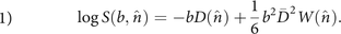
Equation 1 describes the behavior of the (log of the) normalized dMRI signal from tissue with diffusion described by a 3 × 3 diffusion tensor D, and kurtosis is described by the 4D tensor W. As written here, the signal is measured along a direction n̂ with diffusion weighting b. The first term on the right-hand side is the DTI signal term, so DTI is fully contained (and actually improved8) in DKI. The second term on the right-hand side of Equation 1 is the kurtosis term, with the kurtosis tensor W describing the non-Gaussian properties of the diffusion, which are not contained in D. As written here, D(n̂)is the apparent diffusivity and W(n̂) is the apparent kurtosis, both observed along the diffusion gradient direction n̂. Note that sometimes the apparent kurtosis is referred to as K(n̂), where K(n̂) = W(n̂)D̄2/D(n̂)2 (D̄ is mean diffusivity, see below). Simply put, the kurtosis term accounts for the signal deviation from log-linear DTI behavior along the diffusion-encoding direction n̂.

The wealth of information available from DTI and DKI is contained in the diffusion tensor D and the kurtosis tensor W. From D, a number of parameters are available, with typical reported metrics being the apparent diffusion coefficient, ADC = D(n̂); mean diffusivity (D̄ ≡ Tr(D)/3 = (λ1 + λ2 + λ3)/3, where λ1−3 is the diffusion tensor eigenvalues—that is, D(x̂′), D(ŷ′), D(ẑ′) in the tensor eigenframe); radial and axial diffusivities; and fractional anisotropy (FA).9 In white matter, the direction with the highest diffusivity (axial diffusivity) largely identifies the main fiber direction.10⇓–12 The pronounced anisotropy of brain white matter is due to the myelin sheath, the axonal membrane itself, and the cytoskeleton inside the axon,13 which collectively cause WM FA to be high (typically >0.6, however see the example below). In DKI, D and W are used together to provide more parameters. These tensors will behave very differently: For example, in a single fiber system, the observed diffusion is high along the fiber direction and low across the fiber direction. In contrast, the kurtosis would be higher across than along the fiber direction, mostly due to restriction effects. This added information improves the capability of dMRI to detect changes in brain and body organ microstructure.14,15 From DKI, typically reported metrics are analogously the mean kurtosis (MK) and radial and axial kurtosis, and, for very aligned WM, a set of biomarkers from the WM tract integrity framework.16 More recently, the KFA has been introduced.17,18 These parameters, their estimation, and varying definitions are reviewed in Hansen and Jespersen.19

A comprehensive review of DKI in neuroimaging is outside the scope of this commentary, but to illustrate the potential of DKI, we list key neurologic disorders and neuroradiologic areas where DKI has already proved useful: addiction,20 stroke,21⇓–23 Alzheimer's disease,24 multiple sclerosis,25 Parkinson's disease,26⇓⇓–29 brain cancer (gliomas),30,31 and head trauma/concussion.32⇓⇓⇓–36 While DKI is increasingly used, so far the relatively new KFA has not been given much attention in preclinical and clinical studies. It is, therefore, noteworthy that KFA features prominently in the article of Zhang et al.2 Thus, KFA and its interpretation are the main focus of the remainder of this commentary.

dMRI Anisotropy Measures: FA and KFA

The DKI framework produces 2 scalar anisotropy measures: the FA from the diffusion tensor D and the KFA from the kurtosis tensor W. The FA is defined as Embedded Image 
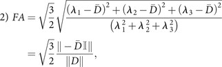
where double bars denote the Euclidean tensor norm (the Frobenius norm), 𝕀 is the 3 × 3 identity matrix, and the remaining parameters are defined above. The numeric front factor ensures that that the FA assumes values in the range from 0 (completely isotropic diffusion) to 1 (fully anisotropic, unidirectional diffusion). Although a few alternative definitions of the kurtosis anisotropy have been proposed (notably the one in Poot et al37), the agreed upon definition today is the one introduced in Hansen et al:18 Embedded Image 
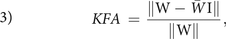
where W is the kurtosis tensor, W̄ is the kurtosis tensor mean,18,38,39 and I is the fully symmetric rank 4 isotropic tensor. Conveniently, this definition of KFA is analogous to the FA definition in Equation 2, with the only difference being due to tensor dimensions. We note the absence of a numeric front factor in Equation 3; KFA naturally assumes values in the 0–1 range. Interested readers are referred to early explorations of KFA in Hansen and Jespersen17 and Glenn et al.40 In these studies, simulations and experiments were used to investigate the information contained in the KFA. It was found that KFA contrast supplements the FA in important ways. This feature is illustrated in the Figure showing side-by-side maps of FA and KFA in the same image plane position in normal human brain (Fig 1A).

FIGURE.
  • Download figure
  • Open in new tab
  • Download powerpoint
FIGURE.

A, Maps of FA and KFA in the same section position in the normal human brain. The color bar on the right applies to both parameter maps. In the FA map in A, a band of low FA values is seen as a dark band in both hemispheres. The red arrow points to this feature. The same white matter region is seen to have KFA values similar to those of the surrounding white matter. B, The fiber arrangement causing this FA behavior is illustrated in a simplified thought experiment. Figure adapted with permission from Hansen and Jespersen.17

The red arrow points to a dark band in the FA map inside the WM. This band is seen in both hemispheres (note the symmetry). In each hemisphere, this band comprises voxels with low FA values, though the voxels are located in anatomic WM. Conversely, in the KFA map on the right in Fig 1A, we see that KFA in WM is high and notably retains its high value in the regions where FA fails as a reporter of anisotropy. These WM voxels assume low FA values because they contain a crossing-fiber arrangement in which WM fibers along 3 orthogonal directions weave between each other. This effectively produces the situation illustrated in the schematic in Fig 1B, which shows 3 separate WM fiber bundles that intersect at right angles. In this example, we ignore extra-axonal diffusion. We see that if diffusivities in the 3 fibers are similar (as they are likely to be), the mean diffusivity will be the same as the eigenvalues (ie, the diffusivities along 3 fiber directions D̄ = (λ1 + λ2 + λ3)/3 ≈ λ1−3) causing the FA to vanish because, in this case, each term in the numerator in the middle expression in Equation 2 will be approximately zero. This happens despite the diffusivity in this fiber arrangement being highly anisotropic. Although not particularly complex, the anisotropy in this fiber arrangement cannot be described by the diffusion tensor, resulting in the low FA constituting the dark band in this WM region.

In the interpretation of these metrics, it is crucial to remember that FA and KFA report on different features of the diffusion process. We, therefore, stress that FA summarizes the spatial variation of diffusion rates, whereas KFA summarizes the directional variation in the degree of non-Gaussian diffusion. Loosely speaking, another difference is that the KFA stems from the kurtosis tensor, which is a 4D tensor with much more “room” to capture, in detail, the spatial variation in kurtosis. Collectively, these factors contribute to the demonstrated behavior in which KFA continues to provide contrast in areas where FA does not. Both metrics can be difficult to interpret in strict terms of tissue properties, but on the basis of the simple example above, we can cautiously state that KFA reflects tissue-diffusion complexity. In the example above, we saw that the 2D diffusion tensor cannot resolve anisotropy in voxels with complex fiber composition. However, we also saw that areas with complex fiber arrangements may be distinguished from genuine low-anisotropy regions using FA and KFA in combination: If FA is low and KFA is high, then likely the diffusion is, in fact, anisotropic but the fiber arrangement is too complex to be captured by the diffusion tensor. Thus, the KFA is a good parameter to include in studies in which subtle remodeling is expected in complex tissue regions. While KFA may therefore prove to be a valuable marker of tissue microstructure, we nevertheless stress that no DKI parameter is specifically bound to a particular microscopic tissue component.

Discussion and Future Perspectives

DKI is sometimes omitted from clinical protocols due to longer acquisition and parameter estimation times than a simple DTI protocol. While strategies exist for fast estimation of most DKI parameters,18,19,38⇓⇓⇓⇓⇓–44 robust estimation of KFA still requires the full kurtosis tensor W to be determined by fitting on a pixel-by-pixel basis.17 This requires multishell dMRI data with typically 30 directions and 2–3 nonzero b-values. With modern dMRI techniques, this is possible in clinically feasible scan times.45 From such datasets, DKI parameter estimation is possible using any one of the many software packages that exist for dMRI data analysis such as MUSC's DKE software package (https://medicine.musc.edu/departments/centers/cbi/dki/dki-data-processing), which also computes the KFA as defined in Equation 3. As noted above, interpretation of DKI findings in terms of biophysical tissue properties is a difficult problem. In preclinical work, subsequent histology may be used to interpret DKI findings,15,46,47 and insights gained from such efforts may aid in interpretation of clinical DKI.

In the discussion of the example in the Figure, we noted that KFA somehow reflects the diffusion anisotropy below the voxel level. Other techniques achieve similar sensitivity48,49 but require nonstandard pulse sequences. Until such techniques reach the clinic in earnest, the KFA may serve as an indicator of the diagnostic potential of such sensitivity.

CONCLUSIONS

Imaging techniques with improved sensitivity to microstructure have utility in many areas of neuroimaging, including diagnosis and monitoring of patients exposed to toxic or poisonous substances. The study by Zhang et al2 uses the microstructurally sensitive DKI framework to assess cerebral damage in CO-poisoned subjects. From DKI, the authors obtain measures of mean diffusivity, FA, MK, and the less explored KFA. These measures are then correlated to neuropsychiatric scores. The work presents a timely contribution to 2 avenues of neuroradiology: 1) the exploration of DKI and KFA in clinical practice, and 2) identification of sensitive markers for diagnostics of CO-intoxicated patients. Insights gained from this study may also benefit and inspire many other areas of neuroimaging where the same techniques could be used. With this commentary, we hope that the KFA will have become more familiar to readers of AJNR so that investigators will consider it a parameter of interest in their future work.

References

  1. 1.↵
    1. Omaye ST
    . Metabolic modulation of carbon monoxide toxicity. Toxicology 2002;180:139–50 doi:10.1016/S0300-483X(02)00387-6 pmid:12324190
    CrossRefPubMed
  2. 2.↵
    1. Zhang Y,
    2. Wang T,
    3. Lei J, et al
    . Cerebral damage after carbon monoxide poisoning: a longitudinal diffusional kurtosis imaging study. AJNR Am J Neuroradiol 2019;40:1630–37
    Abstract/FREE Full Text
  3. 3.↵
    1. Lee CH,
    2. Flint JJ,
    3. Hansen B, et al
    . Investigation of the subcellular architecture of L7 neurons of Aplysia Californica using magnetic resonance microscopy (MRM) at 7.8 microns. Sci Rep 2015;5:11147 doi:10.1038/srep11147 pmid:26059695
    CrossRefPubMed
  4. 4.↵
    1. Flint JJ,
    2. Hansen B,
    3. Portnoy S, et al
    . Magnetic resonance microscopy of human and porcine neurons and cellular processes. Neuroimage 2012;60:1404–11 doi:10.1016/j.neuroimage.2012.01.050 pmid:22281672
    CrossRefPubMed
  5. 5.↵
    1. Flint JJ,
    2. Lee CH,
    3. Hansen B, et al
    . Magnetic resonance microscopy of mammalian neurons. Neuroimage 2009;46:1037–40 doi:10.1016/j.neuroimage.2009.03.009 pmid:19286461
    CrossRefPubMed
  6. 6.↵
    1. Basser PJ,
    2. Mattiello J,
    3. LeBihan D
    . Estimation of the effective self-diffusion tensor from the NMR spin-echo. J Magn Reson B 1994;103:247–54 doi:10.1006/jmrb.1994.1037 pmid:8019776
    CrossRefPubMed
  7. 7.↵
    1. Jensen JH,
    2. Helpern JA,
    3. Ramani A, et al
    . Diffusional kurtosis imaging: the quantification of non-Gaussian water diffusion by means of magnetic resonance imaging. Magn Reson Med 2005;53:1432–40 doi:10.1002/mrm.20508 pmid:15906300
    CrossRefPubMed
  8. 8.↵
    1. Veraart J,
    2. Poot DHJ,
    3. Van Hecke W, et al
    . More accurate estimation of diffusion tensor parameters using diffusion kurtosis imaging. Magn Reson Med 2011;65:138–45 doi:10.1002/mrm.22603 pmid:20878760
    CrossRefPubMed
  9. 9.↵
    1. Basser PJ
    . Inferring microstructural features and the physiological state of tissues from diffusion-weighted images. NMR Biomed 1995;8:333–44 doi:10.1002/nbm.1940080707 pmid:8739270
    CrossRefPubMed
  10. 10.↵
    1. Flint JJ,
    2. Hansen B,
    3. Blackband SJ
    . Diffusion tensor microscopy data (15.6 μm in-plane) of white matter tracts in the human, pig, and rat spinal cord with corresponding tissue histology. Data Brief 2016;9:271–74 doi:10.1016/j.dib.2016.08.020 pmid:27668273
    CrossRefPubMed
  11. 11.↵
    1. Flint JJ,
    2. Hansen B,
    3. Fey M, et al
    . Cellular-level diffusion tensor microscopy and fiber tracking in mammalian nervous tissue with direct histological correlation. Neuroimage 2010;52:556–61 doi:10.1016/j.neuroimage.2010.04.031 pmid:20403443
    CrossRefPubMed
  12. 12.↵
    1. Hansen B,
    2. Flint JJ,
    3. Heon-Lee C, et al
    . Diffusion tensor microscopy in human nervous tissue with quantitative correlation based on direct histological comparison. Neuroimage 2011;57:1458–65 doi:10.1016/j.neuroimage.2011.04.052 pmid:21575730
    CrossRefPubMed
  13. 13.↵
    1. Beaulieu C
    . The basis of anisotropic water diffusion in the nervous system: a technical review. NMR Biomed 2002;15:435–55 doi:10.1002/nbm.782 pmid:12489094
    CrossRefPubMed
  14. 14.↵
    1. Rosenkrantz AB,
    2. Padhani AR,
    3. Chenevert TL, et al
    . Body diffusion kurtosis imaging: basic principles, applications, and considerations for clinical practice. J Magn Reson Imaging 2015;42:1190–202 doi:10.1002/jmri.24985 pmid:26119267
    CrossRefPubMed
  15. 15.↵
    1. Kjølby BF,
    2. Khan AR,
    3. Chuhutin A, et al
    . Fast diffusion kurtosis imaging of fibrotic mouse kidneys. NMR Biomed 2016;29:1709–19 doi:10.1002/nbm.3623 pmid:27731906
    CrossRefPubMed
  16. 16.↵
    1. Fieremans E,
    2. Jensen JH,
    3. Helpern JA
    . White matter characterization with diffusional kurtosis imaging. Neuroimage 2011;58:177–88 doi:10.1016/j.neuroimage.2011.06.006 pmid:21699989
    CrossRefPubMed
  17. 17.↵
    1. Hansen B,
    2. Jespersen SN
    . Kurtosis fractional anisotropy, its contrast and estimation by proxy. Sci Rep 2016;6:2399 doi:10.1038/srep23999 pmid:27041679
    CrossRefPubMed
  18. 18.↵
    1. Hansen B,
    2. Lund TE,
    3. Sangill R, et al
    . Experimentally and computationally fast method for estimation of a mean kurtosis. Magn Reson Med 2013;69:1754–60 doi:10.1002/mrm.24743 pmid:23589312
    CrossRefPubMed
  19. 19.↵
    1. Hansen B,
    2. Jespersen SN
    . Recent developments in fast kurtosis imaging. Front Phys 2017;5 doi:10.3389/fphy.2017.00040
    CrossRef
  20. 20.↵
    1. Garza-Villarreal EA,
    2. Chakravarty MM,
    3. Hansen B, et al
    . The effect of crack cocaine addiction and age on the microstructure and morphology of the human striatum and thalamus using shape analysis and fast diffusion kurtosis imaging. Transl Psychiatry 2017;7:e1122 doi:10.1038/tp.2017.92 pmid:28485734
    CrossRefPubMed
  21. 21.↵
    1. Hui ES,
    2. Fieremans E,
    3. Jensen JH, et al
    . Stroke assessment with diffusional kurtosis imaging. Stroke 2012;43:2968–73 doi:10.1161/STROKEAHA.112.657742 pmid:22933581
    Abstract/FREE Full Text
  22. 22.↵
    1. Weber RA,
    2. Hui ES,
    3. Jensen JH, et al
    . Diffusional kurtosis and diffusion tensor imaging reveal different time-sensitive stroke-induced microstructural changes. Stroke 2015;46:545–50 doi:10.1161/STROKEAHA.114.006782 pmid:25563646
    Abstract/FREE Full Text
  23. 23.↵
    1. Bay V,
    2. Kjolby BF,
    3. Iversen NK, et al
    . Stroke infarct volume estimation in fixed tissue: comparison of diffusion kurtosis imaging to diffusion weighted imaging and histology in a rodent MCAO model. PLos One 2018;13:e019616 doi:10.1371/journal.pone.0196161 pmid:29698450
    CrossRefPubMed
  24. 24.↵
    1. Falangola MF,
    2. Jensen JH,
    3. Tabesh A, et al
    . Non-Gaussian diffusion MRI assessment of brain microstructure in mild cognitive impairment and Alzheimer's disease. Magn Reson Imaging 2013;31:840–46 doi:10.1016/j.mri.2013.02.008 pmid:23602730
    CrossRefPubMed
  25. 25.↵
    1. Inglese M,
    2. Bester M
    . Diffusion imaging in multiple sclerosis: research and clinical implications. NMR Biomed 2010;23:865–72 doi:10.1002/nbm.1515 pmid:20882528
    CrossRefPubMed
  26. 26.↵
    1. Arab A,
    2. Ruda-Kucerova J,
    3. Minsterova A, et al
    . Diffusion kurtosis imaging detects microstructural changes in a methamphetamine-induced mouse model of Parkinson's disease. Neurotox Res 2019 Jun 18. [Epub ahead of print] doi:10.1007/s12640-019-00068-0 pmid:31209787
    CrossRefPubMed
  27. 27.↵
    1. Khairnar A,
    2. Latta P,
    3. Drazanova E, et al
    . Diffusion kurtosis imaging detects microstructural alterations in brain of alpha-synuclein overexpressing transgenic mouse model of parkinson's disease: a pilot study. Neurotox Res 2015;28:281–89 doi:10.1007/s12640-015-9537-9 pmid:26153486
    CrossRefPubMed
  28. 28.↵
    1. Wang JJ,
    2. Lin WY,
    3. Lu CS, et al
    . Parkinson disease: diagnostic utility of diffusion kurtosis imaging. Radiology 2011;261:210–17 doi:10.1148/radiol.11102277 pmid:21771952
    CrossRefPubMed
  29. 29.↵
    1. Zhang G,
    2. Zhang Y,
    3. Zhang C, et al
    . Diffusion kurtosis imaging of substantia nigra is a sensitive method for early diagnosis and disease evaluation in Parkinson's disease. Parkinsons Dis 2015;2015:207624 doi:10.1155/2015/207624 pmid:26770867
    CrossRefPubMed
  30. 30.↵
    1. Van Cauter S,
    2. Veraart J,
    3. Sijbers J, et al
    . Gliomas: diffusion kurtosis MR imaging in grading. Radiology 2012;263:492–501 doi:10.1148/radiol.12110927 pmid:22403168
    CrossRefPubMed
  31. 31.↵
    1. Bai Y,
    2. Lin Y,
    3. Tian J, et al
    . Grading of gliomas by using monoexponential, biexponential, and stretched exponential diffusion-weighted MR imaging and diffusion kurtosis MR imaging. Radiology 2016;278:496–504 doi:10.1148/radiol.2015142173 pmid:26230975
    CrossRefPubMed
  32. 32.↵
    1. Grossman EJ,
    2. Ge Y,
    3. Jensen JH, et al
    . Thalamus and cognitive impairment in mild traumatic brain injury: a diffusional kurtosis imaging study. J Neurotrauma 2012;29:2318–27 doi:10.1089/neu.2011.1763 pmid:21639753
    CrossRefPubMed
  33. 33.↵
    1. Grossman EJ,
    2. Jensen JH,
    3. Babb JS, et al
    . Cognitive impairment in mild traumatic brain injury: a longitudinal diffusional kurtosis and perfusion imaging study. AJNR Am J Neuroradiol 2013;34:951–57 doi:10.3174/ajnr.A3358 pmid:23179649
    Abstract/FREE Full Text
  34. 34.↵
    1. Næss-Schmidt E,
    2. Blicher JU,
    3. Eskildsen SF, et al
    . Microstructural changes in the thalamus after mild traumatic brain injury: a longitudinal diffusion and mean kurtosis tensor MRI study. Brain Injury 2017;31:230–36 doi:10.1080/02699052.2016.1229034 pmid:28055267
    CrossRefPubMed
  35. 35.↵
    1. Østergaard L,
    2. Engedal TS,
    3. Aamand R, et al
    . Capillary transit time heterogeneity and flow-metabolism coupling after traumatic brain injury. J Cereb Blood Flow Metab 2014;34:1585–98 doi:10.1038/jcbfm.2014.131 pmid:25052556
    CrossRefPubMed
  36. 36.↵
    1. Næss-Schmidt E,
    2. Blicher JU,
    3. Tietze A, et al
    . Diffusion MRI findings in patients with extensive and minimal post-concussion symptoms after mTBI and healthy controls: a cross sectional study. Brain Inj 2018;32:91–8 doi:10.1080/02699052.2017.1377352 pmid:29095055
    CrossRefPubMed
  37. 37.↵
    1. Poot DH,
    2. den Dekker AJ,
    3. Achten E, et al
    . Optimal experimental design for diffusion kurtosis imaging. IEEE Trans Med Imaging 2010;29:819–29 doi:10.1109/TMI.2009.2037915 pmid:20199917
    CrossRefPubMed
  38. 38.↵
    1. Hansen B,
    2. Lund TE,
    3. Sangill R, et al
    . Experimental considerations for fast kurtosis imaging. Magn Reson Med 2016;76:1455–68 doi:10.1002/mrm.26055 pmid:26608731
    CrossRefPubMed
  39. 39.↵
    1. Hansen B,
    2. Lund TE,
    3. Sangill R, et al
    . Erratum: Hansen, Lund, Sangill, and Jespersen. Experimentally and computationally fast method for estimation of a mean kurtosis. Mag Reson Med 2014;71:2250
    CrossRef
  40. 40.↵
    1. Glenn GR,
    2. Helpern JA,
    3. Tabesh A, et al
    . Quantitative assessment of diffusional kurtosis anisotropy. NMR Biomed 2015;28:448–59 doi:10.1002/nbm.3271 pmid:25728763
    CrossRefPubMed
  41. 41.↵
    1. Alstrup AKO,
    2. Munk OL,
    3. Jensen TH, et al
    . Magnetic resonance imaging and computed tomography as tools for the investigation of sperm whale (Physeter macrocephalus) teeth and eye. Acta Vet Scand 2017;59:38 doi:10.1186/s13028-017-0307-y pmid:28606113
    CrossRefPubMed
  42. 42.↵
    1. Hansen B,
    2. Jespersen SN
    . Data for evaluation of fast kurtosis strategies, b-value optimization and exploration of diffusion MRI contrast. Sci Data 2016;3:160072 doi:10.1038/sdata.2016.72 pmid:27576023
    CrossRefPubMed
  43. 43.↵
    1. Hansen B,
    2. Khan AR,
    3. Shemesh N, et al
    . White matter biomarkers from fast protocols using axially symmetric diffusion kurtosis imaging. NMR Biomed 2017;30 doi:10.1002/nbm.3741 pmid:28543843
    CrossRefPubMed
  44. 44.↵
    1. Hansen B,
    2. Shemesh N,
    3. Jespersen SN
    . Fast imaging of mean, axial and radial diffusion kurtosis. Neuroimage 2016;142:381–93 doi:10.1016/j.neuroimage.2016.08.022 pmid:27539807
    CrossRefPubMed
  45. 45.↵
    1. Setsompop K,
    2. Fan Q,
    3. Stockmann J, et al
    . High-resolution in vivo diffusion imaging of the human brain with generalized slice dithered enhanced resolution: simultaneous multislice (gSlider-SMS). Magn Reson Med 2018;79:141–51 doi:10.1002/mrm.26653 pmid:28261904
    CrossRefPubMed
  46. 46.↵
    1. Khan AR,
    2. Chuhutin A,
    3. Wiborg O, et al
    . Biophysical modeling of high field diffusion MRI demonstrates microstructural aberration in chronic mild stress rat brain. Neuroimage 2016;142:421–30 doi:10.1016/j.neuroimage.2016.07.001 pmid:27389790
    CrossRefPubMed
  47. 47.↵
    1. Khan AR,
    2. Hansen B,
    3. Wiborg O, et al
    . Diffusion MRI and MR spectroscopy reveal microstructural and metabolic brain alterations in chronic mild stress exposed rats: a CMS recovery study. Neuroimage 2018;167:342–53 doi:10.1016/j.neuroimage.2017.11.053 pmid:29196269
    CrossRefPubMed
  48. 48.↵
    1. Lasič S,
    2. Szczepankiewicz F,
    3. Eriksson S, et al
    . Microanisotropy imaging: quantification of microscopic diffusion anisotropy and orientational order parameter by diffusion MRI with magic-angle spinning of the q-vector. Front Phys 2014;2 doi:10.3389/fphy.2014.00011
    CrossRef
  49. 49.↵
    1. Avram AV,
    2. Ozarslan E,
    3. Sarlls JE, et al
    . In vivo detection of microscopic anisotropy using quadruple pulsed-field gradient (qPFG) diffusion MRI on a clinical scanner. Neuroimage 2013;64:229–39 doi:10.1016/j.neuroimage.2012.08.048 pmid:22939872
    CrossRefPubMed
  • © 2019 by American Journal of Neuroradiology
PreviousNext
Back to top

In this issue

American Journal of Neuroradiology: 40 (10)
American Journal of Neuroradiology
Vol. 40, Issue 10
1 Oct 2019
  • Table of Contents
  • Index by author
  • Complete Issue (PDF)
Advertisement
Print
Download PDF
Email Article

Thank you for your interest in spreading the word on American Journal of Neuroradiology.

NOTE: We only request your email address so that the person you are recommending the page to knows that you wanted them to see it, and that it is not junk mail. We do not capture any email address.

Enter multiple addresses on separate lines or separate them with commas.
An Introduction to Kurtosis Fractional Anisotropy
(Your Name) has sent you a message from American Journal of Neuroradiology
(Your Name) thought you would like to see the American Journal of Neuroradiology web site.
CAPTCHA
This question is for testing whether or not you are a human visitor and to prevent automated spam submissions.
Cite this article
B. Hansen
An Introduction to Kurtosis Fractional Anisotropy
American Journal of Neuroradiology Oct 2019, 40 (10) 1638-1641; DOI: 10.3174/ajnr.A6235

Citation Manager Formats

  • BibTeX
  • Bookends
  • EasyBib
  • EndNote (tagged)
  • EndNote 8 (xml)
  • Medlars
  • Mendeley
  • Papers
  • RefWorks Tagged
  • Ref Manager
  • RIS
  • Zotero
0 Responses
Respond to this article
Share
Bookmark this article
An Introduction to Kurtosis Fractional Anisotropy
B. Hansen
American Journal of Neuroradiology Oct 2019, 40 (10) 1638-1641; DOI: 10.3174/ajnr.A6235
del.icio.us logo Twitter logo Facebook logo Mendeley logo
  • Tweet Widget
  • Facebook Like
  • Google Plus One
Purchase

Jump to section

  • Article
    • CONCLUSIONS
    • References
  • Figures & Data
  • Info & Metrics
  • Responses
  • References
  • PDF

Related Articles

  • No related articles found.
  • PubMed
  • Google Scholar

Cited By...

  • Limbic System White Matter in Children and Adolescents with ADHD: A Longitudinal Diffusion MRI Analysis
  • Quantitative Diffusion and Spectroscopic Neuroimaging Combined with a Novel Early-Developmental Assessment Improves Models for 1-Year Developmental Outcomes
  • Diffusion Kurtosis Imaging of neonatal Spinal Cord in clinical routine
  • The Role of the Temporal Pole in Temporal Lobe Epilepsy: A Diffusion Kurtosis Imaging Study
  • Diffusional Kurtosis Imaging in the Diffusion Imaging in Python Project
  • Crossref (8)
  • Google Scholar

This article has been cited by the following articles in journals that are participating in Crossref Cited-by Linking.

  • Diffusional Kurtosis Imaging in the Diffusion Imaging in Python Project
    Rafael Neto Henriques, Marta M. Correia, Maurizio Marrale, Elizabeth Huber, John Kruper, Serge Koudoro, Jason D. Yeatman, Eleftherios Garyfallidis, Ariel Rokem
    Frontiers in Human Neuroscience 2021 15
  • Microstructural and Cerebral Blood Flow Abnormalities in Subjective Cognitive Decline Plus: Diffusional Kurtosis Imaging and Three-Dimensional Arterial Spin Labeling Study
    Zhongxian Yang, Yu Rong, Zhen Cao, Yi Wu, Xinzhu Zhao, Qiuxia Xie, Min Luo, Yubao Liu
    Frontiers in Aging Neuroscience 2021 13
  • The role of neuronal plasticity in cervical spondylotic myelopathy surgery: functional assessment and prognostic implication
    Lapo Bonosi, Sofia Musso, Luigi Maria Cusimano, Massimiliano Porzio, Evier Andrea Giovannini, Umberto Emanuele Benigno, Giuseppe Roberto Giammalva, Rosa Maria Gerardi, Lara Brunasso, Roberta Costanzo, Federica Paolini, Andrea Sciortino, Benedetta Maria Campisi, Kevin Giardina, Gianluca Scalia, Domenico Gerardo Iacopino, Rosario Maugeri
    Neurosurgical Review 2023 46 1
  • Amygdala structural and functional reorganization as an indicator of affective dysfunction in patients with tinnitus
    Yan Huang, Qian Chen, Han Lv, Zhaodi Wang, Xinghao Wang, Chunli Liu, Yuyou Huang, Pengfei Zhao, Zhenghan Yang, Shusheng Gong, Zhenchang Wang
    Human Brain Mapping 2024 45 8
  • Detection of recurrent high-grade glioma using microstructure characteristics of distinct metabolic compartments in a multimodal and integrative 18F-FET PET/fast-DKI approach
    Johannes Lohmeier, Helena Radbruch, Winfried Brenner, Bernd Hamm, Brian Hansen, Anna Tietze, Marcus R. Makowski
    European Radiology 2023 34 4
  • Diffusion Kurtosis Imaging of Neonatal Spinal Cord in Clinical Routine
    Rosella Trò, Monica Roascio, Domenico Tortora, Mariasavina Severino, Andrea Rossi, Julien Cohen-Adad, Marco Massimo Fato, Gabriele Arnulfo
    Frontiers in Radiology 2022 2
  • Quantitative Diffusion and Spectroscopic Neuroimaging Combined with a Novel Early-Developmental Assessment Improves Models for 1-Year Developmental Outcomes
    H.G. Moss, L.G. Wolf, P. Coker-Bolt, V. Ramakrishnan, T. Aljuhani, M. Yazdani, T.R. Brown, J.H. Jensen, D.D. Jenkins
    American Journal of Neuroradiology 2022 43 1
  • Comparison of diffusion MRI methods in the study of structural reorganization of the brain in the early post-stroke period
    Yu. A. Stankevich, I. S. Karabanov, V. V. Popov, O. B. Bogomyakova, A. A. Tulupov
    Сибирский научный медицинский журнал 2024 44 1

More in this TOC Section

Adult Brain

  • Diagnostic Neuroradiology of Monoclonal Antibodies
  • Cerebral ADC Changes in Fabry Disease
  • ML for Glioma Molecular Subtype Prediction
Show more Adult Brain

Commentary

  • Evidence for a Link of COVID-19-Associated Long-Term Neurologic Symptoms and Altered Brain Integrity?
  • Neonatal Intracranial Bleeds Around Birth
  • Cerebral Veins: A New “New Frontier”
Show more Commentary

Similar Articles

Advertisement

Indexed Content

  • Current Issue
  • Accepted Manuscripts
  • Article Preview
  • Past Issues
  • Editorials
  • Editor's Choice
  • Fellows' Journal Club
  • Letters to the Editor
  • Video Articles

Cases

  • Case Collection
  • Archive - Case of the Week
  • Archive - Case of the Month
  • Archive - Classic Case

More from AJNR

  • Trainee Corner
  • Imaging Protocols
  • MRI Safety Corner
  • Book Reviews

Multimedia

  • AJNR Podcasts
  • AJNR Scantastics

Resources

  • Turnaround Time
  • Submit a Manuscript
  • Submit a Video Article
  • Submit an eLetter to the Editor/Response
  • Manuscript Submission Guidelines
  • Statistical Tips
  • Fast Publishing of Accepted Manuscripts
  • Graphical Abstract Preparation
  • Imaging Protocol Submission
  • Evidence-Based Medicine Level Guide
  • Publishing Checklists
  • Author Policies
  • Become a Reviewer/Academy of Reviewers
  • News and Updates

About Us

  • About AJNR
  • Editorial Board
  • Editorial Board Alumni
  • Alerts
  • Permissions
  • Not an AJNR Subscriber? Join Now
  • Advertise with Us
  • Librarian Resources
  • Feedback
  • Terms and Conditions
  • AJNR Editorial Board Alumni

American Society of Neuroradiology

  • Not an ASNR Member? Join Now

© 2025 by the American Society of Neuroradiology All rights, including for text and data mining, AI training, and similar technologies, are reserved.
Print ISSN: 0195-6108 Online ISSN: 1936-959X

Powered by HighWire