
Article Figures & Data
Figures
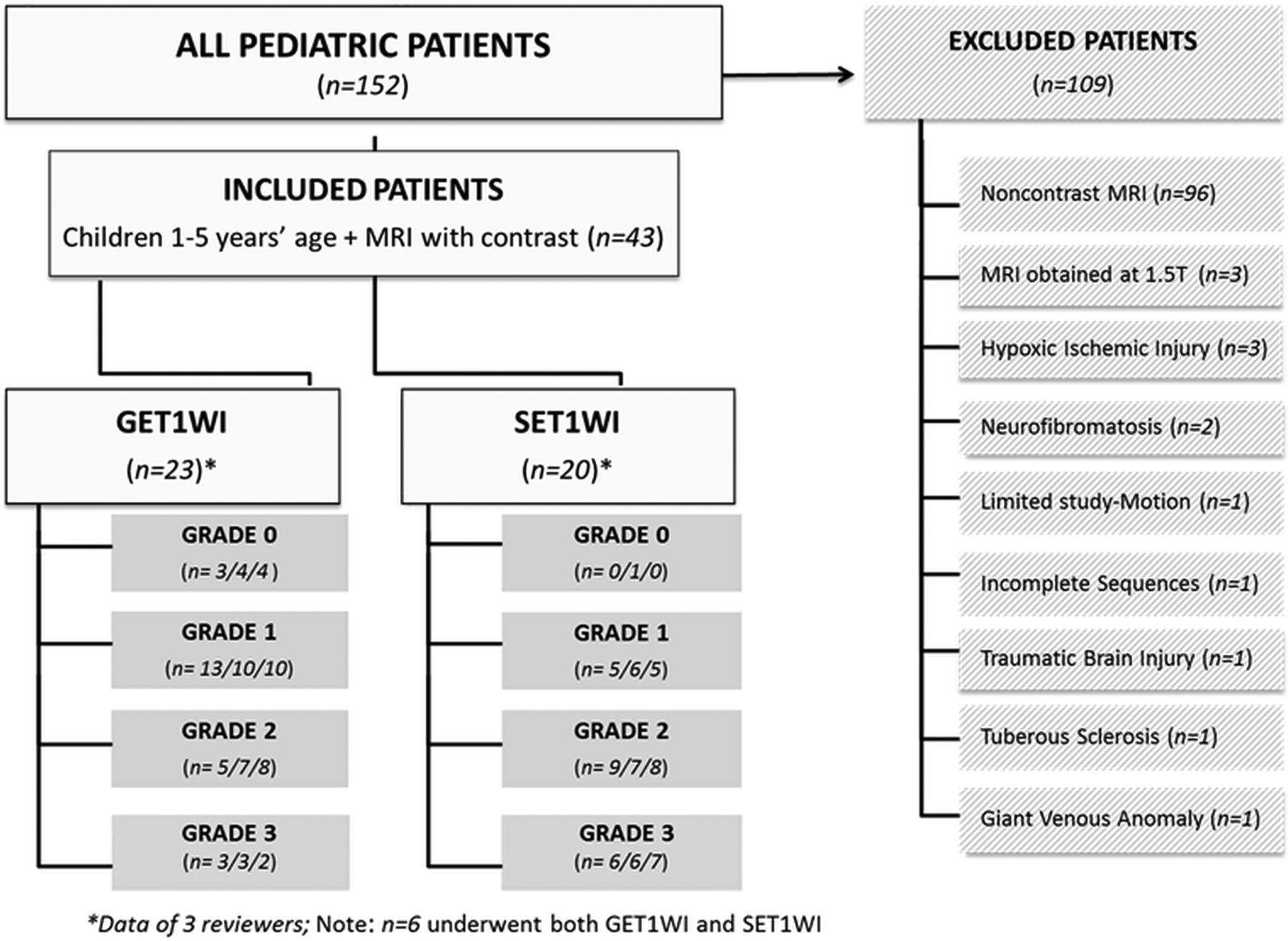
- Fig 1.
Organization chart showing the makeup of 43 children included in this study and grades per reviewer.
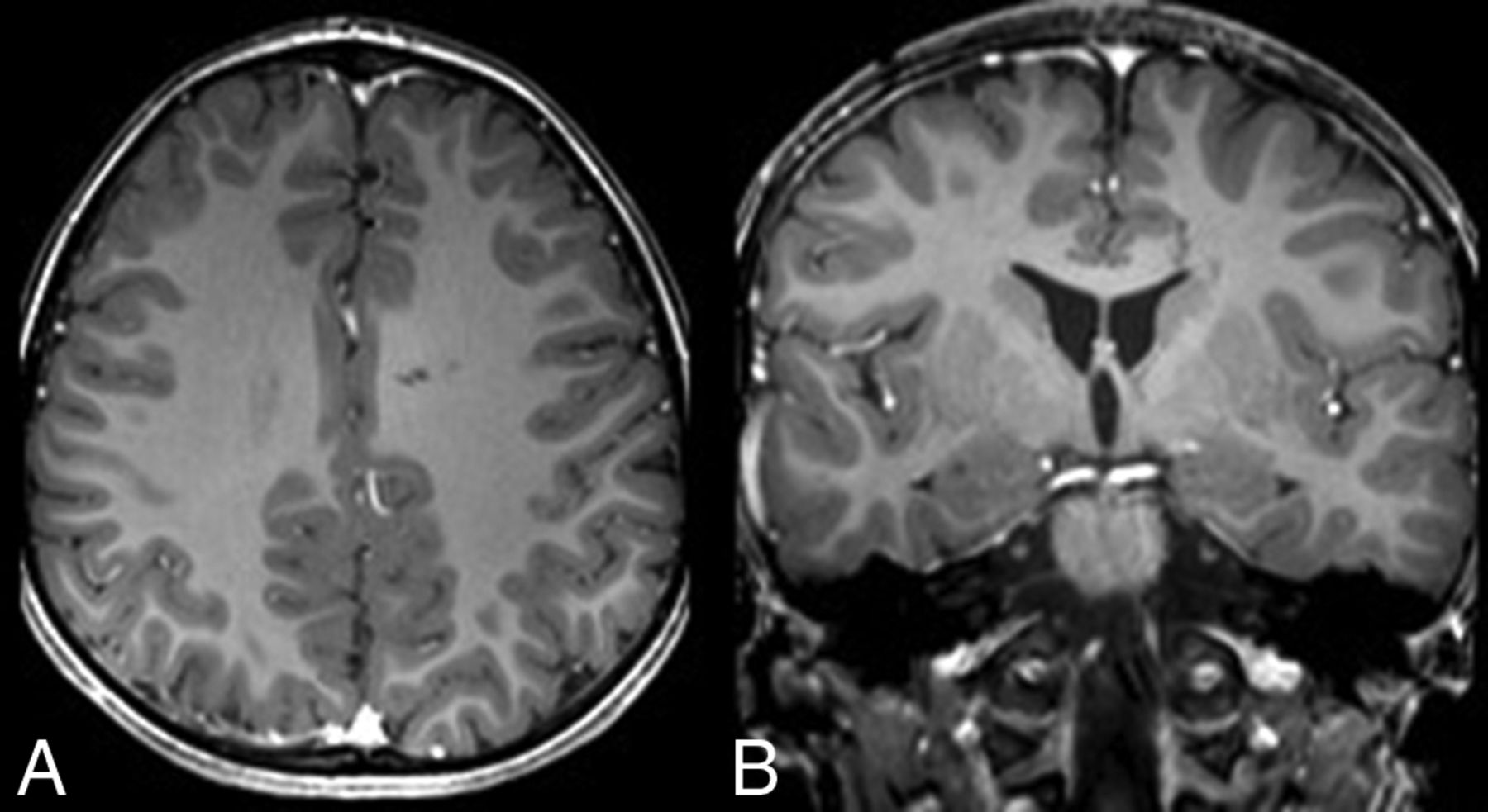
- Fig 2.
Grade 0 pseudo-LMCE in a 2-year-old girl post-trauma. Axial (A) and coronal (B) GE TIWI shows only minimal vasculature within the sulci. This grade of enhancement was present only on GE TIWI in 13%–17%, while no patients were graded as 0 on SE TIWI.
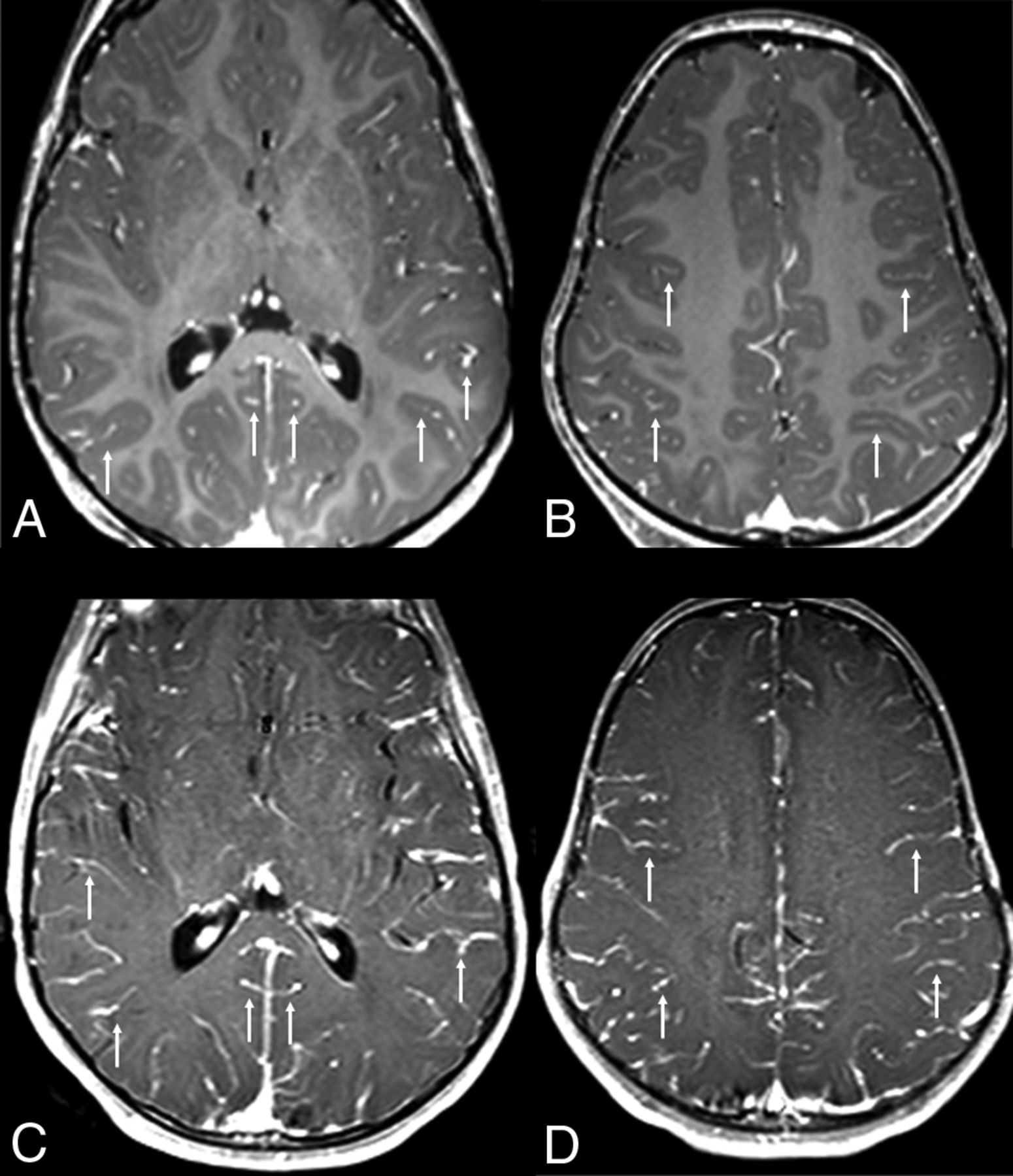
- Fig 3.
Grade 1 pseudo-LMCE on both sequences in a 3-year-old girl with seizures. Pseudo-LMCE appears as small vascular structures (arrows) within the depths of the sulci on GE TIWI axial (A) and coronal (B) images and on SE TIWI axial (C) and coronal (D) images. This grade was more frequent on GE TIWI (43%–57%) than on SE TIWI (25%–30%).
- Fig 4.
Examples of grade 2 pseudo-LMCE, demonstrated on both GE TIWI and SE TIWI in 2 different patients. In a 3-year-old girl with weakness, grade 2 pseudo-LMCE appears as smooth and slightly thickened enhancement (arrows) throughout the depths of the sulci on axial (A) and coronal (B) GE TIWI. In a 5-year-old boy with headaches, there is mildly thickened vasculature diffusely throughout the sulci (arrows) on axial (C) and coronal (D) SE TIWI. Note that grade 2 enhancement was slightly more frequent on SE TIWI (35%–45%) than on GE TIWI (22%–35%).
- Fig 5.
Discrepancy of the LMCE grade between sequences: grade 3 pseudo-LMCE on SE TIWI versus grade 2 on GE TIWI in a 3-year-old boy with fever. A and B, Axial GE TIWI depicts irregular enhancement (arrows) throughout many of the sulci, being slightly thickened, consistent with grade 2 pseudo-LMCE. C and D, Axial SE TIWI in the same patient demonstrates thicker pseudo-LMCE (arrows), appearing nearly nodular or parenchymal in some locations. This case demonstrates how such pseudo-LMCE is typically more prominent on SE TWI because grade 3 LMCE was much more frequent on SE TIWI (30%–35%) than on GE TIWI (8%–13%).


Tables
Parameter Range Mean SD Age (yr) 1.3–5.0 3.1 1.4 Weight (kg) 5.7–24.6 15.3 4.5 Propofol dose (mcg/kg/min) 73–303 192 52 Sedation duration (min) 43–110 66.6 13.8 TTI GE T1WI (min) 8.0–17.0 12.6 2.2 TTI SE T1WI (min) 8.0–17.0 11.0 2.1 LMCE score on SE T1WI 1.9–2.1 2.0 0.8 LMCE score on GE T1WI 1.2–1.4 1.2 0.8 Correlation TTI Overall (SE and GE T1WI) TTI SE T1WI Only TTI GE T1WI Only Weight (kg) Age (yr) Dose/Weight (mg/kg) Duration of Sedation (min) LMCE (ρ) −0.232 to −0.302 −0.358 to −.475 0.016–0.190 −0.366 to −0.418 −0.315 to −0.418 0.103–0.210 0.023–0.147 P value .051–.130 .036b–.122c .371–.940 .003–.011b .004–.032b .151–.484 .318–.875


