
Article Figures & Data
Figures
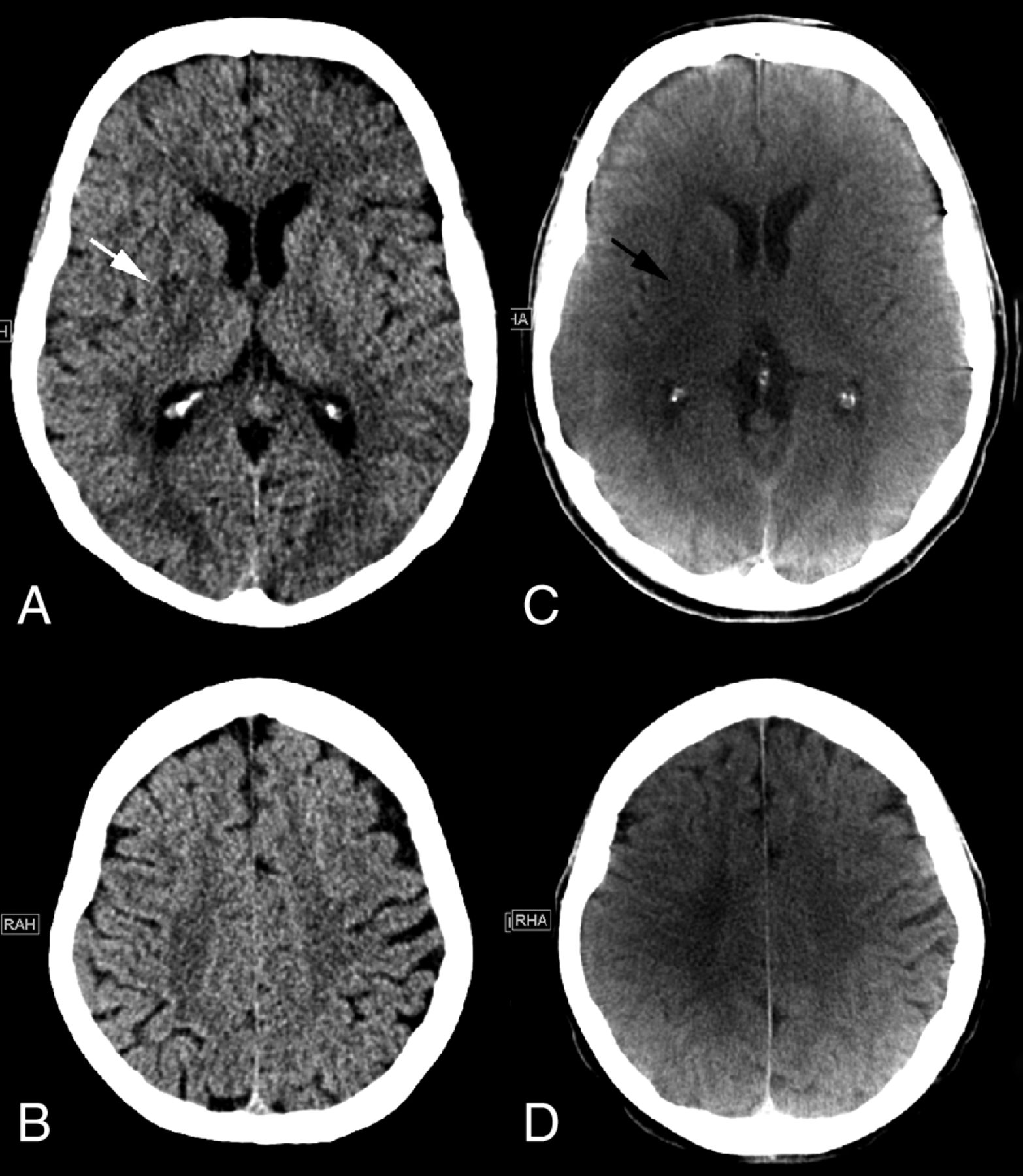
- Fig 1.
Initial MDCT in the peripheral stroke center shows hypodensity of the right lentiform nucleus (white arrow) and no other early ischemic signs on the ganglionic (A) or supraganglionic (B) level. An ASPECTS of 9 was rated on MDCT images. C, FDCT acquired in our comprehensive stroke center 172 minutes after the initial CT shows hypodensities of the right lentiform nucleus (black arrow) and insula. No early ischemic signs were detected on the supraganglionic level, resulting in an ASPECTS of 8.
- Fig 2.
A, Initial MDCT in the peripheral stroke center shows hypodensities of the left striatum, insula, internal capsule, M1, M2, and M3 segments. B, In the supraganglionic levels, we observe early signs in the M4, M5, and M6 segments, resulting in an ASPECTS of 0. C and D, FDCT acquired in our comprehensive stroke center 94 minutes after the initial CT shows early ischemic signs in the left anterior cerebral artery and MCA territories, resulting in an ASPECTS of 2. The M3 and M6 segments are not classified as ischemic on FDCT images.
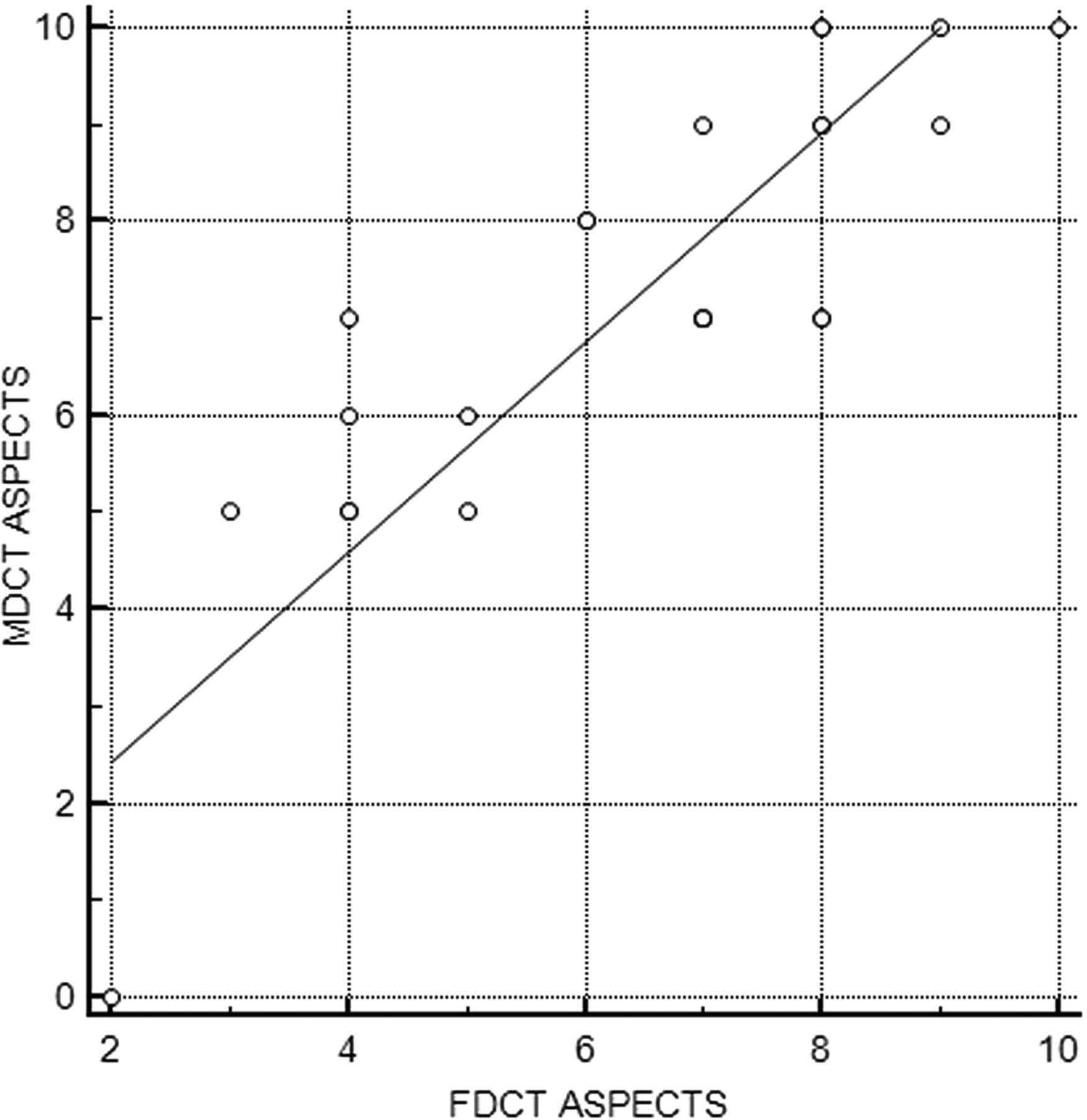
- Fig 3.
Pearson correlation between MDCT and FDCT ASPECTS.
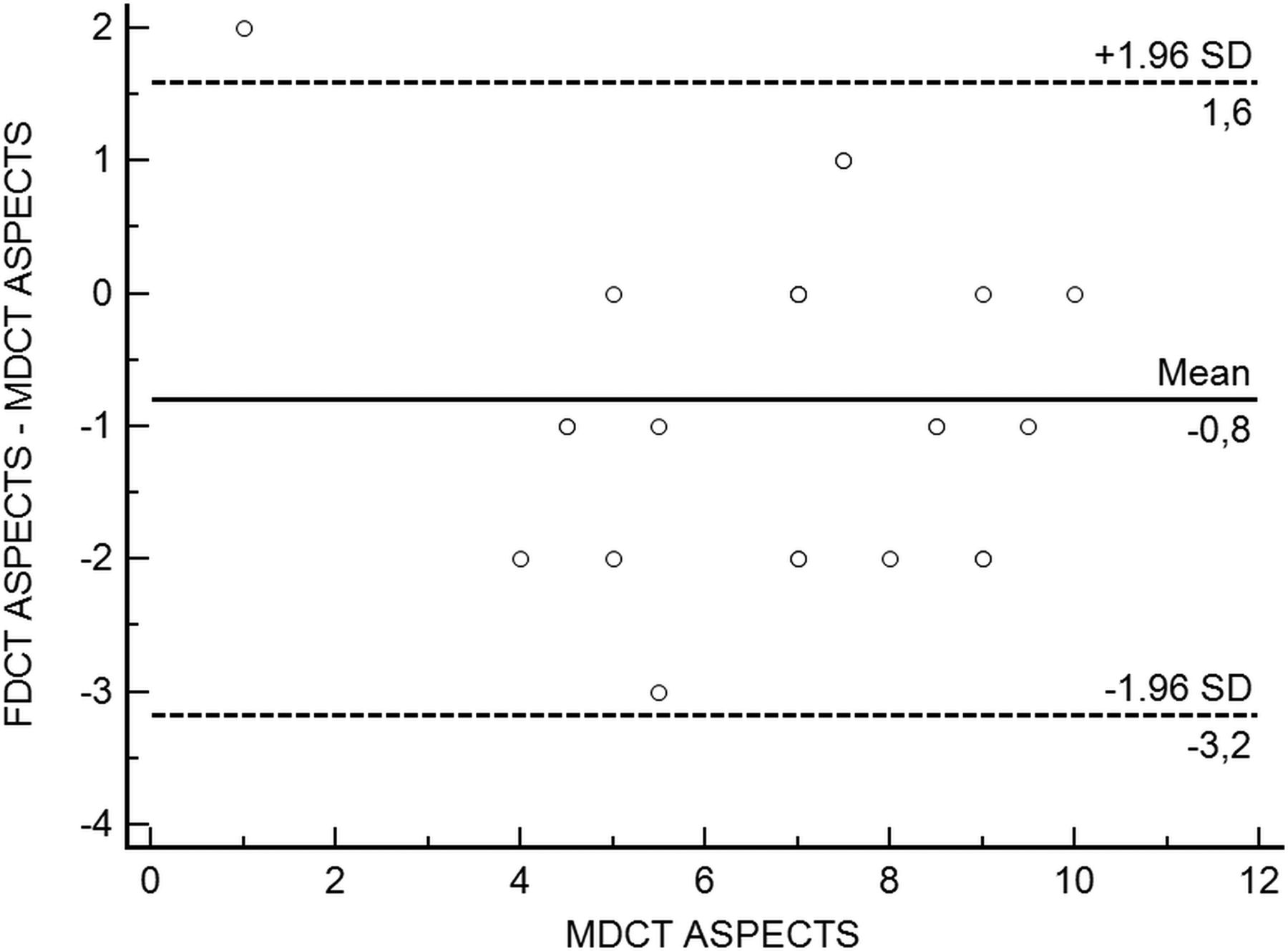
- Fig 4.
Bland-Altman plot of MDCT and FDCT ASPECTS.
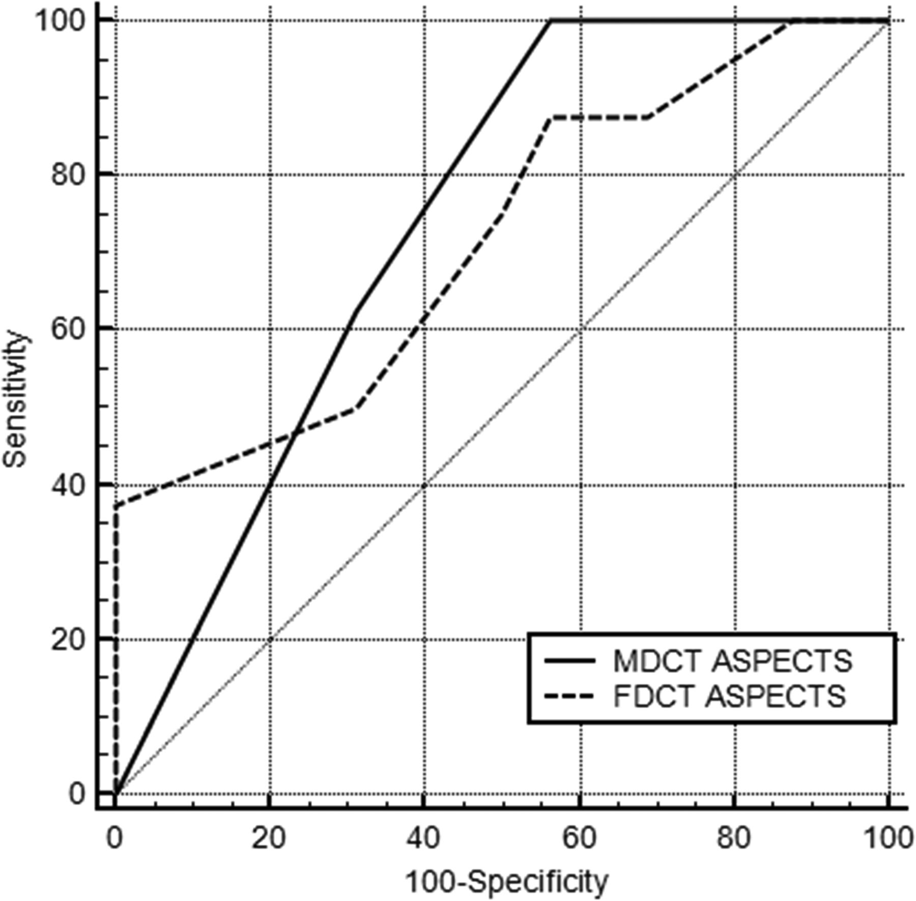
- Fig 5.
Area under the receiver operating curve analysis for the predictive value of MDCT and FDCT ASPECTS for favorable functional outcome (mRS ≤ 2).


Tables
Parameter (n = 25) Age (mean) (yr) 77.3 ± 10.3 Sex (male) (%) 10 (40) Occluded vessel Proximal ICA (No.) (%) 1 (4) Carotid terminus (No.) (%) 11 (44) M1 (No.) (%) 11 (44) M2 (No.) (%) 2 (8) IV thrombolysis (No.) (%) 15 (60) Transfer patientsa Drip and ship (No.) (%) 14 (56) Ship and drip (No.) (%) 1 (4) Just ship (No.) (%) 10 (40) General anesthesia at the time of FDCT (No.) (%) 3 (12) Arterial hypertension (No.) (%) 19 (76) Hyperlipoproteinemia (No.) (%) 11 (44) Diabetes mellitus (No.) (%) 9 (36) Atrial fibrillation (No.) (%) 8 (32) Coronary artery disease (No.) (%) 9 (36) Chronic kidney failure (No.) (%) 7 (28) NIHSS score on admission (median) (IQR) 17 (14–19) mRS score on admission (median) (IQR) 5 (4–5) Successful reperfusion (No.) (%) 22 (88) Symptom-to-groin time (mean) (min) 249.3 ± 57.1 Symptom-to-reperfusion time (n = 22) (mean) (min) 294.6 ± 66.3 Door-to-groin time (mean) (min) 25.2 ± 8.9 Door-to-reperfusion time (n = 22) (mean) (min) 77.3 ± 30 Symptom-to-MDCT time (mean) (min) 99.4 ± 52.6 Symptom-to-FDCT time (mean) (min) 234.9 ± 55.2 MDCT-to-FDCT time (mean) (min) 143.6 ± 49.5 MDCT ASPECTS (median) (IQR) 7 (5.5–9) FDCT ASPECTS (median) (IQR) 7 (4.25–8) ↵a Drip and ship: transfer patients with IV thrombolysis started in a peripheral stroke center; ship and drip: transfer patients with IV thrombolysis started in our tertiary stroke center; just ship: transfer patients without IV thrombolysis.
- Table 3:
Number of patients with early ischemic signs in different regions according to the ASPECTS on MDCT vs FDCT (n = 24)
ASPECTS Early Ischemic Signs on MDCT Early Ischemic Signs on FDCT Early Ischemic Signs on MDCT and FDCT Sensitivity Specificity M1 (No.) (%) 2 (8.3) 3 (12.5) 1 (4.2) 50 90.1 M2 (No.) (%) 2 (8.3) 7 (29.2) 2 (8.3) 100 77.3 M3 (No.) (%) 1 (4.2) 1 (4.2) 0 (0) 0 95.8 C (No.) (%) 14 (58.3) 14 (58.3) 13 (54.2) 92.9 90 L (No.) (%) 15 (62.5) 21 (58.3) 14 (58.3) 93.2 22.2 IC (No.) (%) 3 (12.5) 4 (16.7) 1 (4.2) 33.3 85.7 I (No.) (%) 18 (75) 22 (91.7) 18 (75) 100 33.3 M4 (No.) (%) 5 (20.8) 8 (33.3) 5 (20.8) 100 84.2 M5 (No.) (%) 4 (16.7) 6 (25) 3 (12.5) 75 85 M6 (No.) (%) 3 (12.5) 0 (0) 0 (0) NA NA Overall (No.) (% of 240 locations) 67 (27.9) 86 (35.8) 57 (23.8) 85.1 83.2 Dense media sign (No. positive) (%) 18 (75) 15 (62.5) 13 (54.2) 72.2 66.7 Note:—Anatomic regions according to the Alberta Stroke Program Early CT Score: M1–M6 indicate cortical regions of the medial cerebral artery territory; C, caudate nucleus; L, lentiform nucleus; IC, internal capsule; I, insula; NA, not applicable.


Jump to section
Related Articles
Cited By...
- Motion artifact correction for cone beam CT stroke imaging: a prospective series
- Direct to angiosuite strategy versus standard workflow triage for endovascular therapy: systematic review and meta-analysis
- The butterfly effect: improving brain cone-beam CT image artifacts for stroke assessment using a novel dual-axis trajectory
- Evaluation of Sine Spin flat detector CT imaging compared with multidetector CT
- Direct Transfer to Angiosuite in Acute Stroke: Why, When, and How?