
Article Figures & Data
Figures
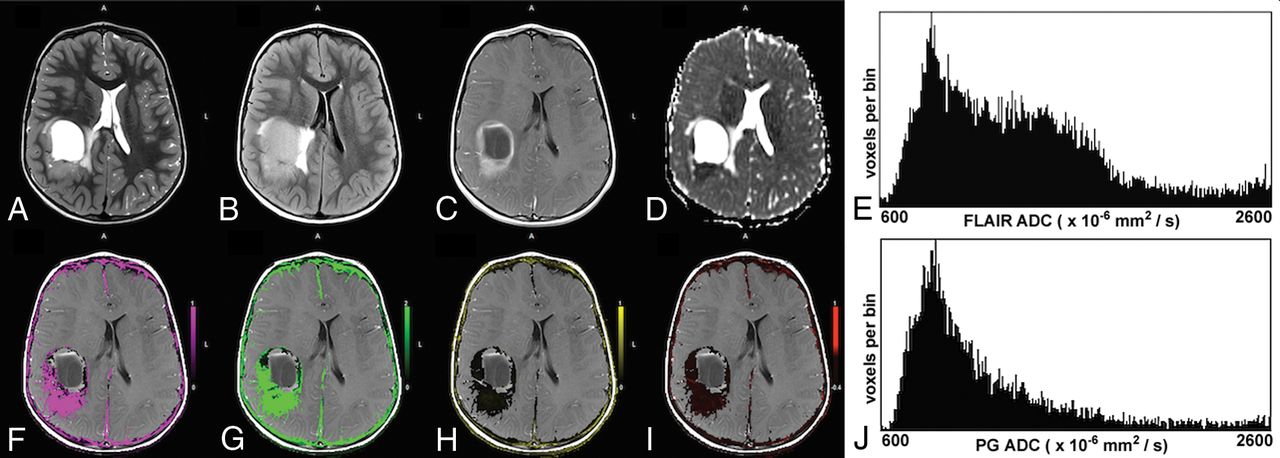
- Fig 1.
A 6-year-old boy with a low-grade pilocytic astrocytoma. The upper row shows the T2-weighted image (A), T2-FLAIR image (B), postcontrast T1-weighted image (C), ADC map (D), and the ADC histogram of the FLAIR tumor volume (E), respectively. The lower row shows the Ktrans map (F), Kep map (G), ve map (H), vp map (I), and the ADC histogram of the enhancing tumor volume (J), respectively. The enhancing tumor volume was defined as the nodular enhancement of the solid component for pilocytic astrocytomas. Note the low Ktrans and Kep and the high ve values in this low-grade tumor. The mean ADC of the FLAIR tumor volume is 1821 × 10−6 mm2/s and 1808 × 10−6 mm2/s for the enhancing tumor volume.
- Fig 2.
A 5-year-old boy with parietal anaplastic ependymoma. The upper row shows the T2-weighted image (A), T2-FLAIR image (B), postcontrast T1-weighted image (C), ADC map (D), and the ADC histogram of the FLAIR tumor volume (E), respectively. The lower row shows the Ktrans map (F), Kep map (G), ve map (H), vp map (I), and the ADC histogram of the enhancing tumor volume (J), respectively. Note the high Ktrans and Kep and the lower ve values in this high-grade tumor. The histogram of the FLAIR tumor volume is bimodal. The mean ADC of the lower peak of the FLAIR tumor volume is 867 × 10−6 mm2/s and 1262 × 10−6 mm2/s for the enhancing tumor volume.
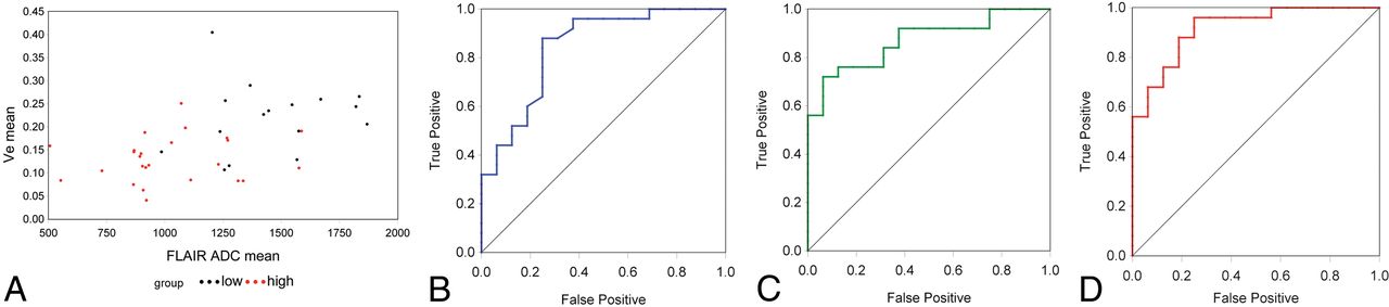
- Fig 3.
A, Scatterplot of ve versus FL_ADC_mean for all 41 cases analyzed. High-grade tumors are shown in red, and low-grade, in black. B, ROC curve for ve (AROC = 0.843). C, ROC curve for FL_ADC_mean (AROC = 0.870). D, ROC curve when ve and FL_ADC_mean are combined (AROC = 0.918). Note the improved accuracy shown for the multiparametric predictor, reflected in the increased AROC.


Tables
Variable Low-Grade (Mean) High-Grade (Mean) Mean Diff Mean SE Lower 95% CL Upper 95% CL P Value (T Test) χ2 (Wilcoxon) P Value (Wilcoxon) Ktrans (min−1) 0.14 1.50 1.36 0.45 0.46 2.27 .004 16.72 <.001 Kep (min−1) 1.18 8.85 7.67 1.99 3.64 11.70 <.001 18.98 <.001 ve 0.22 0.13 −0.09 0.02 −0.13 −0.05 <.001 13.41 <.001 vp 0.05 0.05 0.00 0.01 −0.02 0.02 .764 0.23 .630 FL_ADC_mean (×10−6 mm2/s) 1458 1022 −436 84.47 −607 −265 <.001 15.65 <.001 FL_ADC_SD (×10−6 mm2/s) 336.8 285.0 −51.8 36.01 −125 21.06 .158 .56 .454 FL_ADC_skew 0.45 1.24 0.80 0.26 0.27 1.32 .004 7.55 .006 FL_ADC_kurt 1.28 1.97 0.70 0.73 −0.79 2.18 .347 1.58 .208 PG_ADC_mean (×10−6 mm2/s) 1472 1066 −406 100.7 −610 −203 <.001 11.70 <.001 PG_ADC_SD (×10−6 mm2/s) 261.0 308.4 47.46 40.47 −34.4 129.3 .248 1.93 .165 PG_ADC_skew 0.69 1.30 0.61 0.30 0.01 1.21 .047 2.26 .133 PG_ADC_kurt 2.51 2.31 −0.20 0.93 −2.09 1.70 .835 0.34 .560 Note:—Diff indicates difference; SE, standard error; CL, confidence limit; skew, skewness; kurt, kurtosis.
Predictor AROC SE 95% Lower CL 95% Upper CL T Value P Value Ktrans 0.883 0.052 0.781 0.984 7.40 <.001 Kep 0.908 0.047 0.815 1.0 8.59 <.001 Ve 0.843 0.066 0.713 0.972 5.19 <.001 FL_ADC_mean 0.870 0.055 0.762 0.978 6.74 <.001 PG_ADC_mean 0.820 0.066 0.691 0.949 4.86 <.001 Ve + FL_ADC_mean 0.918 0.043 0.834 1.0 9.81 <.001 Note:—CL indicates confidence limit.


