Advertisement

Main menu

  • Home
  • Content
    • Current Issue
    • Accepted Manuscripts
    • Article Preview
    • Past Issue Archive
    • Video Articles
    • AJNR Case Collection
    • Case of the Week Archive
    • Case of the Month Archive
    • Classic Case Archive
  • Special Collections
    • AJNR Awards
    • Low-Field MRI
    • Alzheimer Disease
    • ASNR Foundation Special Collection
    • Photon-Counting CT
    • View All
  • Multimedia
    • AJNR Podcasts
    • AJNR SCANtastic
    • Trainee Corner
    • MRI Safety Corner
    • Imaging Protocols
  • For Authors
    • Submit a Manuscript
    • Submit a Video Article
    • Submit an eLetter to the Editor/Response
    • Manuscript Submission Guidelines
    • Statistical Tips
    • Fast Publishing of Accepted Manuscripts
    • Graphical Abstract Preparation
    • Imaging Protocol Submission
    • Author Policies
  • About Us
    • About AJNR
    • Editorial Board
    • Editorial Board Alumni
  • More
    • Become a Reviewer/Academy of Reviewers
    • Subscribers
    • Permissions
    • Alerts
    • Feedback
    • Advertisers
    • ASNR Home

User menu

  • Alerts
  • Log in

Search

  • Advanced search
American Journal of Neuroradiology

American Journal of Neuroradiology

ASHNR American Society of Functional Neuroradiology ASHNR American Society of Pediatric Neuroradiology ASSR
  • Alerts
  • Log in

Advanced Search

  • Home
  • Content
    • Current Issue
    • Accepted Manuscripts
    • Article Preview
    • Past Issue Archive
    • Video Articles
    • AJNR Case Collection
    • Case of the Week Archive
    • Case of the Month Archive
    • Classic Case Archive
  • Special Collections
    • AJNR Awards
    • Low-Field MRI
    • Alzheimer Disease
    • ASNR Foundation Special Collection
    • Photon-Counting CT
    • View All
  • Multimedia
    • AJNR Podcasts
    • AJNR SCANtastic
    • Trainee Corner
    • MRI Safety Corner
    • Imaging Protocols
  • For Authors
    • Submit a Manuscript
    • Submit a Video Article
    • Submit an eLetter to the Editor/Response
    • Manuscript Submission Guidelines
    • Statistical Tips
    • Fast Publishing of Accepted Manuscripts
    • Graphical Abstract Preparation
    • Imaging Protocol Submission
    • Author Policies
  • About Us
    • About AJNR
    • Editorial Board
    • Editorial Board Alumni
  • More
    • Become a Reviewer/Academy of Reviewers
    • Subscribers
    • Permissions
    • Alerts
    • Feedback
    • Advertisers
    • ASNR Home
  • Follow AJNR on Twitter
  • Visit AJNR on Facebook
  • Follow AJNR on Instagram
  • Join AJNR on LinkedIn
  • RSS Feeds

AJNR Awards, New Junior Editors, and more. Read the latest AJNR updates

Research ArticlePediatric Neuroimaging

Multiparametric Analysis of Permeability and ADC Histogram Metrics for Classification of Pediatric Brain Tumors by Tumor Grade

S. Vajapeyam, D. Brown, P.R. Johnston, K.I. Ricci, M.W. Kieran, H.G.W. Lidov and T.Y. Poussaint
American Journal of Neuroradiology March 2018, 39 (3) 552-557; DOI: https://doi.org/10.3174/ajnr.A5502
S. Vajapeyam
aFrom the Departments of Radiology (S.V., D.B., P.R.J., T.Y.P.)
eHarvard Medical School (S.V., M.W.K., H.G.W.L., T.Y.P.), Boston, Massachusetts.
  • Find this author on Google Scholar
  • Find this author on PubMed
  • Search for this author on this site
  • ORCID record for S. Vajapeyam
D. Brown
aFrom the Departments of Radiology (S.V., D.B., P.R.J., T.Y.P.)
  • Find this author on Google Scholar
  • Find this author on PubMed
  • Search for this author on this site
  • ORCID record for D. Brown
P.R. Johnston
aFrom the Departments of Radiology (S.V., D.B., P.R.J., T.Y.P.)
  • Find this author on Google Scholar
  • Find this author on PubMed
  • Search for this author on this site
  • ORCID record for P.R. Johnston
K.I. Ricci
cCancer Center (K.I.R.), Massachusetts General Hospital, Boston, Massachusetts
  • Find this author on Google Scholar
  • Find this author on PubMed
  • Search for this author on this site
  • ORCID record for K.I. Ricci
M.W. Kieran
dDivision of Pediatric Oncology (M.W.K.), Dana-Farber Boston Children's Cancer and Blood Disorders Center, Boston, Massachusetts
eHarvard Medical School (S.V., M.W.K., H.G.W.L., T.Y.P.), Boston, Massachusetts.
  • Find this author on Google Scholar
  • Find this author on PubMed
  • Search for this author on this site
  • ORCID record for M.W. Kieran
H.G.W. Lidov
bPathology (H.G.W.L.), Boston Children's Hospital, Boston, Massachusetts
eHarvard Medical School (S.V., M.W.K., H.G.W.L., T.Y.P.), Boston, Massachusetts.
  • Find this author on Google Scholar
  • Find this author on PubMed
  • Search for this author on this site
  • ORCID record for H.G.W. Lidov
T.Y. Poussaint
aFrom the Departments of Radiology (S.V., D.B., P.R.J., T.Y.P.)
eHarvard Medical School (S.V., M.W.K., H.G.W.L., T.Y.P.), Boston, Massachusetts.
  • Find this author on Google Scholar
  • Find this author on PubMed
  • Search for this author on this site
  • ORCID record for T.Y. Poussaint
  • Article
  • Figures & Data
  • Info & Metrics
  • Responses
  • References
  • PDF
Loading

Abstract

BACKGROUND AND PURPOSE: Accurate tumor grading is essential for treatment planning of pediatric brain tumors. We hypothesized that multiparametric analyses of a combination of permeability metrics and ADC histogram metrics would differentiate high- and low-grade tumors with high accuracy.

MATERIALS AND METHODS: DTI and dynamic contrast-enhanced MR imaging using T1-mapping with flip angles of 2°, 5°, 10°, and 15°, followed by a 0.1-mmol/kg body weight gadolinium-based bolus was performed on all patients in addition to standard MR imaging. Permeability data were processed and transfer constant from the blood plasma into the extracellular extravascular space, rate constant from the extracellular extravascular space back into blood plasma, extravascular extracellular volume fraction, and fractional blood plasma volume were calculated from 3D tumor volumes. Apparent diffusion coefficient histogram metrics were calculated for 3 separate tumor volumes derived from T2-FLAIR sequences, T1 contrast-enhanced sequences, and permeability maps, respectively.

RESULTS: Results from 41 patients (0.3–16.76 years of age; mean, 6.22 years) with newly diagnosed contrast-enhancing brain tumors (16 low-grade; 25 high-grade) were included in the institutional review board–approved retrospective analysis. Wilcoxon tests showed a higher transfer constant from blood plasma into extracellular extravascular space and rate constant from extracellular extravascular space back into blood plasma, and lower extracellular extravascular volume fraction (P < .001) in high-grade tumors. The mean ADCs of FLAIR and enhancing tumor volumes were significantly lower in high-grade tumors (P < .001). ROC analysis showed that a combination of extravascular volume fraction and mean ADC of FLAIR volume differentiated high- and low-grade tumors with high accuracy (area under receiver operating characteristic curve = 0.918).

CONCLUSIONS: ADC histogram metrics combined with permeability metrics differentiate low- and high-grade pediatric brain tumors with high accuracy.

ABBREVIATIONS:

AROC
area under receiver operating characteristic curve
DCE-MRI
dynamic contrast-enhanced MR imaging
FL
3D tumor volume from T2-FLAIR images
EES
extracellular extravascular space
Kep
rate constant from EES back into blood plasma
Ktrans
transfer constant from blood plasma into EES
PG
3D tumor volume from T1-contrast-enhanced images
ROC
receiver operating characteristic
ve
extracellular extravascular volume fraction
vp
fractional blood plasma volume

Primary brain tumors have an incidence rate of 5.67 per 100,0001 in children younger than 19 years of age and remain the most common cause of disease-related death in children. Accurate assessment of tumor grade is critical for diagnosis and treatment planning, especially with recent advances in genomic and molecular subtyping of tumors and targeted treatments for different subtypes. Conventional MR imaging remains the technique of choice for diagnosis and treatment; however, advanced imaging techniques such as perfusion and diffusion imaging have also proved useful for physiologic characterization of tumors.

Dynamic contrast-enhanced MR imaging (DCE-MRI) provides information on the vessel permeability and microvasculature in brain tumors and has been used extensively in adults in tumor grading,2⇓⇓–5 but less frequently in children.6,7 DTI provides information about tumor cellularity and is useful for characterization of tissue, tumor grading, tumor response to treatment, and distinction of tissue types.8 ADC histogram analysis provides a comprehensive analysis of the volumetric diffusion characteristics of the tumor and has been used to predict survival in both adults9,10 and children.11

We retrospectively analyzed DCE-MR imaging and DTI data from children presenting with a brain mass during 3 years at Boston Children's Hospital and examined the permeability and ADC histogram metrics as predictors of tumor grade. We hypothesized that a combination of DCE-MR imaging and ADC histogram metrics would improve the accuracy of tumor grading on MR imaging.

Materials and Methods

Subjects

Children who presented with a brain mass from 2013 to 2017 who had DCE-MR imaging and DTI studies were included. The study was performed with the approval of the institutional review board of the Dana Farber Cancer Institute.

MR Imaging Acquisition

All MR imaging studies were performed on a 3T scanner (Skyra; Siemens, Erlangen, Germany). Standard MR imaging in all patients consisted of sagittal T1, axial T2, precontrast axial T2-FLAIR, axial DTI (TR = 6500 ms, TE = 88 ms, slice thickness = 2 mm, b-values of 0 and 1000 s/mm2, 30 gradient directions), and postcontrast T1 images. All patients underwent a DCE-MR imaging protocol as follows: 1) variable flip angle echo-spoiled gradient-echo T1-mapping sequences using flip angles of 15°, 10°, 5°, and 2°; TR = 5 seconds; TE = minimum; FOV = 240 mm; section thickness = 5 mm; and 2) DCE-MR imaging sequence consisting of 50 phases, 7 seconds apart, flip angle = 15°, TR = 4 seconds, TE = minimum. FOV, section thickness, and scan locations were identical to those in the T1-mapping sequences. A single bolus of gadobutrol (0.1-mL/kg body weight) was injected 20 seconds after the start of scanning at an injection rate of 2 mL/s.

DCE-MR Imaging Postprocessing

DCE-MR images were transferred to a VersaVue workstation (In vivo, Gainesville, Florida) for automated processing using OmniLook software. Voxelwise T1 maps were generated from the variable flip angle images, and the 2-compartment Tofts model12 was used to generate voxelwise values of the pharmacokinetic parameters, namely the transfer constant from the blood plasma into the extracellular extravascular space (Ktrans), rate constant from extracellular extravascular space back into blood plasma (Kep), extracellular extravascular volume fraction (ve), and fractional blood plasma volume (vp).

The model of Weinmann et al13 was used for blood plasma concentration. ROIs were drawn on each section of the tumor around contrast-enhancing portions of the tumor by an imaging data analyst or by a PhD scientist and verified by a Certificate of Added Qualification–certified neuroradiologist, and the mean (over voxels) and SDs of each of the variables were recorded. Only enhancing tumors were included in the analysis.

ADC Histogram Analysis

Due to their superior anatomic contrast, axial B0 images from the DTI sequence were registered to the axial T2-FLAIR images using the mutual information algorithm in FSL (http://www.fmrib.ox.ac.uk/fsl)14 and the same transformation matrix used on the ADC maps to register them to the T2-FLAIR images. Axial postcontrast images were also separately registered to T2-FLAIR using the same methodology. 3D ROIs comprising the 3D tumor volume from T2-FLAIR images (FL) and the 3D tumor volume from T1-contrast-enhanced images (PG) were automatically generated using the thresholding feature in Fiji (http://fiji.sc/),15 an Open Source (https://opensource.org/) distribution of Java modules along with ImageJ software (National Institutes of Health, Bethesda, Maryland). The ADC values of the voxels in the 3D tumor volume from T2-FLAIR images (FL), and PG ROIs were then used to generate the FL and PG ADC volumes, respectively. These volumes were thresholded using a uniform range of 600–2600 × 10−6 mm2/s to automatically exclude cyst, necrosis, and hemorrhage. FL and PG ADC histograms were then generated using a uniform bin width of 7.8 mm2/s to facilitate comparison of shape descriptors of the histograms. Histogram metrics used for statistical analysis were the number of peaks (unimodal or bimodal), mean, SD, skewness, and kurtosis of these histograms. In the case of tumors showing bimodal ADC histograms, the lower peak was measured for mean and SD, and skewness and kurtosis were not recorded.

DCE-MR imaging and ADC histogram analyses were blinded to tumor grading results.

Statistical Methods

Low- and high-grade tumor groups were compared for permeability variables and ADC histogram metrics using the Wilcoxon test and t test using the NPAR1WAY and t test procedures of SAS (SAS Institute, Cary, North, Carolina).16 Corresponding to the t test, estimates and 95% confidence intervals for the difference between means were calculated.

Receiver operating characteristic (ROC) curves and their areas (AROC) were estimated for highly statistically significant variables in the permeability set (ve, Ktrans, Kep), ADC histogram set (FL_ADC_mean, PG_ADC_mean), and the 6 pairs of variables comprising 1 variable from each set. Focus was on the ve and FL_ADC_mean pair because these variables and their relationship were considered the most stable.

The ROC curve shows the trade-off between the true-positive rate (classifying the tumor of a patient with a high-grade tumor as high-grade) and false-positive rate (classifying the tumor of a patient with a low-grade tumor as high-grade). The AROC estimates the average true-positive rate over all possible false-positive rates and is also an estimate of the probability of correctly classifying a random pair of patients, 1 from the low-grade group and 1 from the high-grade group.

In addition to point estimates, Wald-based tests (corresponding to the null hypothesis that the true AROC is 0.5) and 95% confidence intervals for the AROC were calculated. All estimates, confidence intervals, and tests were nonparametric and were calculated using the LOGISTIC procedure of SAS.

Results

Sixty-three patients were identified. Six patients had final diagnoses that were not brain tumors, 10 had nonenhancing tumors and were excluded from DCE-MR imaging analysis, and 6 patients were excluded due to motion or susceptibility artifacts. The remaining 41 patients were included.

Twenty-three children were boys, and 18, girls (age range, 0.30–16.76 years; median age, 4.76 years; mean age, 6.22 years). There were 31 infratentorial and 10 supratentorial tumors, and all except 2 were newly diagnosed.

Forty patients had enhancing biopsy-confirmed tumors, and 1 had a diffuse midline glioma located in the pons presumed to be high-grade. World Health Organization grades 1–2 were considered low-grade, whereas grades 3–4 were considered high-grade. Of these tumors, 16 were confirmed low-grade (7 pilocytic astrocytomas, 5 low-grade gliomas, 1 mature teratoma, 1 atypical meningioma, 1 low-grade ganglioglioma, and 1 low-grade mixed germ cell tumor). Twenty-five tumors were high-grade (12 medulloblastomas, 4 glioblastomas, 4 anaplastic ependymomas, and 1 each of atypical teratoid/rhabdoid tumor, embryonal tumor not otherwise specified, choroid plexus carcinoma, embryonal tumor with rhabdoid features, and diffuse midline glioma). Not all final diagnoses were given as the World Health Organization 2016 classification because some cases did not fit well into this classification at the time of diagnosis.

Molecular subtyping was successful in 11 of the 12 patients with medulloblastoma, yielding 3 sonic hedgehog/SHH-activated, 2 wingless/WNT-activated, and 6 classified as group 3 or 4.

There was no statistically significant difference in mean ages between the low-grade (mean, 7.28 years; median, 5.31 years) and the high-grade cohort (mean, 5.54 years; median, 4.57 years) (P = .552).

Group comparisons of all permeability and ADC histogram variables are shown in Table 1. Of the pharmacokinetic variables analyzed, Ktrans and Kep were significantly higher in high-grade tumors compared with low-grade tumors (P < .001), and ve was significantly lower in the high-grade group (P < .001).

View this table:
  • View inline
  • View popup
Table 1:

Group comparisons: low- versus high-grade tumor

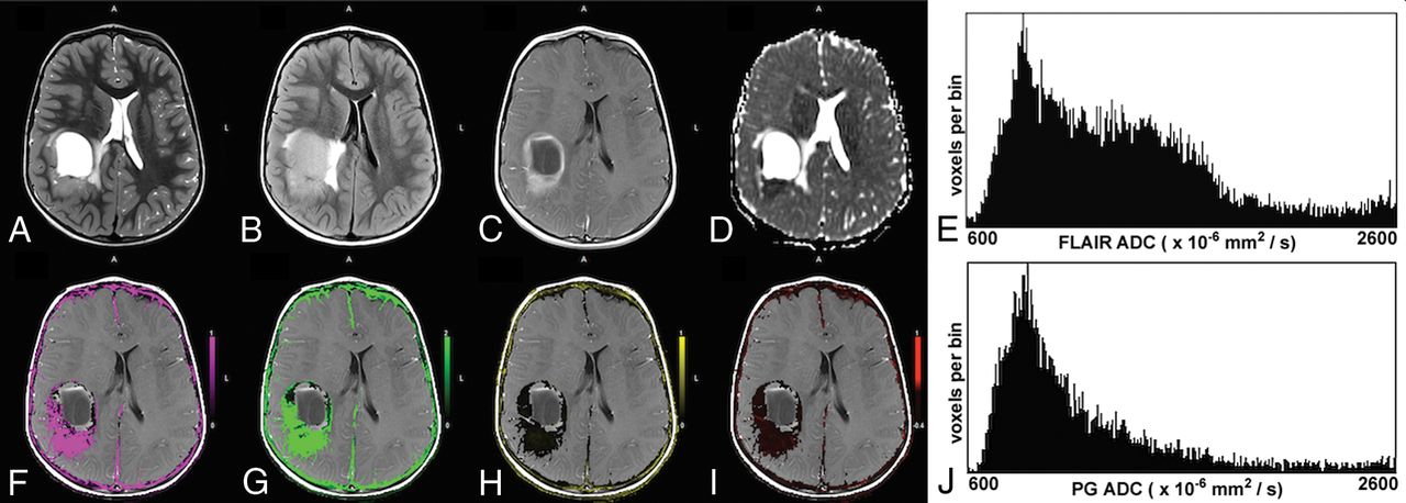
Of the ADC histogram metrics analyzed, the mean ADC of the T2-FLAIR tumor volume (FL_ADC_mean) and the PG tumor volume (PG_ADC_mean) were significantly higher for low-grade tumors (Fig 1) compared with high-grade tumors (P < .001) (Fig 2). Low-grade tumors also showed a tendency for lower skewness for T2-FLAIR tumor volume (FL_ADC_skew, P = .006). None of the other variables showed statistically significant group differences between low- and high-grade tumors. Six high-grade tumors showed bimodal T2-FLAIR ADC histograms, and 4 showed bimodal PG ADC histograms, while all the ADC histograms for low-grade tumors had just a single peak.

Fig 1.
  • Download figure
  • Open in new tab
  • Download powerpoint
Fig 1.

A 6-year-old boy with a low-grade pilocytic astrocytoma. The upper row shows the T2-weighted image (A), T2-FLAIR image (B), postcontrast T1-weighted image (C), ADC map (D), and the ADC histogram of the FLAIR tumor volume (E), respectively. The lower row shows the Ktrans map (F), Kep map (G), ve map (H), vp map (I), and the ADC histogram of the enhancing tumor volume (J), respectively. The enhancing tumor volume was defined as the nodular enhancement of the solid component for pilocytic astrocytomas. Note the low Ktrans and Kep and the high ve values in this low-grade tumor. The mean ADC of the FLAIR tumor volume is 1821 × 10−6 mm2/s and 1808 × 10−6 mm2/s for the enhancing tumor volume.

Fig 2.
  • Download figure
  • Open in new tab
  • Download powerpoint
Fig 2.

A 5-year-old boy with parietal anaplastic ependymoma. The upper row shows the T2-weighted image (A), T2-FLAIR image (B), postcontrast T1-weighted image (C), ADC map (D), and the ADC histogram of the FLAIR tumor volume (E), respectively. The lower row shows the Ktrans map (F), Kep map (G), ve map (H), vp map (I), and the ADC histogram of the enhancing tumor volume (J), respectively. Note the high Ktrans and Kep and the lower ve values in this high-grade tumor. The histogram of the FLAIR tumor volume is bimodal. The mean ADC of the lower peak of the FLAIR tumor volume is 867 × 10−6 mm2/s and 1262 × 10−6 mm2/s for the enhancing tumor volume.

No group differences were found among the molecular subtypes in the medulloblastoma cohort (P ≥ .294).

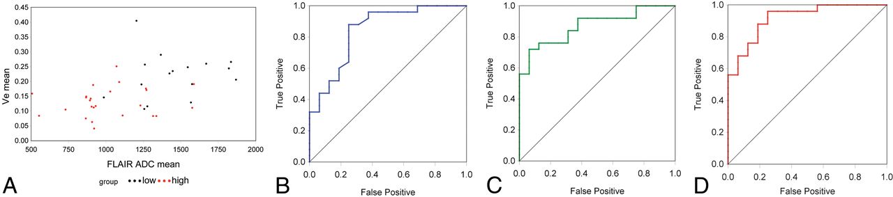
ROC analysis of the 5 highly statistically significant variables (Ktrans, Kep, ve, FL_ADC_mean, and PG_ADC_mean) showed that all displayed high sensitivity and specificity in discriminating low- and high-grade tumors, with AROC values above 0.82 (P < .001) as seen in Table 2. AROC values for ve and FL_ADC_mean were 0.843 and 0.870, respectively, while the AROC estimate for the 2-variable combination of these metrics was 0.918 (Fig 3). Focus was on this pair because the individual variables and their relationship were considered the most stable.

View this table:
  • View inline
  • View popup
Table 2:

Predictors of AROC

Fig 3.
  • Download figure
  • Open in new tab
  • Download powerpoint
Fig 3.

A, Scatterplot of ve versus FL_ADC_mean for all 41 cases analyzed. High-grade tumors are shown in red, and low-grade, in black. B, ROC curve for ve (AROC = 0.843). C, ROC curve for FL_ADC_mean (AROC = 0.870). D, ROC curve when ve and FL_ADC_mean are combined (AROC = 0.918). Note the improved accuracy shown for the multiparametric predictor, reflected in the increased AROC.

The Spearman correlation between ve and FL_ADC_mean across the 41 tumors was 0.49 (P = .001). Correlation within the low- and high-grade groups was not significant.

Discussion

Multiparametric imaging of pediatric brain tumors provides physiologic information beyond standard MR imaging. Koob et al7 recently reported increased accuracy in differentiating grades I and IV pediatric brain tumors using a combination of diffusion and DSC perfusion metrics. Similarly, Morana et al17 showed improved prognostic capability by combining DWI, arterial spin-labeling, and PET data in pediatric diffuse astrocytic tumors. Other studies have demonstrated the advantage of combinations of DWI, DSC perfusion, MR spectroscopy, and PET data in studying pediatric brain tumors.18⇓–20 To our knowledge, this is the first study to demonstrate a multiparametric approach using DCE and ADC histogram metrics in pediatric brain tumors.

While DSC and arterial spin-labeling perfusion metrics have been reported for pediatric brain tumor grading,21⇓–23 there are fewer studies using DCE-MR imaging.6 Our results show that among the DCE-MR imaging parameters, Ktrans, Kep, and ve all successfully distinguished low- and high-grade tumors, and this result is consistent with our report from a smaller cohort.6 High-grade tumors exhibited higher values of Ktrans and Kep, indicating leaky vessels. This finding is consistent with the angiogenesis seen in high-grade tumors caused by neoangiogenic proteins such as vascular endothelial growth factor. Our finding of lower ve in high-grade tumors differs from reports in adult glioblastomas,3,24,25 likely due to the multiple different high-grade tumor types in children. Jensen et al,26 however, found that adult patients with gliomas with lower ve in peritumoral edema had significantly shorter overall survival rates, supporting our hypothesis that lower ve corresponds to a higher grade and more aggressive tumor.

Of the ADC histogram metrics we tested, FL_ADC_mean and PG_ADC_mean showed significant differences between low- and high-grade tumors, with lower ADC corresponding to high-grade tumors. Lower ADC indicates high tumor cellularity and has been associated with shorter survival as well.9⇓–11 ADC histogram skewness and kurtosis did not show the same significant differences. Although the number of ADC histograms with bimodal peaks was too small for statistical analysis (6 for FL_ADC_mean and 4 for PG_ADC_mean), all tumors exhibiting bimodal peaks were high-grade. High-grade tumors often yield bimodal ADC distributions due to the presence of peritumoral edema.9⇓–11

We did not detect any group differences among the molecular subtypes of medulloblastoma, which may be due to the small sample size. Recent studies have reported other distinguishing MR imaging features of some of these subtypes,27⇓–29 and a larger future sample size may show differences in permeability and ADC histogram metrics among the subtypes. We would postulate that the group 3 tumors, which are ill-defined and have the poorest prognosis,30 would have high Ktrans and Kep and low ve with low mean ADC histogram values.

Because ve is a measure of the extracellular extravascular space and ADC is also indicative of extracellular space, we tested for a correlation between ADC and ve. ADC and ve were highly correlated across the entire cohort (P = .001). Although a previous study was unable to find voxelwise correlations between ve and ADC in adult gliomas,31 our findings suggest a link between the mean values in pediatric brain tumors, possibly due to the different tumor types seen in children.

Permeability metrics assess tumor microvasculature, whereas ADC histogram metrics assess tumor cellularity. We hypothesized that these modalities may provide complementary information to assist in tumor grading and tested combinations of permeability and ADC variables as predictors of tumor grade using ROC curves. A combination of ve and FL_ADC_mean showed a higher AROC (0.918) compared with ve (0.843) or FL_ADC_mean (0.870) alone. Thus, multiparametric analysis may yield the best predictors of tumor grade in pediatric brain tumors.

One limitation of the study is the relatively small number and heterogeneity of tumors studied. Larger numbers of individual tumor types are necessary to validate these findings across all tumor types. The addition of PET19 and MR spectroscopy7,18 has also shown promise and may benefit future analyses. Future prospective work could include other multiparametric approaches incorporating ADC histogram and T1 permeability to differentiate molecular and genomic subtypes of pediatric brain tumors.

Conclusions

The permeability metrics Ktrans, Kep, and ve and the ADC histogram metrics FL_ADC_mean and PG_ADC_mean were able to distinguish low- and high-grade pediatric brain tumors with a high degree of accuracy. A multiparametric approach combining permeability and ADC histogram metrics improves the accuracy of pediatric brain tumor grading with high sensitivity and specificity.

Footnotes

  • Disclosures: Tina Young Poussaint—UNRELATED: Grants/Grants Pending: Pediatric Brain Tumor Consortium Neuroimaging Center, Comments: National Institutes of Health grant with subcontract*; Royalties: Springer Science for a book. *Money paid to the institution.

  • Paper previously presented at: Annual Meeting of the American Society of Neuroradiology and the Foundation of the ASNR Symposium, April 22–27, 2017; Long Beach, California.

References

  1. 1.↵
    1. Ostrom QT,
    2. Gittleman H,
    3. Xu J, et al
    . CBTRUS Statistical Report: Primary Brain and Other Central Nervous System Tumors Diagnosed in the United States in 2009–2013. Neuro Oncol 2016;18:v1–v75 doi:10.1093/neuonc/now207 pmid:28475809
    CrossRefPubMed
  2. 2.↵
    1. Falk A,
    2. Fahlström M,
    3. Rostrup E, et al
    . Discrimination between glioma grades II and III in suspected low-grade gliomas using dynamic contrast-enhanced and dynamic susceptibility contrast perfusion MR imaging: a histogram analysis approach. Neuroradiology 2014;56:1031–38 doi:10.1007/s00234-014-1426-z pmid:25204450
    CrossRefPubMed
  3. 3.↵
    1. Li X,
    2. Zhu Y,
    3. Kang H, et al
    . Glioma grading by microvascular permeability parameters derived from dynamic contrast-enhanced MRI and intratumoral susceptibility signal on susceptibility weighted imaging. Cancer Imaging 2015;15:4 doi:10.1186/s40644-015-0039-z pmid:25889239
    CrossRefPubMed
  4. 4.↵
    1. Roberts HC,
    2. Roberts TP,
    3. Ley S, et al
    . Quantitative estimation of microvascular permeability in human brain tumors: correlation of dynamic Gd-DTPA–enhanced MR imaging with histopathologic grading. Acad Radiol 2002;9:S151–55 doi:10.1016/S1076-6332(03)80425-7 pmid:12019855
    CrossRefPubMed
  5. 5.↵
    1. Zhang N,
    2. Zhang L,
    3. Qiu B, et al
    . Correlation of volume transfer coefficient Ktrans with histopathologic grades of gliomas. J Magn Reson Imaging 2012;36:355–63 doi:10.1002/jmri.23675 pmid:22581762
    CrossRefPubMed
  6. 6.↵
    1. Vajapeyam S,
    2. Stamoulis C,
    3. Ricci K, et al
    . Automated processing of dynamic contrast-enhanced MRI: correlation of advanced pharmacokinetic metrics with tumor grade in pediatric brain tumors. AJNR Am J Neuroradiol 2017;38:170–75 doi:10.3174/ajnr.A4949 pmid:27633807
    Abstract/FREE Full Text
  7. 7.↵
    1. Koob M,
    2. Girard N,
    3. Ghattas B, et al
    . The diagnostic accuracy of multiparametric MRI to determine pediatric brain tumor grades and types. J Neurooncol 2016;127:345–53 doi:10.1007/s11060-015-2042-4 pmid:26732081
    CrossRefPubMed
  8. 8.↵
    1. Poussaint TY,
    2. Rodriguez D
    . Advanced neuroimaging of pediatric brain tumors: MR diffusion, MR perfusion, and MR spectroscopy. Neuroimaging Clin N Am 2006;16:169–92, ix doi:10.1016/j.nic.2005.11.005 pmid:16543091
    CrossRefPubMed
  9. 9.↵
    1. Pope WB,
    2. Qiao XJ,
    3. Kim HJ, et al
    . Apparent diffusion coefficient histogram analysis stratifies progression-free and overall survival in patients with recurrent GBM treated with bevacizumab: a multi-center study. J Neurooncol 2012;108:491–98 doi:10.1007/s11060-012-0847-y pmid:22426926
    CrossRefPubMed
  10. 10.↵
    1. Rahman R,
    2. Hamdan A,
    3. Zweifler R, et al
    . Histogram analysis of apparent diffusion coefficient within enhancing and nonenhancing tumor volumes in recurrent glioblastoma patients treated with bevacizumab. J Neurooncol 2014;119:149–58 doi:10.1007/s11060-014-1464-8 pmid:24805151
    CrossRefPubMed
  11. 11.↵
    1. Poussaint TY,
    2. Vajapeyam S,
    3. Ricci KI, et al
    . Apparent diffusion coefficient histogram metrics correlate with survival in diffuse intrinsic pontine glioma: a report from the Pediatric Brain Tumor Consortium. Neuro Oncol 2016;18:725–34 doi:10.1093/neuonc/nov256 pmid:26487690
    CrossRefPubMed
  12. 12.↵
    1. Tofts PS
    . Modeling tracer kinetics in dynamic Gd-DTPA MR imaging. J Magn Reson Imaging 1997;7:91–101 doi:10.1002/jmri.1880070113 pmid:9039598
    CrossRefPubMed
  13. 13.↵
    1. Weinmann HJ,
    2. Laniado M,
    3. Mützel W
    . Pharmacokinetics of GdDTPA/dimeglumine after intravenous injection into healthy volunteers. Physiol Chem Phys Med NMR 1984;16:167–72 pmid:6505043
    PubMed
  14. 14.↵
    1. Jenkinson M,
    2. Beckmann CF,
    3. Behrens TE, et al
    . FSL. Neuroimage 2012;62:782–90 doi:10.1016/j.neuroimage.2011.09.015 pmid:21979382
    CrossRefPubMed
  15. 15.↵
    1. Schindelin J,
    2. Arganda-Carreras I,
    3. Frise E, et al
    . Fiji: an open-source platform for biological-image analysis. Nat Methods 2012;9:676–82 doi:10.1038/nmeth.2019 pmid:22743772
    CrossRefPubMed
  16. 16.↵
    SAS/STAT 14.1 User's Guide. July 2015. http://support.sas.com/documentation/cdl/en/statug/68162/HTML/default/viewer.htm#titlepage.htm. Updated November 2, 2015
  17. 17.↵
    1. Morana G,
    2. Piccardo A,
    3. Tortora D, et al
    . Grading and outcome prediction of pediatric diffuse astrocytic tumors with diffusion and arterial spin labeling perfusion MRI in comparison with 18F-DOPA PET. Eur J Nucl Med Mol Imaging 2017;44:2084–89 doi:10.1007/s00259-017-3777-2 pmid:28752225
    CrossRefPubMed
  18. 18.↵
    1. Tzika AA,
    2. Astrakas LG,
    3. Zarifi MK, et al
    . Multiparametric MR assessment of pediatric brain tumors. Neuroradiology 2003;45:1–10 doi:10.1007/s00234-002-0865-0 pmid:12525947
    CrossRefPubMed
  19. 19.↵
    1. Zukotynski KA,
    2. Fahey FH,
    3. Vajapeyam S, et al
    . Exploratory evaluation of MR permeability with 18F-FDG PET mapping in pediatric brain tumors: a report from the Pediatric Brain Tumor Consortium. J Nucl Med 2013;54:1237–43 doi:10.2967/jnumed.112.115782 pmid:23801675
    Abstract/FREE Full Text
  20. 20.↵
    1. Tensaouti F,
    2. Ducassou A,
    3. Chaltiel L, et al
    ; radiotherapy committee of the French Society for Childhood Cancer (SFCE). Prognostic and predictive values of diffusion and perfusion MRI in paediatric intracranial ependymomas in a large national study. Br J Radiol 2016;89:20160537 doi:10.1259/bjr.20160537 pmid:27550423
    CrossRefPubMed
  21. 21.↵
    1. Ho CY,
    2. Cardinal JS,
    3. Kamer AP, et al
    . Relative cerebral blood volume from dynamic susceptibility contrast perfusion in the grading of pediatric primary brain tumors. Neuroradiology 2015;57:299–306 doi:10.1007/s00234-014-1478-0 pmid:25504266
    CrossRefPubMed
  22. 22.↵
    1. Ho CY,
    2. Cardinal JS,
    3. Kamer AP, et al
    . Contrast leakage patterns from dynamic susceptibility contrast perfusion MRI in the grading of primary pediatric brain tumors. AJNR Am J Neuroradiol 2016;37:544–51 doi:10.3174/ajnr.A4559 pmid:26564438
    Abstract/FREE Full Text
  23. 23.↵
    1. Yeom KW,
    2. Mitchell LA,
    3. Lober RM, et al
    . Arterial spin-labeled perfusion of pediatric brain tumors. AJNR Am J Neuroradiol 2014;35:395–401 doi:10.3174/ajnr.A3670 pmid:23907239
    Abstract/FREE Full Text
  24. 24.↵
    1. Abe T,
    2. Mizobuchi Y,
    3. Nakajima K, et al
    . Diagnosis of brain tumors using dynamic contrast-enhanced perfusion imaging with a short acquisition time. Springerplus 2015;4:88 doi:10.1186/s40064-015-0861-6 pmid:25793147
    CrossRefPubMed
  25. 25.↵
    1. Choi HS,
    2. Kim AH,
    3. Ahn SS, et al
    . Glioma grading capability: comparisons among parameters from dynamic contrast-enhanced MRI and ADC value on DWI. Korean J Radiol 2013;14:487–92 doi:10.3348/kjr.2013.14.3.487 pmid:23690718
    CrossRefPubMed
  26. 26.↵
    1. Jensen RL,
    2. Mumert ML,
    3. Gillespie DL, et al
    . Preoperative dynamic contrast-enhanced MRI correlates with molecular markers of hypoxia and vascularity in specific areas of intratumoral microenvironment and is predictive of patient outcome. Neuro Oncol 2014;16:280–91 doi:10.1093/neuonc/not148 pmid:24305704
    CrossRefPubMed
  27. 27.↵
    1. Perreault S,
    2. Ramaswamy V,
    3. Achrol AS, et al
    . MRI surrogates for molecular subgroups of medulloblastoma. AJNR Am J Neuroradiol 2014;35:1263–69 doi:10.3174/ajnr.A3990 pmid:24831600
    Abstract/FREE Full Text
  28. 28.↵
    1. Lastowska M,
    2. Jurkiewicz E,
    3. Trubicka J, et al
    . Contrast enhancement pattern predicts poor survival for patients with non-WNT/SHH medulloblastoma tumours. J Neurooncol 2015;123:65–73 doi:10.1007/s11060-015-1779-0 pmid:25862008
    CrossRefPubMed
  29. 29.↵
    1. Yeom KW,
    2. Mobley BC,
    3. Lober RM, et al
    . Distinctive MRI features of pediatric medulloblastoma subtypes. AJR Am J Roentgenol 2013;200:895–903 doi:10.2214/AJR.12.9249 pmid:23521467
    CrossRefPubMed
  30. 30.↵
    1. Ramaswamy V,
    2. Northcott PA,
    3. Taylor MD
    . FISH and chips: the recipe for improved prognostication and outcomes for children with medulloblastoma. Cancer Genet 2011;204:577–88 doi:10.1016/j.cancergen.2011.11.001 pmid:22200083
    CrossRefPubMed
  31. 31.↵
    1. Mills SJ,
    2. Soh C,
    3. Rose CJ, et al
    . Candidate biomarkers of extravascular extracellular space: a direct comparison of apparent diffusion coefficient and dynamic contrast-enhanced MR imaging–derived measurement of the volume of the extravascular extracellular space in glioblastoma multiforme. AJNR Am J Neuroradiol 2010;31:549–53 doi:10.3174/ajnr.A1844 pmid:19850765
    Abstract/FREE Full Text
  • Received August 18, 2017.
  • Accepted after revision October 30, 2017.
  • © 2018 by American Journal of Neuroradiology
View Abstract
PreviousNext
Back to top

In this issue

American Journal of Neuroradiology: 39 (3)
American Journal of Neuroradiology
Vol. 39, Issue 3
1 Mar 2018
  • Table of Contents
  • Index by author
  • Complete Issue (PDF)
Advertisement
Print
Download PDF
Email Article

Thank you for your interest in spreading the word on American Journal of Neuroradiology.

NOTE: We only request your email address so that the person you are recommending the page to knows that you wanted them to see it, and that it is not junk mail. We do not capture any email address.

Enter multiple addresses on separate lines or separate them with commas.
Multiparametric Analysis of Permeability and ADC Histogram Metrics for Classification of Pediatric Brain Tumors by Tumor Grade
(Your Name) has sent you a message from American Journal of Neuroradiology
(Your Name) thought you would like to see the American Journal of Neuroradiology web site.
CAPTCHA
This question is for testing whether or not you are a human visitor and to prevent automated spam submissions.
Cite this article
S. Vajapeyam, D. Brown, P.R. Johnston, K.I. Ricci, M.W. Kieran, H.G.W. Lidov, T.Y. Poussaint
Multiparametric Analysis of Permeability and ADC Histogram Metrics for Classification of Pediatric Brain Tumors by Tumor Grade
American Journal of Neuroradiology Mar 2018, 39 (3) 552-557; DOI: 10.3174/ajnr.A5502

Citation Manager Formats

  • BibTeX
  • Bookends
  • EasyBib
  • EndNote (tagged)
  • EndNote 8 (xml)
  • Medlars
  • Mendeley
  • Papers
  • RefWorks Tagged
  • Ref Manager
  • RIS
  • Zotero
0 Responses
Respond to this article
Share
Bookmark this article
Multiparametric Analysis of Permeability and ADC Histogram Metrics for Classification of Pediatric Brain Tumors by Tumor Grade
S. Vajapeyam, D. Brown, P.R. Johnston, K.I. Ricci, M.W. Kieran, H.G.W. Lidov, T.Y. Poussaint
American Journal of Neuroradiology Mar 2018, 39 (3) 552-557; DOI: 10.3174/ajnr.A5502
del.icio.us logo Twitter logo Facebook logo Mendeley logo
  • Tweet Widget
  • Facebook Like
  • Google Plus One
Purchase

Jump to section

  • Article
    • Abstract
    • ABBREVIATIONS:
    • Materials and Methods
    • Results
    • Discussion
    • Conclusions
    • Footnotes
    • References
  • Figures & Data
  • Info & Metrics
  • Responses
  • References
  • PDF

Related Articles

  • No related articles found.
  • PubMed
  • Google Scholar

Cited By...

  • Differentiating Low-Grade from High-Grade Intracranial Ependymomas: Comparison of Dynamic Contrast-Enhanced MRI and Diffusion-Weighted Imaging
  • ADC Histogram Analysis of Pediatric Low-Grade Glioma Treated with Selumetinib: A Report from the Pediatric Brain Tumor Consortium
  • Crossref (14)
  • Google Scholar

This article has been cited by the following articles in journals that are participating in Crossref Cited-by Linking.

  • Comparison of Feature Selection Methods and Machine Learning Classifiers for Radiomics Analysis in Glioma Grading
    Pan Sun, Defeng Wang, Vincent Ct Mok, Lin Shi
    IEEE Access 2019 7
  • Targeting PI3K/Akt/mTOR signaling pathway by polyphenols: Implication for cancer therapy
    Mohammad Mirza-Aghazadeh-Attari, Elyad Mohammadi Ekrami, Seyyed Ali Mousavi Aghdas, Ainaz Mihanfar, Shahin Hallaj, Bahman Yousefi, Amin Safa, Maryam Majidinia
    Life Sciences 2020 255
  • Multimodal Disentangled Variational Autoencoder With Game Theoretic Interpretability for Glioma Grading
    Jianhong Cheng, Min Gao, Jin Liu, Hailin Yue, Hulin Kuang, Jun Liu, Jianxin Wang
    IEEE Journal of Biomedical and Health Informatics 2022 26 2
  • Prediction of Glioma Grade Using Intratumoral and Peritumoral Radiomic Features From Multiparametric MRI Images
    Jianhong Cheng, Jin Liu, Hailin Yue, Harrison Bai, Yi Pan, Jianxin Wang
    IEEE/ACM Transactions on Computational Biology and Bioinformatics 2022 19 2
  • Posterior fossa tumors in children: Radiological tips &amp; tricks in the age of genomic tumor classification and advance MR technology
    Basile Kerleroux, Jean Philippe Cottier, Kévin Janot, Antoine Listrat, Dominique Sirinelli, Baptiste Morel
    Journal of Neuroradiology 2020 47 1
  • Quantitative Assessment of Bladder Cancer Reflects Grade and Recurrence: Comparing of Three Methods of Positioning Region of Interest for ADC Measurements at Diffusion-weighted MR Imaging
    Hongyi Li, Lin Liu, Lei Ding, Zhenming Zhang, Mengchao Zhang
    Academic Radiology 2019 26 9
  • The role of artificial intelligence in paediatric neuroradiology
    Catherine Pringle, John-Paul Kilday, Ian Kamaly-Asl, Stavros Michael Stivaros
    Pediatric Radiology 2022 52 11
  • Application of Apparent Diffusion Coefficient Histogram Metrics for Differentiation of Pediatric Posterior Fossa Tumors
    Fabrício Guimarães Gonçalves, Alireza Zandifar, Jorge Du Ub Kim, Luis Octavio Tierradentro-García, Adarsh Ghosh, Dmitry Khrichenko, Savvas Andronikou, Arastoo Vossough
    Clinical Neuroradiology 2022 32 4
  • Texture Analysis to Differentiate Malignant Renal Tumors in Children Using Gray-Scale Ultrasonography Images
    Hyun Joo Shin, Jin Young Kwak, Eunjung Lee, Mi-Jung Lee, Haesung Yoon, Kyunghwa Han, Myung-Joon Kim
    Ultrasound in Medicine & Biology 2019 45 8
  • ADC Histogram Analysis of Pediatric Low-Grade Glioma Treated with Selumetinib: A Report from the Pediatric Brain Tumor Consortium
    S. Vajapeyam, D. Brown, A. Ziaei, S. Wu, G. Vezina, J.S. Stern, A. Panigrahy, Z. Patay, B. Tamrazi, J.Y. Jones, S.S. Haque, D.S. Enterline, S. Cha, B.V. Jones, K.W. Yeom, A. Onar-Thomas, I.J. Dunkel, M. Fouladi, J.R. Fangusaro, T.Y. Poussaint
    American Journal of Neuroradiology 2022 43 3

More in this TOC Section

  • Comparing MRI Perfusion in Pediatric Brain Tumors
  • Frontal Paraventricular Cysts
  • Sodium MRI in Pediatric Brain Tumors
Show more PEDIATRIC NEUROIMAGING

Similar Articles

Advertisement

Indexed Content

  • Current Issue
  • Accepted Manuscripts
  • Article Preview
  • Past Issues
  • Editorials
  • Editor's Choice
  • Fellows' Journal Club
  • Letters to the Editor
  • Video Articles

Cases

  • Case Collection
  • Archive - Case of the Week
  • Archive - Case of the Month
  • Archive - Classic Case

More from AJNR

  • Trainee Corner
  • Imaging Protocols
  • MRI Safety Corner
  • Book Reviews

Multimedia

  • AJNR Podcasts
  • AJNR Scantastics

Resources

  • Turnaround Time
  • Submit a Manuscript
  • Submit a Video Article
  • Submit an eLetter to the Editor/Response
  • Manuscript Submission Guidelines
  • Statistical Tips
  • Fast Publishing of Accepted Manuscripts
  • Graphical Abstract Preparation
  • Imaging Protocol Submission
  • Evidence-Based Medicine Level Guide
  • Publishing Checklists
  • Author Policies
  • Become a Reviewer/Academy of Reviewers
  • News and Updates

About Us

  • About AJNR
  • Editorial Board
  • Editorial Board Alumni
  • Alerts
  • Permissions
  • Not an AJNR Subscriber? Join Now
  • Advertise with Us
  • Librarian Resources
  • Feedback
  • Terms and Conditions
  • AJNR Editorial Board Alumni

American Society of Neuroradiology

  • Not an ASNR Member? Join Now

© 2025 by the American Society of Neuroradiology All rights, including for text and data mining, AI training, and similar technologies, are reserved.
Print ISSN: 0195-6108 Online ISSN: 1936-959X

Powered by HighWire