Advertisement

Main menu

  • Home
  • Content
    • Current Issue
    • Accepted Manuscripts
    • Article Preview
    • Past Issue Archive
    • Video Articles
    • AJNR Case Collection
    • Case of the Week Archive
    • Case of the Month Archive
    • Classic Case Archive
  • Special Collections
    • AJNR Awards
    • Low-Field MRI
    • Alzheimer Disease
    • ASNR Foundation Special Collection
    • Photon-Counting CT
    • View All
  • Multimedia
    • AJNR Podcasts
    • AJNR SCANtastic
    • Trainee Corner
    • MRI Safety Corner
    • Imaging Protocols
  • For Authors
    • Submit a Manuscript
    • Submit a Video Article
    • Submit an eLetter to the Editor/Response
    • Manuscript Submission Guidelines
    • Statistical Tips
    • Fast Publishing of Accepted Manuscripts
    • Graphical Abstract Preparation
    • Imaging Protocol Submission
    • Author Policies
  • About Us
    • About AJNR
    • Editorial Board
    • Editorial Board Alumni
  • More
    • Become a Reviewer/Academy of Reviewers
    • Subscribers
    • Permissions
    • Alerts
    • Feedback
    • Advertisers
    • ASNR Home

User menu

  • Alerts
  • Log in

Search

  • Advanced search
American Journal of Neuroradiology

American Journal of Neuroradiology

ASHNR American Society of Functional Neuroradiology ASHNR American Society of Pediatric Neuroradiology ASSR
  • Alerts
  • Log in

Advanced Search

  • Home
  • Content
    • Current Issue
    • Accepted Manuscripts
    • Article Preview
    • Past Issue Archive
    • Video Articles
    • AJNR Case Collection
    • Case of the Week Archive
    • Case of the Month Archive
    • Classic Case Archive
  • Special Collections
    • AJNR Awards
    • Low-Field MRI
    • Alzheimer Disease
    • ASNR Foundation Special Collection
    • Photon-Counting CT
    • View All
  • Multimedia
    • AJNR Podcasts
    • AJNR SCANtastic
    • Trainee Corner
    • MRI Safety Corner
    • Imaging Protocols
  • For Authors
    • Submit a Manuscript
    • Submit a Video Article
    • Submit an eLetter to the Editor/Response
    • Manuscript Submission Guidelines
    • Statistical Tips
    • Fast Publishing of Accepted Manuscripts
    • Graphical Abstract Preparation
    • Imaging Protocol Submission
    • Author Policies
  • About Us
    • About AJNR
    • Editorial Board
    • Editorial Board Alumni
  • More
    • Become a Reviewer/Academy of Reviewers
    • Subscribers
    • Permissions
    • Alerts
    • Feedback
    • Advertisers
    • ASNR Home
  • Follow AJNR on Twitter
  • Visit AJNR on Facebook
  • Follow AJNR on Instagram
  • Join AJNR on LinkedIn
  • RSS Feeds

AJNR Awards, New Junior Editors, and more. Read the latest AJNR updates

Research ArticleNeurointervention

Quantitative Analysis of Conebeam CT for Delineating Stents in Stent-Assisted Coil Embolization

T. Kuriyama, N. Sakai, M. Beppu, C. Sakai, H. Imamura, K. Masago, N. Katakami and H. Isoda
American Journal of Neuroradiology March 2018, 39 (3) 488-493; DOI: https://doi.org/10.3174/ajnr.A5533
T. Kuriyama
aFrom the Department of Radiological and Medical Laboratory Sciences (T.K., H. Isoda), Nagoya University Graduate School of Medicine, Nagoya, Japan
bDivisions of Radiological Technology (T.K.)
  • Find this author on Google Scholar
  • Find this author on PubMed
  • Search for this author on this site
  • ORCID record for T. Kuriyama
N. Sakai
cNeuroendovascular Therapy (N.S., C.S.)
eDivision of Neurosurgery (N.S., M.B., H. Imamura), Kobe City Medical Center General Hospital, Kobe, Japan
  • Find this author on Google Scholar
  • Find this author on PubMed
  • Search for this author on this site
  • ORCID record for N. Sakai
M. Beppu
eDivision of Neurosurgery (N.S., M.B., H. Imamura), Kobe City Medical Center General Hospital, Kobe, Japan
  • Find this author on Google Scholar
  • Find this author on PubMed
  • Search for this author on this site
  • ORCID record for M. Beppu
C. Sakai
cNeuroendovascular Therapy (N.S., C.S.)
  • Find this author on Google Scholar
  • Find this author on PubMed
  • Search for this author on this site
  • ORCID record for C. Sakai
H. Imamura
eDivision of Neurosurgery (N.S., M.B., H. Imamura), Kobe City Medical Center General Hospital, Kobe, Japan
  • Find this author on Google Scholar
  • Find this author on PubMed
  • Search for this author on this site
  • ORCID record for H. Imamura
K. Masago
fDepartment of Pathology and Molecular Diagnostics (K.M.), Aichi Cancer Center, Nagoya, Japan
  • Find this author on Google Scholar
  • Find this author on PubMed
  • Search for this author on this site
  • ORCID record for K. Masago
N. Katakami
dIntegrated Oncology (N.K.), Institute of Biomedical Research and Innovation, Kobe, Japan
  • Find this author on Google Scholar
  • Find this author on PubMed
  • Search for this author on this site
  • ORCID record for N. Katakami
H. Isoda
aFrom the Department of Radiological and Medical Laboratory Sciences (T.K., H. Isoda), Nagoya University Graduate School of Medicine, Nagoya, Japan
gBrain & Mind Research Center (H. Isoda), Nagoya University, Nagoya, Japan.
  • Find this author on Google Scholar
  • Find this author on PubMed
  • Search for this author on this site
  • ORCID record for H. Isoda
  • Article
  • Figures & Data
  • Info & Metrics
  • Responses
  • References
  • PDF
Loading

Article Figures & Data

Figures

  • Tables
  • Fig 1.
    • Download figure
    • Open in new tab
    • Download powerpoint
    Fig 1.

    3D-MIP images of phantom stents at various concentrations of contrast medium.

  • Fig 2.
    • Download figure
    • Open in new tab
    • Download powerpoint
    Fig 2.

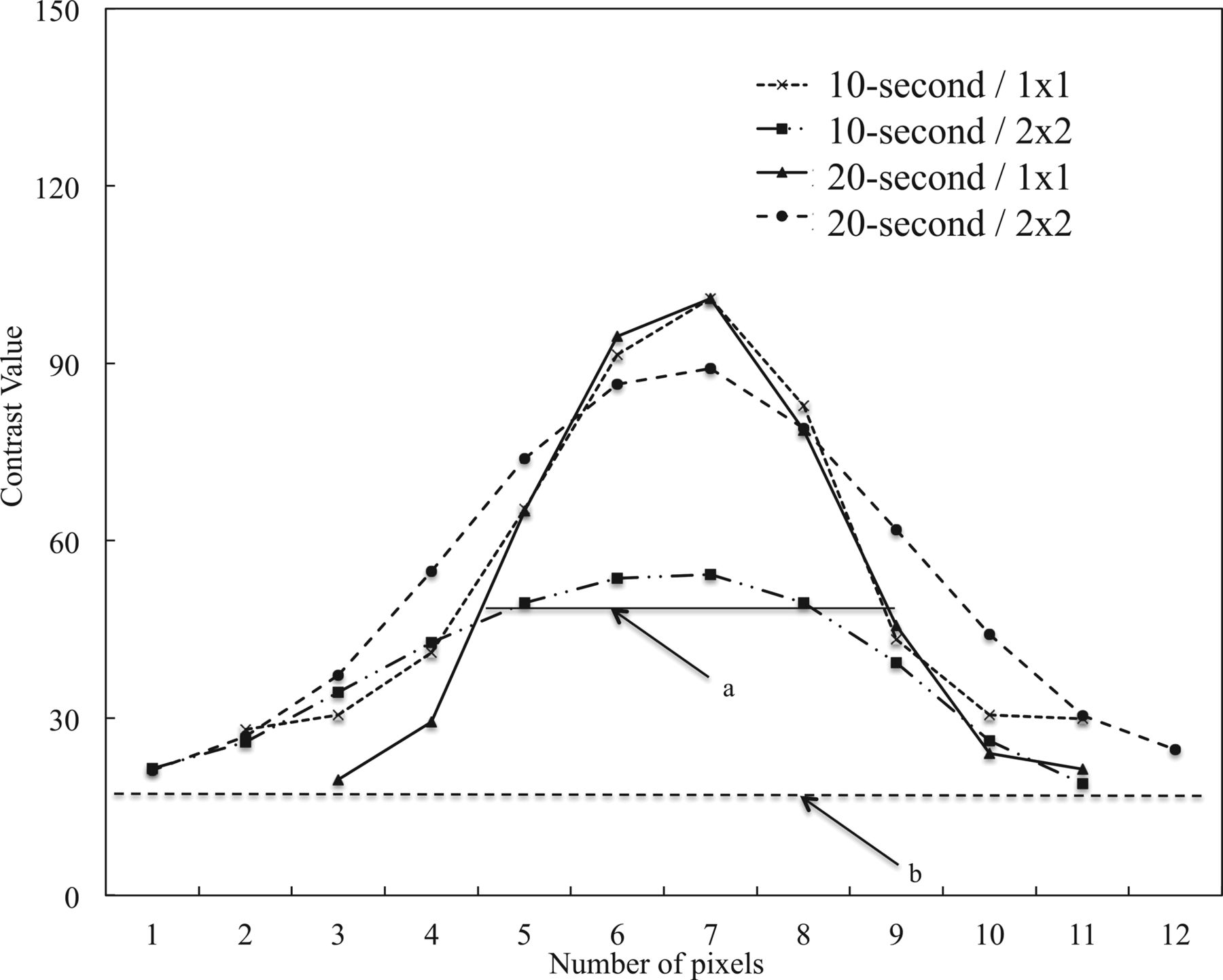
    CNR of a phantom stent imaged at various settings (10- or 20-second acquisition time, 1 × 1 or 2 × 2 binning) plotted against increasing concentrations of contrast medium.

  • Fig 3.
    • Download figure
    • Open in new tab
    • Download powerpoint
    Fig 3.

    FWHM values of the phantom stent images obtained in saline at various settings (10- or 20-second acquisition time; 1 × 1 or 2 × 2 binning).

  • Fig 4.
    • Download figure
    • Open in new tab
    • Download powerpoint
    Fig 4.

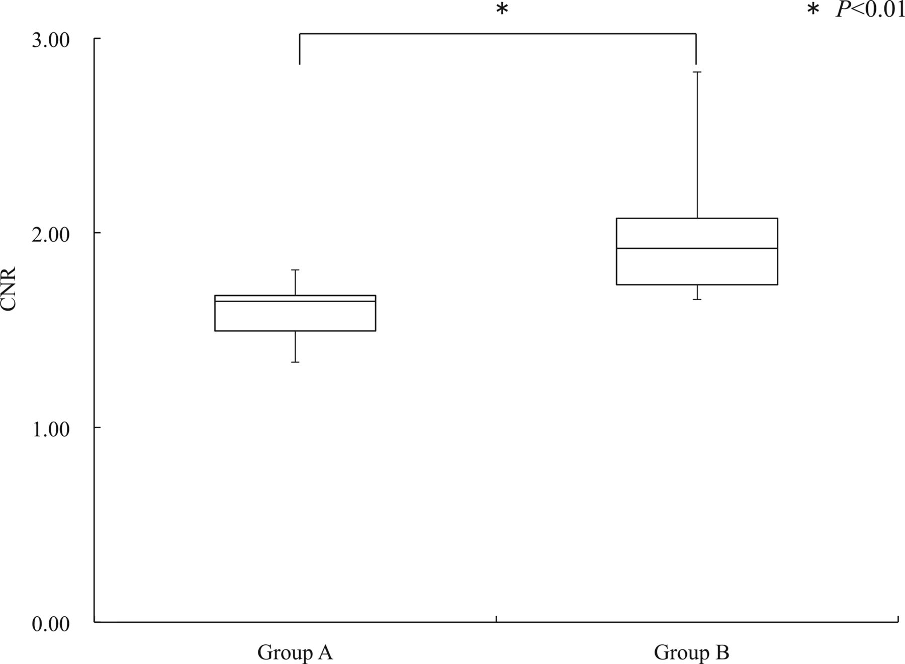
    CNR of stent struts imaged with CBCT with a 15% concentration of contrast medium in groups A and B.
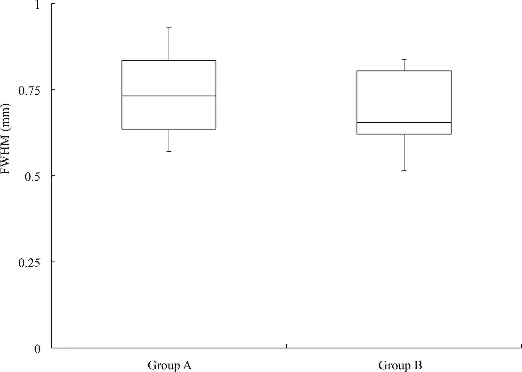
  • Fig 5.
    • Download figure
    • Open in new tab
    • Download powerpoint
    Fig 5.

    FWHM of the stent strut on CBCT images with a 15% concentration of contrast medium in groups A and B.
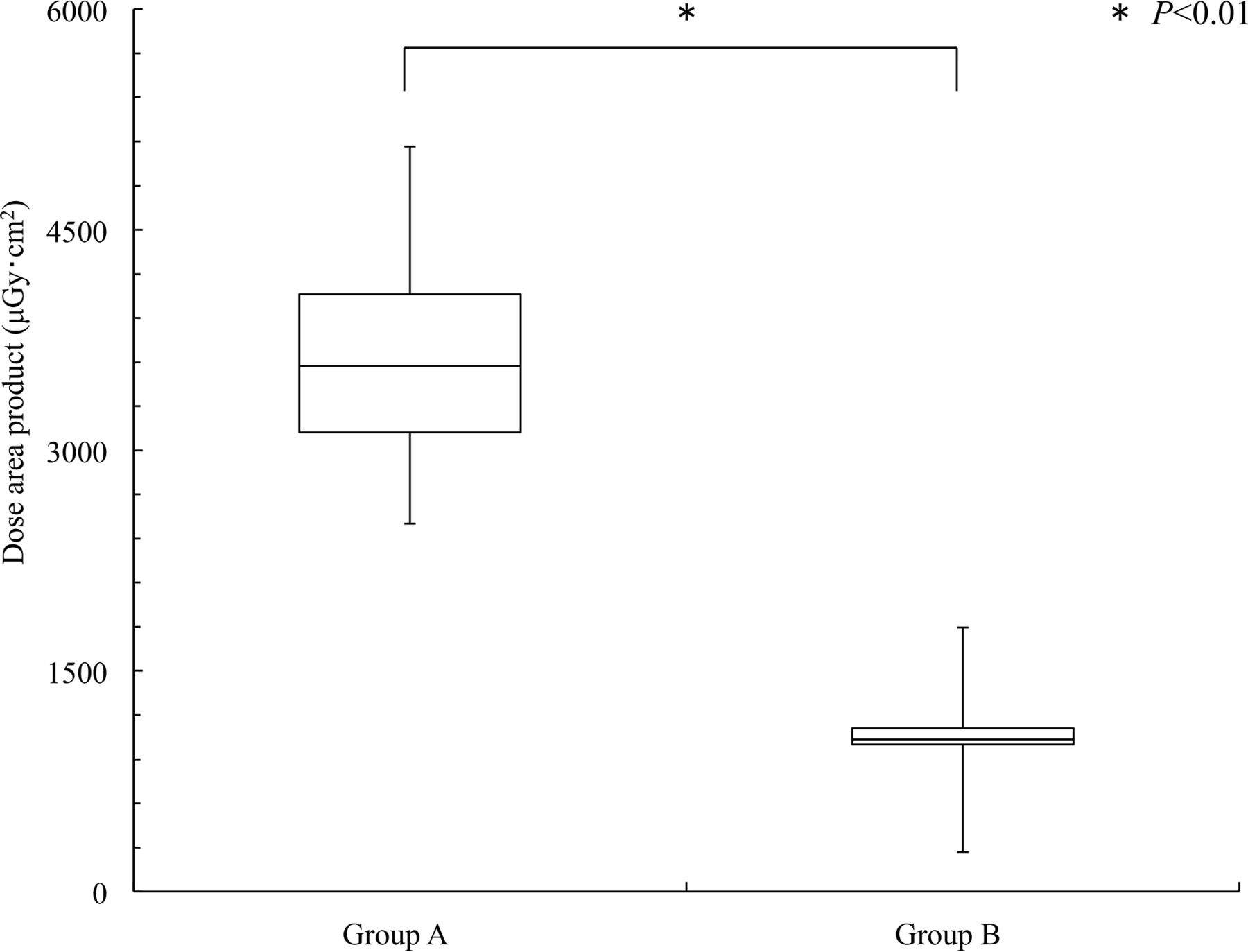
  • Fig 6.
    • Download figure
    • Open in new tab
    • Download powerpoint
    Fig 6.

    The DAP values in groups A and B.
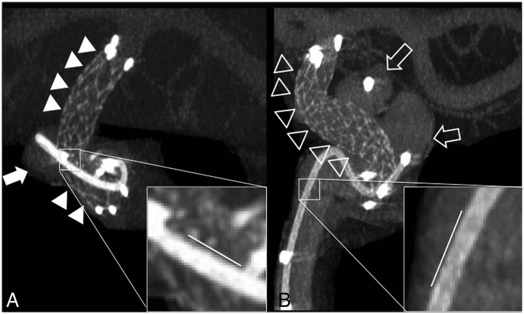
  • Fig 7.
    • Download figure
    • Open in new tab
    • Download powerpoint
    Fig 7.

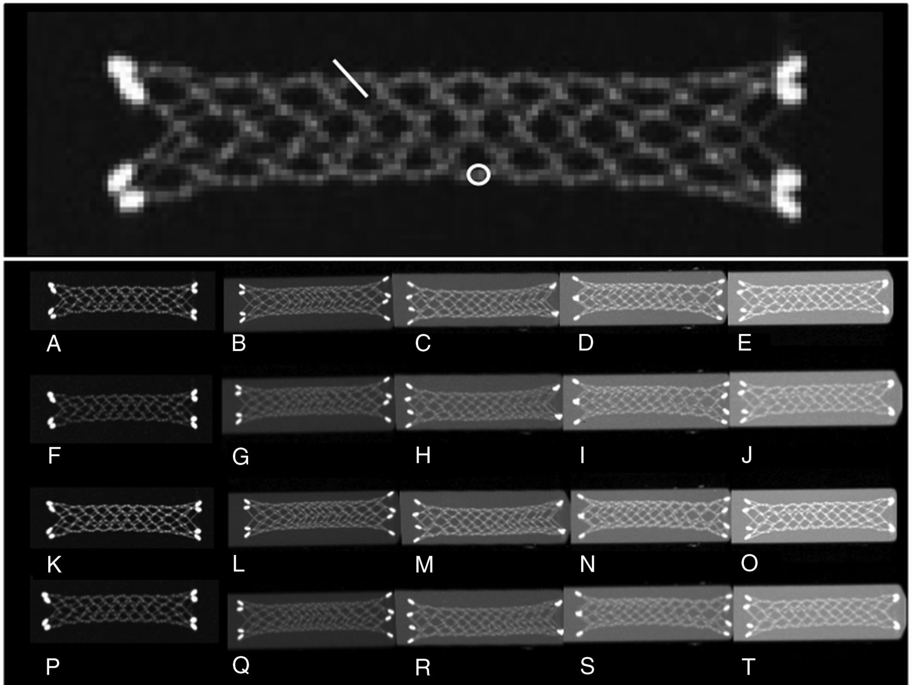
    3D-MIP images of 2 clinical cases imaged with different acquisition times and binning settings and a 15% concentration of contrast medium.

Tables

  • Figures
    • View popup
    Table 1:

    Imaging parameters and conditions for CBCT

    10-Second CBCT20-Second CBCT
    Exposure parameters
        Tube voltage70 kV70 kV
        Pulse width12.5 ms12.5 ms
        Radiation exposure1.20 μGy/pulse1.20 μGy/pulse
        Binning/focus size/FOV1 × 1/0.4 mm/22 cm
    2 × 2/0.6 mm/42 cm
    Acquisition parameters
        Angle step0.8°/frame0.4°/frame
        Angle200°200°
        Total frame250500
        Matrix size/binning9602/2 × 2
    10242/1 × 1
    Injection condition
        Injection rate1.0 mL/s1.0 mL/s
        Concentration of contrast medium14%14%
        X-ray delay time4.5 seconds4.5 seconds
        Injection time of contrast medium14.5 seconds24.5 seconds
    • View popup
    Table 2:

    P values for differences in contrast-to-noise ratios of phantom stent struts by acquisition time/binning level at various contrast medium concentrations

    Acquisition Combination (Sec/Binning vs Sec/Binning)Concentration of Contrast Medium
    Just Saline10%15%20%
    10/1 × 1 vs 10/2 × 2.9328.5888.2733.2734
    10/1 × 1 vs 20/1 × 1.9419.9828.2595.4224
    10/1 × 1 vs 20/2 × 2.0019.9413.6592.2044
    10/2 × 2 vs 20/1 × 1.0257.1573.0088.0145
    10/2 × 2 vs 20/2 × 2.9985.8009.2948.5754
    20/1 × 1 vs 20/2 × 2.0568.9481.092.0113
    • All numbers represent P values.

PreviousNext
Back to top

In this issue

American Journal of Neuroradiology: 39 (3)
American Journal of Neuroradiology
Vol. 39, Issue 3
1 Mar 2018
  • Table of Contents
  • Index by author
  • Complete Issue (PDF)
Advertisement
Print
Download PDF
Email Article

Thank you for your interest in spreading the word on American Journal of Neuroradiology.

NOTE: We only request your email address so that the person you are recommending the page to knows that you wanted them to see it, and that it is not junk mail. We do not capture any email address.

Enter multiple addresses on separate lines or separate them with commas.
Quantitative Analysis of Conebeam CT for Delineating Stents in Stent-Assisted Coil Embolization
(Your Name) has sent you a message from American Journal of Neuroradiology
(Your Name) thought you would like to see the American Journal of Neuroradiology web site.
CAPTCHA
This question is for testing whether or not you are a human visitor and to prevent automated spam submissions.
Cite this article
T. Kuriyama, N. Sakai, M. Beppu, C. Sakai, H. Imamura, K. Masago, N. Katakami, H. Isoda
Quantitative Analysis of Conebeam CT for Delineating Stents in Stent-Assisted Coil Embolization
American Journal of Neuroradiology Mar 2018, 39 (3) 488-493; DOI: 10.3174/ajnr.A5533

Citation Manager Formats

  • BibTeX
  • Bookends
  • EasyBib
  • EndNote (tagged)
  • EndNote 8 (xml)
  • Medlars
  • Mendeley
  • Papers
  • RefWorks Tagged
  • Ref Manager
  • RIS
  • Zotero
0 Responses
Respond to this article
Share
Bookmark this article
Quantitative Analysis of Conebeam CT for Delineating Stents in Stent-Assisted Coil Embolization
T. Kuriyama, N. Sakai, M. Beppu, C. Sakai, H. Imamura, K. Masago, N. Katakami, H. Isoda
American Journal of Neuroradiology Mar 2018, 39 (3) 488-493; DOI: 10.3174/ajnr.A5533
del.icio.us logo Twitter logo Facebook logo Mendeley logo
  • Tweet Widget
  • Facebook Like
  • Google Plus One
Purchase

Jump to section

  • Article
    • Abstract
    • ABBREVIATIONS:
    • Materials and Methods
    • Results
    • Discussion
    • Conclusions
    • Footnotes
    • References
  • Figures & Data
  • Info & Metrics
  • Responses
  • References
  • PDF

Related Articles

  • No related articles found.
  • PubMed
  • Google Scholar

Cited By...

  • Visualization of stent apposition after stent-assisted coiling of intracranial aneurysms using high resolution 3D fusion images acquired by C-arm CT
  • Crossref (9)
  • Google Scholar

This article has been cited by the following articles in journals that are participating in Crossref Cited-by Linking.

  • Visualization of stent apposition after stent-assisted coiling of intracranial aneurysms using high resolution 3D fusion images acquired by C-arm CT
    Naoki Kato, Ichiro Yuki, Toshihiro Ishibashi, Ayako Ikemura, Issei Kan, Kengo Nishimura, Tomonobu Kodama, Syougo Kaku, Yukiko Abe, Katharina Otani, Yuichi Murayama
    Journal of NeuroInterventional Surgery 2020 12 2
  • Application of High-Resolution Flat Detector Computed Tomography in Stent Implantation for Intracranial Atherosclerotic Stenosis
    Tengfei Li, Yuting Wang, Ji Ma, Michael Levitt, Mahmud Mossa-Basha, Chengcheng Shi, Yuncai Ran, Jianzhuang Ren, Xinwei Han, Chengcheng Zhu
    Frontiers in Neuroscience 2021 15
  • Flow Diverter Apposition in Patients with Large or Giant Intracranial Aneurysms Evaluated on Three-Dimensional Fusion Images Acquired by High-Resolution Cone-Beam Computed Tomography and Digital Subtraction Angiography
    Naoki Kato, Ichiro Yuki, Katharina Otani, Toshihiro Ishibashi, Shota Kakizaki, Gota Nagayama, Fumiaki Maruyama, Ayako Ikemura, Issei Kan, Tomonobu Kodama, Yukiko Abe, Yuichi Murayama
    World Neurosurgery 2021 147
  • Visualization of flow diverter stent wall apposition during intracranial aneurysm treatment using a virtually diluted cone beam CT technique (Vessel ASSIST)
    Ameer E. Hassan, Jim Wise, Elizabeth M. Burke, Wondwossen G. Tekle
    Neuroradiology 2021 63 1
  • Clinical evaluation of high-resolution cone-beam computed tomography for the implantation of flow-diverter stents in intracranial aneurysms
    Sun Yu, Cheng-Cheng Shi, Ji Ma, Ye Wang, Ming Zhu, Bao-Ma, Jian-Zhuang Ren, Xin-Wei Han, Teng-Fei Li
    Journal of Clinical Neuroscience 2022 103
  • Silk + flow-diverter stent for the treatment of intracranial aneurysms associated with balloon angioplasty: A retrospective study
    José Alberto Almeida Filho, Dan Zimelewicz Oberman, Diogo Gonçalves Freitas, Rodrigo Azeredo Costa, Thiago Dantas S. Brandão, Orlando Teixeira Maia Junior
    Surgical Neurology International 2023 14
  • Diluted contrast media combined with high-resolution C-arm CT for evaluating apposition of flow diversions in a porcine model
    Shuhai Long, Sun Yu, Chengcheng Shi, Ji Ma, Zhen Li, Jingliang Cheng, Yuncai Ran, Xinwei Han, Tengfei Li
    Interventional Neuroradiology 2024
  • Three-dimensional fusion imaging to assess apposition of low-profile visualized intraluminal support stent for intracranial aneurysm coiling
    Naoki Kato, Toshihiro Ishibashi, Katharina Otani, Yukiko Abe, Tohru Sano, Gota Nagayama, Michiyasu Fuga, Shunsuke Hataoka, Issei Kan, Yuichi Murayama
    World Neurosurgery: X 2024 23
  • Virtual diluted cone beam CT for device apposition assessment during endovascular treatment of intracranial aneurysm: A technical note
    Patricio Muszynski, Jean François Hak, Basile Kerleroux, Benjamin Gory, René Anxionnat, François Zhu
    Journal of Neuroradiology 2024 51 2

More in this TOC Section

  • A Retrospective Study in Tentorial DAVFs
  • Proximal Protection Devices for Carotid Stenting
  • Rescue Reentry in Carotid Near-Occlusion
Show more NEUROINTERVENTION

Similar Articles

Advertisement

Indexed Content

  • Current Issue
  • Accepted Manuscripts
  • Article Preview
  • Past Issues
  • Editorials
  • Editor's Choice
  • Fellows' Journal Club
  • Letters to the Editor
  • Video Articles

Cases

  • Case Collection
  • Archive - Case of the Week
  • Archive - Case of the Month
  • Archive - Classic Case

More from AJNR

  • Trainee Corner
  • Imaging Protocols
  • MRI Safety Corner
  • Book Reviews

Multimedia

  • AJNR Podcasts
  • AJNR Scantastics

Resources

  • Turnaround Time
  • Submit a Manuscript
  • Submit a Video Article
  • Submit an eLetter to the Editor/Response
  • Manuscript Submission Guidelines
  • Statistical Tips
  • Fast Publishing of Accepted Manuscripts
  • Graphical Abstract Preparation
  • Imaging Protocol Submission
  • Evidence-Based Medicine Level Guide
  • Publishing Checklists
  • Author Policies
  • Become a Reviewer/Academy of Reviewers
  • News and Updates

About Us

  • About AJNR
  • Editorial Board
  • Editorial Board Alumni
  • Alerts
  • Permissions
  • Not an AJNR Subscriber? Join Now
  • Advertise with Us
  • Librarian Resources
  • Feedback
  • Terms and Conditions
  • AJNR Editorial Board Alumni

American Society of Neuroradiology

  • Not an ASNR Member? Join Now

© 2025 by the American Society of Neuroradiology All rights, including for text and data mining, AI training, and similar technologies, are reserved.
Print ISSN: 0195-6108 Online ISSN: 1936-959X

Powered by HighWire