
Article Figures & Data
Figures
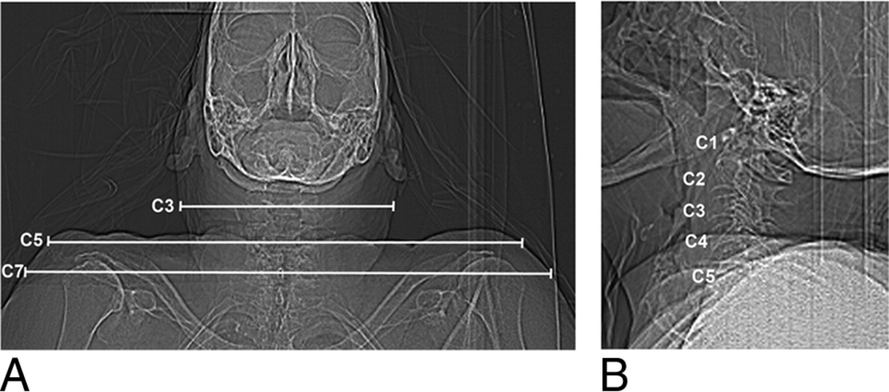
- Fig 1.
Lateral body width was measured on each anteroposterior topogram of the cadaveric specimens at 3 different heights: C3, C5, and C7 (A). On the lateral topogram, the shoulder level of this cadaveric specimen was C5 (B).
- Fig 2.
Quantitative and qualitative image noise were evaluated on 4 different cervical spine levels (A, dashed lines). Axial image (B) at level C5–6 shows 4 ROIs of 100 mm2 each in the extracorporeal air for quantitative noise measurements.
- Fig 3.
Lateral topogram (A) of this cadaveric specimen reveals shoulder height at the C5 level. Sagittal reformatted CT images (B–E; window level/width, 600/2000) of the cervical spine at 45, 105, 195, and 355 mAs reconstructed with sonogram-affirmed iterative reconstruction (strength level, 3) using bone convolution kernels show a decreasing image noise with increasing tube currents, but still sufficient image quality at 105 mAs compared with 355 mAs.
- Fig 4.
Qualitative image noise for iterative reconstructed images was evaluated on each cervical spine level using a noise score scale (1, no noise; 2, minor noise acceptable [dashed line, A–D]; 3, major noise, unacceptable). The median image noise was at least acceptable at levels C1–2 (A) with each tube current and at 75 mAs at levels C3–4 (B), but unacceptable at levels C5–T1 (C and D), except for 355 mAs at C5–6 (C). Data are median and 25th to 75th percentiles.
- Fig 5.
Grouped analysis of qualitative image noise shows significant differences at ≤150 mAs with 355 mAs for all cervical spine levels (45 mAs, P < .001; 75 mAs, P < .001; 105 mAs, P = .0006; 135 mAs, P = .0019; 150 mAs, P = .039) (A). Significant differences in image quality for levels C1–4, which were not superimposed by the shoulder girdle, were found for scans with ≤75 mAs (45 mAs, P < .001; 75 mAs, P < .001) compared with 355 mAs (B). The dashed lines in A and B represent the lowest tube currents (165 mAs for C1–T1 and 105 mAs for C1–4) that are not significantly different with respect to qualitative image noise compared with 355 mAs. Asterisks indicate values that are significantly different compared with 355 mAs (P < .05). Dots and bars indicate median and 25th to 75th percentiles.
- Fig 6.
Analysis of morphologic characteristics for iterative reconstructed images shows significant impaired image quality with 75 mAs (P = .0249) and 45 mAs (P = .0002) compared with 355 mAs for all cervical spine levels (A). The dashed line (A) at 105 mAs represents the lowest value that is not significantly different in image quality compared with 355 mAs and can be recommended as the lowest tube current value with sufficient image quality. Analysis of cervical spine levels 1–4, which are not superimposed by the shoulders, shows no significant difference in image quality with any tube current compared with 355 mAs (B). Asterisks indicate values that are significantly different compared with 355 mAs (P < .05). Dots and bars indicate median and 25th to 75th percentiles.


Tables
Cadaveric Specimen Body Weight (kg) Height (m) BMI (kg/m2) Shoulder Level Body Width (cm) at Level C7 C5 C3 1 63 1.63 23.7 C5 38.3 34.8 15.7 2 93 1.68 33.0 C5 43.4 36.0 19.0 3 62 1.48 28.3 C5 37.3 32.3 15.4 4 94 1.62 35.6 C5 46.4 39.4 16.3 Note:—BMI indicates body mass index.
↵a Cadavers' body weights included approximately 10–15 L formalin fixation.
Assessment, Localization Score Criteria Cortex Vertebral body (sag/ax) 0 Not visible 1 Visible, but not analyzable 2 Clearly visible Facet joint (sag) 0 Not visible 1 Visible, but not analyzable 2 Clearly visible Trabeculae Vertebral body (sag) 0 Not visible 1 Clearly visible Integrity Anterior vertebral body line (sag) 0 Not visible 1 Clearly visible Posterior vertebral body line (sag) 0 Not visible 1 Clearly visible Alignment Vertebral body (sag) 0 Not visible 1 Clearly visible Facet joint (sag) 0 Not visible 1 Clearly visible Maximal sum 9 Note:—sag indicates sagittal reformations; ax, axial reformations.
↵a Cortex, trabeculae, and integrity were assessed on each cervical vertebral segment (eg, C3); alignment was assessed on each cervical level (eg, C3–4). The least visible cortices of each vertebral body and facet joint were used for this analysis.
Applied Tube Current (mAs) Effective Tube Current (mAs) C1–2 C3–4 C5–6 C7–T1 45 37 ± 4 46 ± 3 45 ± 3 43 ± 2 75 62 ± 7 78 ± 4 76 ± 2 70 ± 3 105 88 ± 8 110 ± 6 106 ± 5 99 ± 3 135 113 ± 13 139 ± 8 133 ± 9 127 ± 6 150 126 ± 13 155 ± 10 149 ± 6 142 ± 6 165 138 ± 14 170 ± 9 160 ± 6 157 ± 5 195 163 ± 16 204 ± 11 190 ± 6 186 ± 7 275 234 ± 23 280 ± 18 270 ± 19 257 ± 12 355 331 ± 27 344 ± 49 343 ± 14 336 ± 13 ↵a Data represent mean ± SD at the respective cervical spine levels.
- Table 4:
Minimum required dose for at least sufficient qualitative image quality of the cervical spine in IR and FBP images compared with 355 mAs (IR)a
Levels C1–T1 Levels C1–4 IR FBP IR FBP Image noise score 165 mAs 275 mAs 105 mAs 150 mAs DLP = 250 mGy × cm DLP = 412 mGy × cm DLP = 160 mGy × cm DLP = 225 mGy × cm ED = 1.3 mSv ED = 2.1 mSv ED = 0.8 mSv ED = 1.1 mSv Morphologic characteristics score 105 mAs 105 mAs 45 mAs 45 mAs DLP = 160 mGy × cm DLP = 160 mGy × cm DLP = 65 mGy × cm DLP = 65 mGy × cm ED = 0.8 mSv ED = 0.8 mSv ED = 0.3 mSv ED = 0.3 mSv Note:—ED indicates effective dose.
↵a Data presented are the lowest applied tube current–time product at which scans showed no statistically significant differences in image quality compared with scans at the highest applied tube current–time product (355 mAs IR). Dose-length product and estimated effective dose are shown for each applied tube current–time product.


Jump to section
Related Articles
Cited By...
- No citing articles found.