
Article Figures & Data
Figures
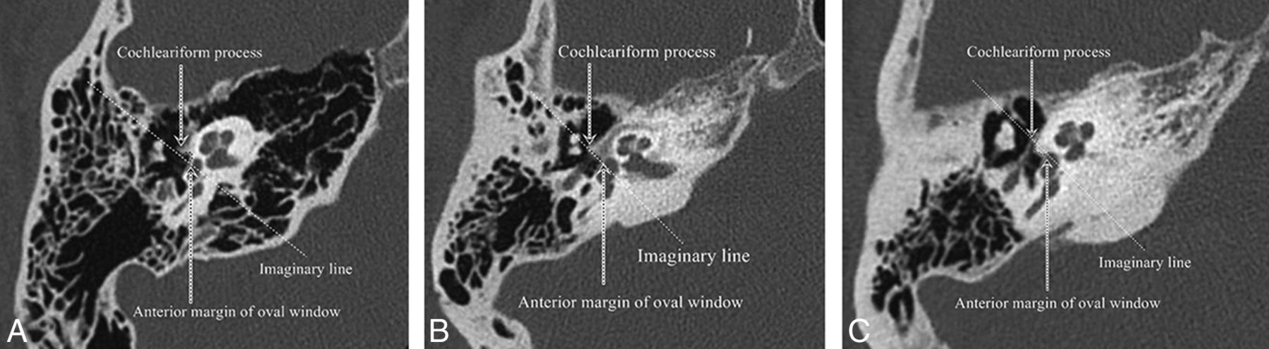
- Fig 1.
Qualitative description of the otic capsule contour relative to an imaginary line drawn from the anterior margin of the oval window to the cochleariform process. A, Normal temporal bone with a concave contour. B, Lucent phase of fenestral and retrofenestral otosclerosis with a flattened contour. C, Sclerotic phase of fenestral otosclerosis with a bulging or convex contour.
- Fig 2.
Measurement of otic capsule thickness from the posterolateral margin of the cochlea at the junction between the basal and middle turns to the most convex contour. A, Axial images parallel to the plane of the lateral semicircular canal (LSCC) are created. B, The axial slice at the level of the cochleariform process and anterior margin of the oval window is chosen. C, Measurement from the posterolateral margin of the cochlea at the junction of the basal and middle turns to the apex of the convex contour of the otic capsule is performed.
- Fig 3.
Measurement of otic capsule thickness in a patient without a convex contour of the otic capsule. A, Axial images parallel to the plane of the lateral semicircular canal (LSCC) are created. B, The axial slice at the level of the cochleariform process and anterior margin of the oval window is chosen. The sulcus between the cochleariform process and the anterior margin of the oval window is shown (white dashed line). C, Measurement from the posterolateral margin of the cochlea at the junction of the basal and middle turns to the depth of the sulcus anterior to the oval window is performed.


Tables
Patient Characteristics Otosclerosis Normal Hearing No. of Patients 58 54 No. of Ears 104 108 Sex Male (%) 21 (36.2) 14 (25.9) Female (%) 37 (63.8) 40 (74.1) P value .31 Mean age (yr) 49.7 38.5 Age (SD) (yr) 13.9 13.7 P value <.001 CT modalities MDCT (%) 35 (60.3) 44 (81.5) CBCT (%) 23 (39.7) 10 (18.5) P value .02 Otosclerosis involvement (%) Fenestral only 71 (68.3) – Fenestral and retrofenestral 31 (29.8) – Isolated round window 2 (1.9) – Otosclerosis phase of disease (%) Sclerotic disease 25 (24.0) – Mixed sclerotic lucent disease 34 (32.7) – Lucent disease 45 (43.3) – Note:—–indicates not applicable.
Contour Otosclerosis Normal Hearing Total Bulging/convex 71 2 73 Flattened/concave 33 106 139 Total 104 108 212 Two-tailed P value <.001 Diagnostic Performance Bulging/Convex Contoura Otic Capsule Thickness >2.3 mmb Sensitivity (%) 68.3 96.2 Specificity (%) 98.1 100 Positive predictive value (%) 97.3 100 Negative predictive value (%) 76.3 96.4 ↵a Bulging/convex contour across an imaginary line between the anterior margin of the oval window and the cochleariform process.
↵b Otic capsule thickness measured from the posterolateral margin of the cochlea closest to the middle ear (junction of the basal and middle turns) to the most convex contour.
Study Groups Radiologist 1 Radiologist 2 Overall Otosclerosis (n = 104) 3.09 (0.92) 3.06 (0.97) 3.08 (0.93) Normal hearing (n = 108) 1.87 (0.32) 1.78 (0.33) 1.82 (0.31) - Table 5:
Overall mean otic capsule thickness in millimeters (2 SDs) and percentage of patients with a bulging/convex contour by phase of otosclerosis
Phase of Otosclerosis Mean Thickness Bulging/Convex Contour (%) Flattened/Concave Contour (%) Sclerotic (n = 25) 3.02 (0.82) 64.0 36.0 Mixed (n = 34) 3.20 (1.08) 70.6 29.4 Lucent (n = 45) 3.06 (0.82) 68.9 31.1 P value .26a .86b


