
Article Figures & Data
Figures
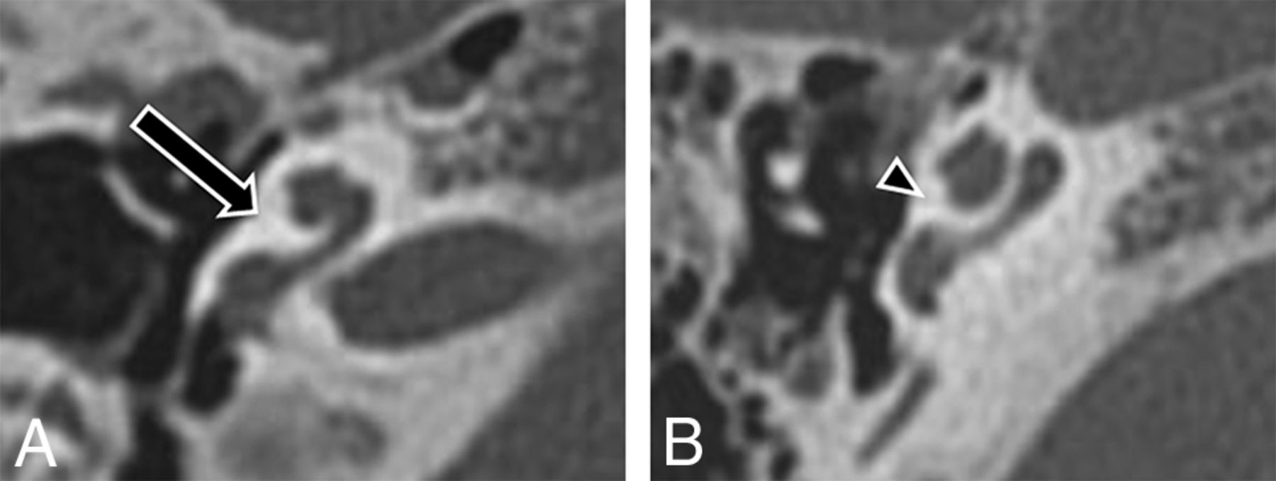
- Fig 1.
Unwound cochlea. A, Axial CT image through the right cochlea of a patient with BOR demonstrates characteristic unwound dysmorphology with anteromedial rotation and displacement of the middle turn away from a tapered basal turn (arrow). B, Axial CT image through a normal right cochlea demonstrates normal apposition of the middle and basal turns (arrowhead).
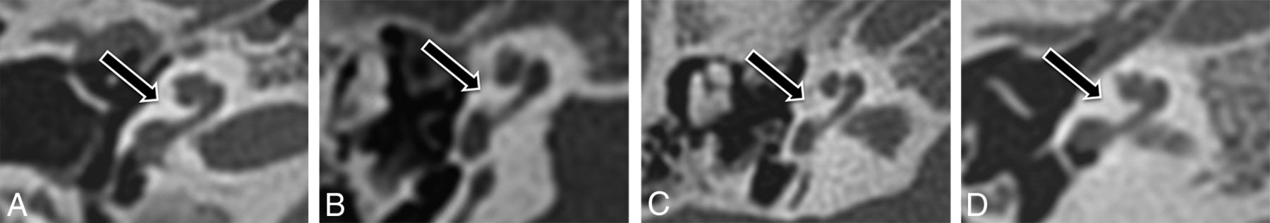
- Fig 2.
Cochlear morphology in patients with branchio-oto-renal syndrome. Axial CT images through the right temporal bone in 4 representative patients (A–D) with unwound cochleae.
- Fig 3.
Branchio-oto-renal syndrome without the unwound cochlea. Axial CT image through the right temporal bone demonstrates a truncated basal turn with complete absence of the middle and apical turns of the cochlea.
- Fig 4.
Interreader disagreement. Axial CT images through the right temporal bone in the single patient with BOR with interreader discrepancy. This cochlea was considered unwound at consensus re-review.


Tables
Patients with BOR Hearing Loss Unwound Cochlea (Reader 1) Unwound Cochlea (Reader 2) Other Inner Ear Abnormalities 1 B Mixed Bilateral Bilateral R vestibular aqueduct enlargement; B medialized facial nerve; B funnel IAC 2 B Mixed Bilateral Bilateral B hypoplastic apical turn/modiolus; right hypoplastic posterior semicircular canal; B funnel IAC 3 B Conductive Bilateral Bilateral B medialized facial nerve; B funnel IAC 4 B Conductive Bilateral Bilateral B vestibular aqueduct enlargement; B funnel IAC 5 B Conductive Bilateral Bilateral L vestibular aqueduct enlargement; B hypoplastic lateral semicircular canal; L medialized facial nerve; B funnel IAC 6 B Sensorineural Bilateral Bilateral B hypoplastic apical turn/modiolus; B cochlear aperture stenosis; B vestibular aqueduct enlargement; B medialized facial nerve; L funnel IAC 7 B Sensorineural No No B cochlear hypoplasia with absent middle and apical turns; B medialized facial nerve 8 B Mixed Bilateral No B medialized facial nerve 9 B Sensorineural Bilateral Bilateral B hypoplastic apical turn/modiolus; B vestibular aqueduct enlargement; B medialized facial nerve; B funnel IAC Note:—IAC indicates internal auditory canal; R, right side; L, left side; B, bilateral.
- Table 2:
Abnormalities of the cochlea and inner ear in control patients with hearing loss not associated with branchio-oto-renal syndrome
Cochlear Abnormality Other Inner Ear Abnormalities Incomplete partition defect type 2 (n = 3) Posterior semicircular canal hypoplasia (n = 1) Cochlear aperture stenosis (n = 1) Posterior semicircular canal dehiscence (n = 1) Labyrinthitis ossificans (n = 1) Vestibular aqueduct enlargement (n = 1) Funnel-shaped internal auditory canal (n = 2) - Table 3:
Test characteristics of the unwound cochlear morphology for a clinical diagnosis of branchio-oto-renal syndromea
Reader Sensitivity Specificity PPV NPV 1 0.89 (0.51–0.99) 1.0 (0.91–1.0) 1.0 (0.60–1.0) 0.98 (0.88–1.0) 2 0.78 (0.40–0.96) 1.0 (0.91–1.0) 1.0 (0.56–1.0) 0.96 (0.86–0.99) Note:—NPV indicates negative predictive value; PPV, positive predictive value.
↵a Numbers in parentheses are 95% CIs.


Jump to section
Related Articles
Cited By...
- Subtle Malformation of the Cochlear Apex and Genetic Abnormalities: Beyond the "Thorny" Cochlea
- The Cochlea in Branchio-Oto-Renal Syndrome: An Objective Method for the Diagnosis of Offset Cochlear Turns
- Re-Examining the Cochlea in Branchio-Oto-Renal Syndrome: Genotype-Phenotype Correlation
- Characteristic Cochlear Hypoplasia in Patients with Walker-Warburg Syndrome: A Radiologic Study of the Inner Ear in {alpha}-Dystroglycan-Related Muscular Disorders