Advertisement

Main menu

  • Home
  • Content
    • Current Issue
    • Accepted Manuscripts
    • Article Preview
    • Past Issue Archive
    • Video Articles
    • AJNR Case Collection
    • Case of the Week Archive
    • Case of the Month Archive
    • Classic Case Archive
  • Special Collections
    • Advancing NeuroMRI with High-Relaxivity Contrast Agents
    • AJNR Awards
    • Low-Field MRI
    • Alzheimer Disease
    • ASNR Foundation Special Collection
    • Photon-Counting CT
    • View All
  • Multimedia
    • AJNR Podcasts
    • AJNR SCANtastic
    • Trainee Corner
    • MRI Safety Corner
    • Imaging Protocols
  • For Authors
    • Submit a Manuscript
    • Submit a Video Article
    • Submit an eLetter to the Editor/Response
    • Manuscript Submission Guidelines
    • Statistical Tips
    • Fast Publishing of Accepted Manuscripts
    • Graphical Abstract Preparation
    • Imaging Protocol Submission
    • Author Policies
  • About Us
    • About AJNR
    • Editorial Board
    • Editorial Board Alumni
  • More
    • Become a Reviewer/Academy of Reviewers
    • Subscribers
    • Permissions
    • Alerts
    • Feedback
    • Advertisers
    • ASNR Home

User menu

  • Alerts
  • Log in

Search

  • Advanced search
American Journal of Neuroradiology

American Journal of Neuroradiology

ASHNR American Society of Functional Neuroradiology ASHNR American Society of Pediatric Neuroradiology ASSR
  • Alerts
  • Log in

Advanced Search

  • Home
  • Content
    • Current Issue
    • Accepted Manuscripts
    • Article Preview
    • Past Issue Archive
    • Video Articles
    • AJNR Case Collection
    • Case of the Week Archive
    • Case of the Month Archive
    • Classic Case Archive
  • Special Collections
    • Advancing NeuroMRI with High-Relaxivity Contrast Agents
    • AJNR Awards
    • Low-Field MRI
    • Alzheimer Disease
    • ASNR Foundation Special Collection
    • Photon-Counting CT
    • View All
  • Multimedia
    • AJNR Podcasts
    • AJNR SCANtastic
    • Trainee Corner
    • MRI Safety Corner
    • Imaging Protocols
  • For Authors
    • Submit a Manuscript
    • Submit a Video Article
    • Submit an eLetter to the Editor/Response
    • Manuscript Submission Guidelines
    • Statistical Tips
    • Fast Publishing of Accepted Manuscripts
    • Graphical Abstract Preparation
    • Imaging Protocol Submission
    • Author Policies
  • About Us
    • About AJNR
    • Editorial Board
    • Editorial Board Alumni
  • More
    • Become a Reviewer/Academy of Reviewers
    • Subscribers
    • Permissions
    • Alerts
    • Feedback
    • Advertisers
    • ASNR Home
  • Follow AJNR on Twitter
  • Visit AJNR on Facebook
  • Follow AJNR on Instagram
  • Join AJNR on LinkedIn
  • RSS Feeds

AJNR Awards, New Junior Editors, and more. Read the latest AJNR updates


Improved Turnaround Times | Median time to first decision: 12 days

Research ArticleHead and Neck Imaging

Isolated Internal Auditory Canal Diverticula: A Normal Anatomic Variant Not Associated with Sensorineural Hearing Loss

D.C. Mihal, Y. Feng, M.L. Kodet, C.M. Lohse, M.L Carlson and J.I. Lane
American Journal of Neuroradiology December 2018, 39 (12) 2340-2344; DOI: https://doi.org/10.3174/ajnr.A5862
D.C. Mihal
aFrom the Departments of Radiology (D.C.M., J.I.L.)
  • Find this author on Google Scholar
  • Find this author on PubMed
  • Search for this author on this site
  • ORCID record for D.C. Mihal
Y. Feng
bOtolaryngology-Head and Neck Surgery (Y.F., M.L.K., M.L.C.)
  • Find this author on Google Scholar
  • Find this author on PubMed
  • Search for this author on this site
  • ORCID record for Y. Feng
M.L. Kodet
bOtolaryngology-Head and Neck Surgery (Y.F., M.L.K., M.L.C.)
  • Find this author on Google Scholar
  • Find this author on PubMed
  • Search for this author on this site
  • ORCID record for M.L. Kodet
C.M. Lohse
cHealth Sciences Research (C.M.L.), Mayo Clinic, Rochester, Minnesota.
  • Find this author on Google Scholar
  • Find this author on PubMed
  • Search for this author on this site
  • ORCID record for C.M. Lohse
M.L Carlson
bOtolaryngology-Head and Neck Surgery (Y.F., M.L.K., M.L.C.)
  • Find this author on Google Scholar
  • Find this author on PubMed
  • Search for this author on this site
  • ORCID record for M.L Carlson
J.I. Lane
aFrom the Departments of Radiology (D.C.M., J.I.L.)
  • Find this author on Google Scholar
  • Find this author on PubMed
  • Search for this author on this site
  • ORCID record for J.I. Lane
  • Article
  • Figures & Data
  • Info & Metrics
  • Responses
  • References
  • PDF
Loading

Article Figures & Data

Figures

  • Tables
  • Fig 1.
    • Download figure
    • Open in new tab
    • Download powerpoint
    Fig 1.

    Axial CT (A and B) and axial 3D FSE T2 MR (C and D) images of right and left anterior wall IAC diverticula (black and white arrows) in the same patient with unilateral hearing loss.
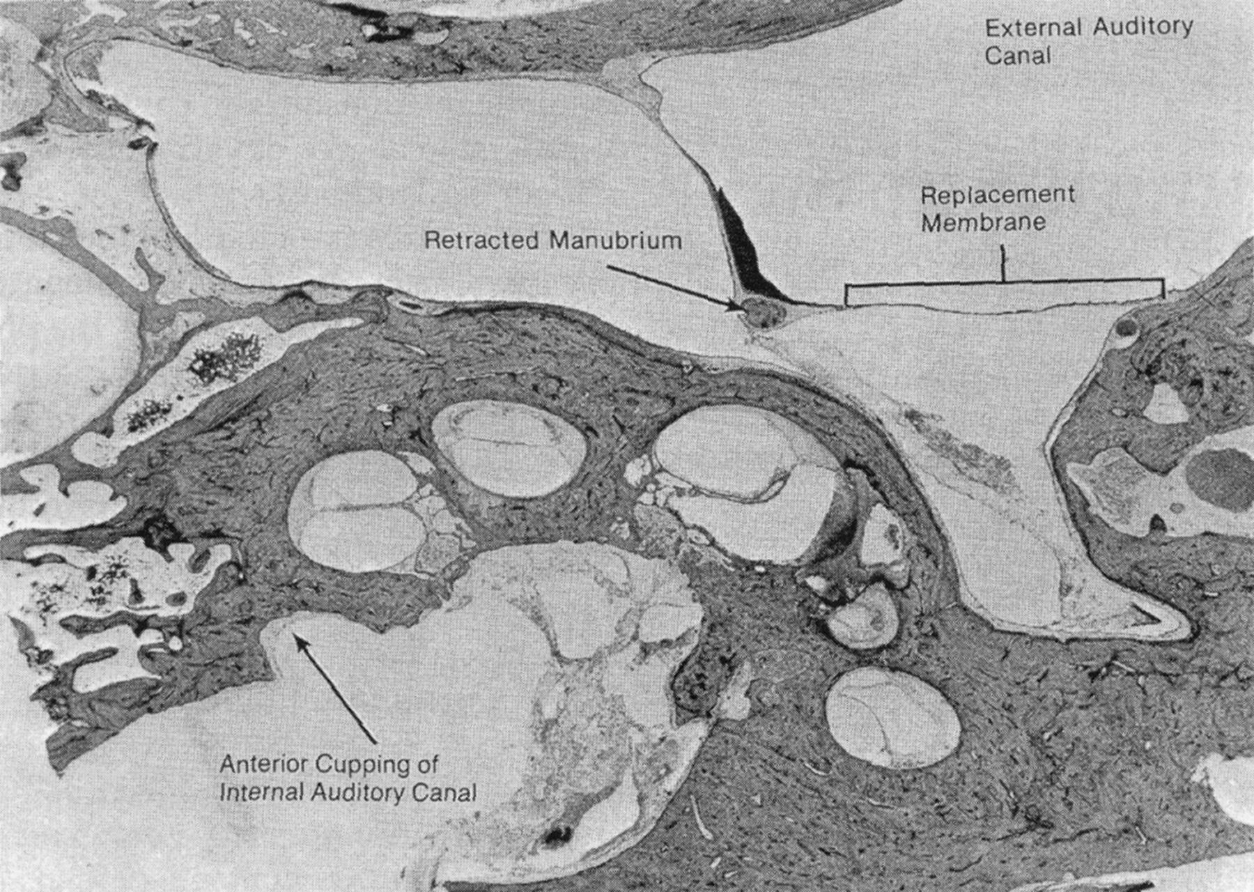
  • Fig 2.
    • Download figure
    • Open in new tab
    • Download powerpoint
    Fig 2.

    Histologic micrograph demonstrating the typical location of IAC diverticulum (ie, IAC cupping). Reproduced with permission from the third edition of Guyla and Schucknecht's Anatomy of the Temporal Bone with Surgical Implications.1
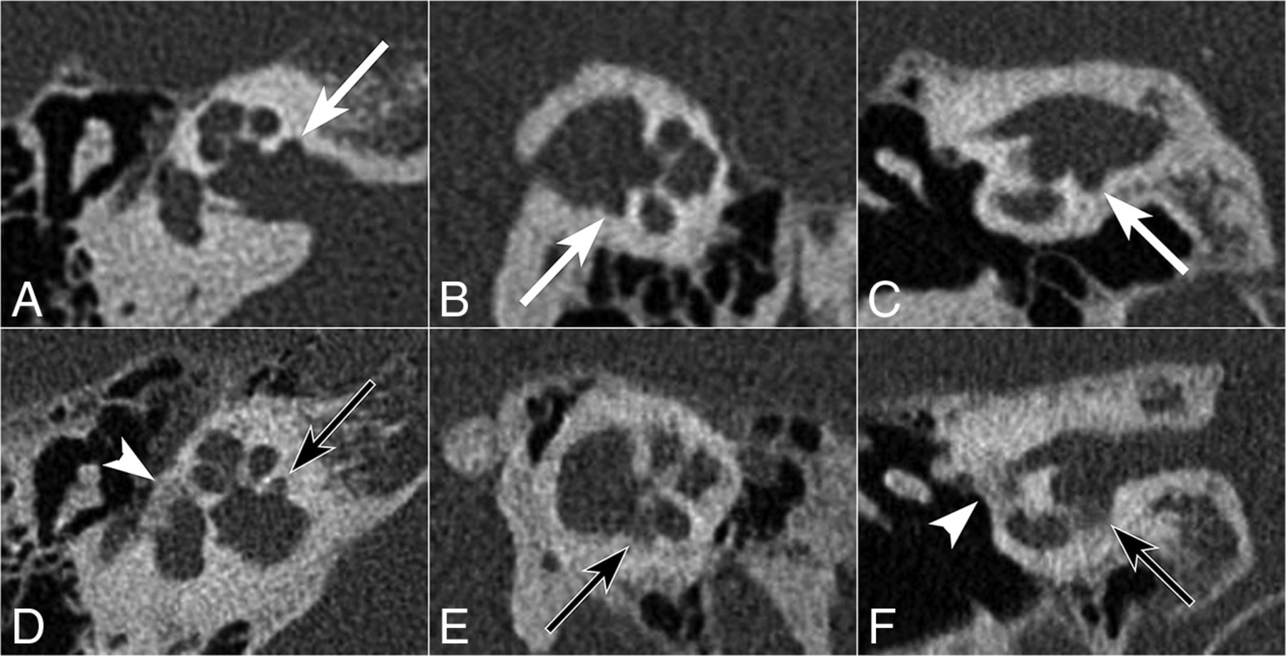
  • Fig 3.
    • Download figure
    • Open in new tab
    • Download powerpoint
    Fig 3.

    Right temporal bone axial (A and D), Pöschl (B and E), and coronal (C and F) views of an IAC diverticulum (upper row) versus cavitary otosclerosis (lower row). Note the well-demarcated margins of a lucent IAC diverticulum (white arrow) compared with the ill-defined spongiotic bone composing the otosclerotic focus along the anterior wall of the IAC (black arrow), with indistinct bony margins between this lesion and the basal turn of the cochlea (E and F). Also note the presence of fenestral otosclerosis (white arrowhead in D and F).

Tables

  • Figures
    • View popup
    Table 1:

    Comparison of the number of patients in each hearing classification category based on audiometric hearing assessment among those with unilateral IAC diverticula in the absence of otosclerosis (N = 22)

    IAC DiverticulumControl Side (No IAC Diverticulum)
    NormalCHLMHLSNHL
    Normal6001
    CHL1110
    MHL1004
    SNHL2023
    • View popup
    Table 2:

    Comparison of audiometric data in ears for patients with unilateral IAC diverticula in the absence of otosclerosis (N = 22)a

    No.bIAC DiverticulumNo IAC DiverticulumDifferencecP Valued
    Bone conduction pure tone average2222.5 (13–41)17 (8–35)1 (−4–17).31
        BC 250 Hz (dB)2017.5 (7.5–25)12.5 (7.5–20)2.5 (−5–15).19
        BC 500 Hz (dB)2120 (10–40)15 (10–25)5 (0–20).14
        BC 1000 Hz (dB)2022.5 (10–40)15 (5–27.5)0 (−2.5–25).17
        BC 2000 Hz (dB)1930 (10–50)20 (10–45)0 (−10–15).50
        BC 3000 Hz (dB)1825 (7.5–47.5)20 (10–55)0 (−10–15).63
        BC 4000 Hz (dB)2125 (10–50)35 (15–60)0 (−5–5).97
    Air conduction pure tone average2231 (20–75)20 (9–41)4 (1–29).06
        AC 250 Hz (dB)2222.5 (15–60)17.5 (10–35)2.5 (−5–20).19
        AC 500 Hz (dB)2225 (15–75)22.5 (10–40)2.5 (−5–20).30
        AC 1000 Hz (dB)2227.5 (15–75)20 (5–40)5 (0–30).06
        AC 2000 Hz (dB)2232.5 (15–70)20 (5–50)10 (0–30).10
        AC 3000 Hz (dB)2037.5 (15–60)20 (12.5–62.5)5 (−5–25).34
        AC 4000 Hz (dB)2240 (20–85)37.5 (15–70)5 (−5–40).22
    Air-bone gap225 (0–12)0 (0–5)2 (0–9).37
        Word recognition (%)17100 (85–100)100 (95–100)0 (−15–0).55
    • Note:—AC indicates air conduction; BC, bone conduction.

    • ↵a Data are median and IQR.

    • ↵b Summarized for a subset of patients with data for both ears.

    • ↵c Calculated as IAC minus non-IAC.

    • ↵d Determined using Wilcoxon signed rank tests.

PreviousNext
Back to top

In this issue

American Journal of Neuroradiology: 39 (12)
American Journal of Neuroradiology
Vol. 39, Issue 12
1 Dec 2018
  • Table of Contents
  • Index by author
  • Complete Issue (PDF)
Advertisement
Print
Download PDF
Email Article

Thank you for your interest in spreading the word on American Journal of Neuroradiology.

NOTE: We only request your email address so that the person you are recommending the page to knows that you wanted them to see it, and that it is not junk mail. We do not capture any email address.

Enter multiple addresses on separate lines or separate them with commas.
Isolated Internal Auditory Canal Diverticula: A Normal Anatomic Variant Not Associated with Sensorineural Hearing Loss
(Your Name) has sent you a message from American Journal of Neuroradiology
(Your Name) thought you would like to see the American Journal of Neuroradiology web site.
CAPTCHA
This question is for testing whether or not you are a human visitor and to prevent automated spam submissions.
Cite this article
D.C. Mihal, Y. Feng, M.L. Kodet, C.M. Lohse, M.L Carlson, J.I. Lane
Isolated Internal Auditory Canal Diverticula: A Normal Anatomic Variant Not Associated with Sensorineural Hearing Loss
American Journal of Neuroradiology Dec 2018, 39 (12) 2340-2344; DOI: 10.3174/ajnr.A5862

Citation Manager Formats

  • BibTeX
  • Bookends
  • EasyBib
  • EndNote (tagged)
  • EndNote 8 (xml)
  • Medlars
  • Mendeley
  • Papers
  • RefWorks Tagged
  • Ref Manager
  • RIS
  • Zotero
0 Responses
Respond to this article
Share
Bookmark this article
Isolated Internal Auditory Canal Diverticula: A Normal Anatomic Variant Not Associated with Sensorineural Hearing Loss
D.C. Mihal, Y. Feng, M.L. Kodet, C.M. Lohse, M.L Carlson, J.I. Lane
American Journal of Neuroradiology Dec 2018, 39 (12) 2340-2344; DOI: 10.3174/ajnr.A5862
del.icio.us logo Twitter logo Facebook logo Mendeley logo
  • Tweet Widget
  • Facebook Like
  • Google Plus One
Purchase

Jump to section

  • Article
    • Abstract
    • ABBREVIATIONS:
    • Materials and Methods
    • Results
    • Discussion
    • Conclusions
    • Acknowledgments
    • Footnotes
    • References
  • Figures & Data
  • Info & Metrics
  • Responses
  • References
  • PDF

Related Articles

  • No related articles found.
  • PubMed
  • Google Scholar

Cited By...

  • Internal Auditory Canal Diverticula among Pediatric Patients: Prevalence and Assessment for Hearing Loss and Anatomic Associations
  • Crossref
  • Google Scholar

This article has not yet been cited by articles in journals that are participating in Crossref Cited-by Linking.

More in this TOC Section

  • Diagnostic Performance of US in Neck Node NIRADS 2
  • Hydrops Herniation into the Semicircular Canals
  • ASL Sensitivity for Head and Neck Paraganglioma
Show more Head and Neck Imaging

Similar Articles

Advertisement

Indexed Content

  • Current Issue
  • Accepted Manuscripts
  • Article Preview
  • Past Issues
  • Editorials
  • Editor's Choice
  • Fellows' Journal Club
  • Letters to the Editor
  • Video Articles

Cases

  • Case Collection
  • Archive - Case of the Week
  • Archive - Case of the Month
  • Archive - Classic Case

More from AJNR

  • Trainee Corner
  • Imaging Protocols
  • MRI Safety Corner
  • Book Reviews

Multimedia

  • AJNR Podcasts
  • AJNR Scantastics

Resources

  • Turnaround Time
  • Submit a Manuscript
  • Submit a Video Article
  • Submit an eLetter to the Editor/Response
  • Manuscript Submission Guidelines
  • Statistical Tips
  • Fast Publishing of Accepted Manuscripts
  • Graphical Abstract Preparation
  • Imaging Protocol Submission
  • Evidence-Based Medicine Level Guide
  • Publishing Checklists
  • Author Policies
  • Become a Reviewer/Academy of Reviewers
  • News and Updates

About Us

  • About AJNR
  • Editorial Board
  • Editorial Board Alumni
  • Alerts
  • Permissions
  • Not an AJNR Subscriber? Join Now
  • Advertise with Us
  • Librarian Resources
  • Feedback
  • Terms and Conditions
  • AJNR Editorial Board Alumni

American Society of Neuroradiology

  • Not an ASNR Member? Join Now

© 2025 by the American Society of Neuroradiology All rights, including for text and data mining, AI training, and similar technologies, are reserved.
Print ISSN: 0195-6108 Online ISSN: 1936-959X

Powered by HighWire