Advertisement

Main menu

  • Home
  • Content
    • Current Issue
    • Accepted Manuscripts
    • Article Preview
    • Past Issue Archive
    • Video Articles
    • AJNR Case Collection
    • Case of the Week Archive
    • Case of the Month Archive
    • Classic Case Archive
  • Special Collections
    • AJNR Awards
    • Low-Field MRI
    • Alzheimer Disease
    • ASNR Foundation Special Collection
    • Photon-Counting CT
    • View All
  • Multimedia
    • AJNR Podcasts
    • AJNR SCANtastic
    • Trainee Corner
    • MRI Safety Corner
    • Imaging Protocols
  • For Authors
    • Submit a Manuscript
    • Submit a Video Article
    • Submit an eLetter to the Editor/Response
    • Manuscript Submission Guidelines
    • Statistical Tips
    • Fast Publishing of Accepted Manuscripts
    • Graphical Abstract Preparation
    • Imaging Protocol Submission
    • Author Policies
  • About Us
    • About AJNR
    • Editorial Board
    • Editorial Board Alumni
  • More
    • Become a Reviewer/Academy of Reviewers
    • Subscribers
    • Permissions
    • Alerts
    • Feedback
    • Advertisers
    • ASNR Home

User menu

  • Alerts
  • Log in

Search

  • Advanced search
American Journal of Neuroradiology

American Journal of Neuroradiology

ASHNR American Society of Functional Neuroradiology ASHNR American Society of Pediatric Neuroradiology ASSR
  • Alerts
  • Log in

Advanced Search

  • Home
  • Content
    • Current Issue
    • Accepted Manuscripts
    • Article Preview
    • Past Issue Archive
    • Video Articles
    • AJNR Case Collection
    • Case of the Week Archive
    • Case of the Month Archive
    • Classic Case Archive
  • Special Collections
    • AJNR Awards
    • Low-Field MRI
    • Alzheimer Disease
    • ASNR Foundation Special Collection
    • Photon-Counting CT
    • View All
  • Multimedia
    • AJNR Podcasts
    • AJNR SCANtastic
    • Trainee Corner
    • MRI Safety Corner
    • Imaging Protocols
  • For Authors
    • Submit a Manuscript
    • Submit a Video Article
    • Submit an eLetter to the Editor/Response
    • Manuscript Submission Guidelines
    • Statistical Tips
    • Fast Publishing of Accepted Manuscripts
    • Graphical Abstract Preparation
    • Imaging Protocol Submission
    • Author Policies
  • About Us
    • About AJNR
    • Editorial Board
    • Editorial Board Alumni
  • More
    • Become a Reviewer/Academy of Reviewers
    • Subscribers
    • Permissions
    • Alerts
    • Feedback
    • Advertisers
    • ASNR Home
  • Follow AJNR on Twitter
  • Visit AJNR on Facebook
  • Follow AJNR on Instagram
  • Join AJNR on LinkedIn
  • RSS Feeds

AJNR Awards, New Junior Editors, and more. Read the latest AJNR updates

Research ArticleAdult Brain

Brain MR Imaging Findings in Woodhouse-Sakati Syndrome

A.H. Abusrair, S. Bohlega, A. Al-Semari, F.S. Al-Ajlan, K. Al-Ahmadi, B. Mohamed and A. AlDakheel
American Journal of Neuroradiology December 2018, 39 (12) 2256-2262; DOI: https://doi.org/10.3174/ajnr.A5879
A.H. Abusrair
aFrom the Departments of Neurosciences (A.H.A., S.B., A.A.-S., F.S.A.-A., B.M., A.A.)
  • Find this author on Google Scholar
  • Find this author on PubMed
  • Search for this author on this site
  • ORCID record for A.H. Abusrair
S. Bohlega
aFrom the Departments of Neurosciences (A.H.A., S.B., A.A.-S., F.S.A.-A., B.M., A.A.)
  • Find this author on Google Scholar
  • Find this author on PubMed
  • Search for this author on this site
  • ORCID record for S. Bohlega
A. Al-Semari
aFrom the Departments of Neurosciences (A.H.A., S.B., A.A.-S., F.S.A.-A., B.M., A.A.)
  • Find this author on Google Scholar
  • Find this author on PubMed
  • Search for this author on this site
  • ORCID record for A. Al-Semari
F.S. Al-Ajlan
aFrom the Departments of Neurosciences (A.H.A., S.B., A.A.-S., F.S.A.-A., B.M., A.A.)
  • Find this author on Google Scholar
  • Find this author on PubMed
  • Search for this author on this site
  • ORCID record for F.S. Al-Ajlan
K. Al-Ahmadi
bRadiology (K.A.-A.), King Faisal Specialist Hospital & Research Center, Riyadh, Saudi Arabia.
  • Find this author on Google Scholar
  • Find this author on PubMed
  • Search for this author on this site
  • ORCID record for K. Al-Ahmadi
B. Mohamed
aFrom the Departments of Neurosciences (A.H.A., S.B., A.A.-S., F.S.A.-A., B.M., A.A.)
  • Find this author on Google Scholar
  • Find this author on PubMed
  • Search for this author on this site
  • ORCID record for B. Mohamed
A. AlDakheel
aFrom the Departments of Neurosciences (A.H.A., S.B., A.A.-S., F.S.A.-A., B.M., A.A.)
  • Find this author on Google Scholar
  • Find this author on PubMed
  • Search for this author on this site
  • ORCID record for A. AlDakheel
  • Article
  • Figures & Data
  • Supplemental
  • Info & Metrics
  • Responses
  • References
  • PDF
Loading

Article Figures & Data

Figures

  • Tables
  • Fig 1.
    • Download figure
    • Open in new tab
    • Download powerpoint
    Fig 1.

    Sagittal T1-weighted MR imaging for patient 22 shows a partially empty sella and a small pituitary gland (arrow).

  • Fig 2.
    • Download figure
    • Open in new tab
    • Download powerpoint
    Fig 2.

    Axial T2-weighted (A), FLAIR (B), and T2* (C) MR images in case 11 show putaminal blooming artifacts (arrows) reflecting iron accumulation. An axial T2* MR image (D) in case 3 shows iron deposition in the substantia nigra (arrowheads).

  • Fig 3.
    • Download figure
    • Open in new tab
    • Download powerpoint
    Fig 3.

    Axial FLAIR MR images show varying degrees of white matter lesions. Patients 1 and 26 (A and B) show a mild degree of white matter lesions, with faint periventricular (A; arrows) and small scattered signal intensities (B; arrows). In patient 4 (C), images show patchy signal intensities (arrows), representing a moderate degree of white matter lesions. A more diffuse, vanishing high signal intensity (arrows) is seen in images (D) for patient 7.
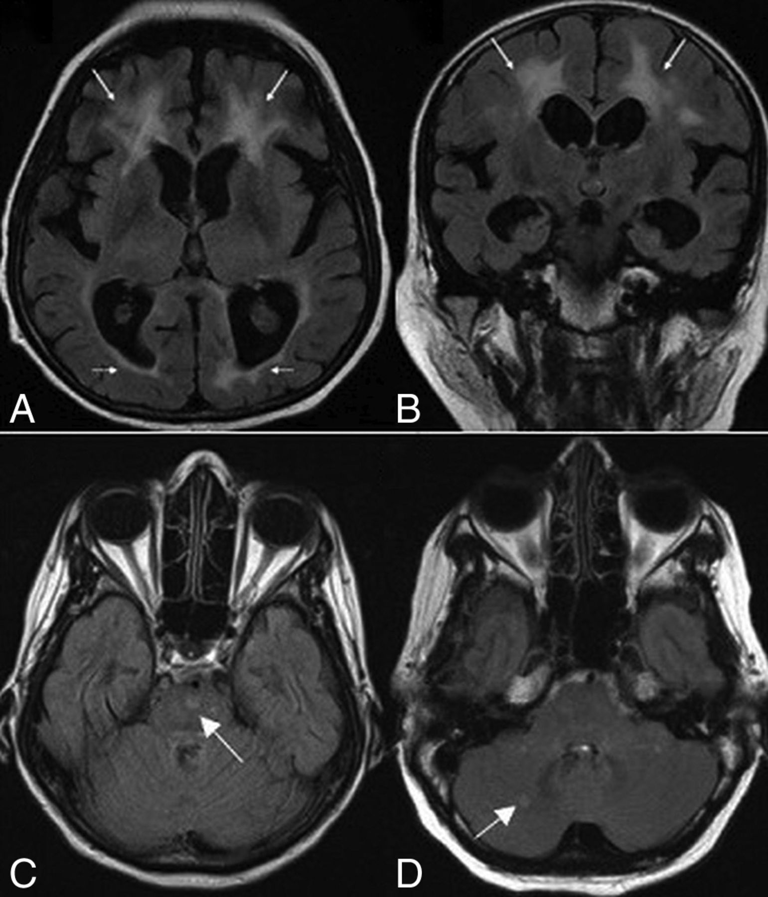
  • Fig 4.
    • Download figure
    • Open in new tab
    • Download powerpoint
    Fig 4.

    Axial (A) and coronal (B) FLAIR MR images for case 24 show white matter changes with frontal predominance (arrows). Axial FLAIR images show signal intensities (arrows) involving the pons (C) in patient 19 and the cerebellum (D) in patient 26.

  • Fig 5.
    • Download figure
    • Open in new tab
    • Download powerpoint
    Fig 5.

    Axial T2 images show type I (A) and type III (B) prominent perivascular spaces.
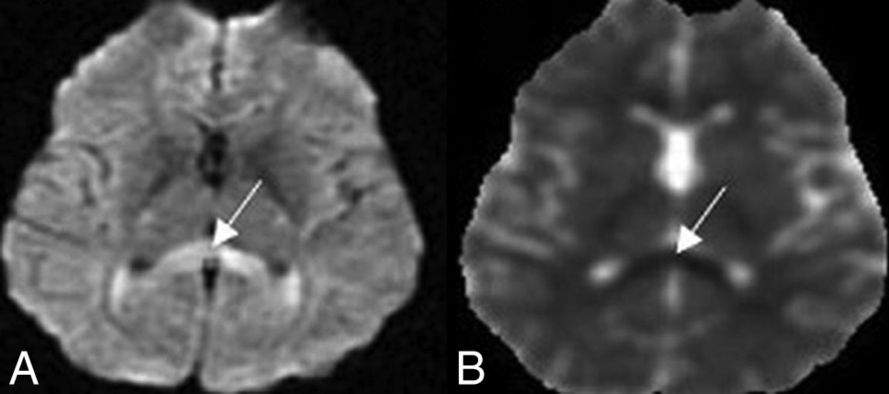
  • Fig 6.
    • Download figure
    • Open in new tab
    • Download powerpoint
    Fig 6.

    Axial DWI (A) and ADC map (B) sequences show diffusion restriction involving the splenium of corpus callosum.

Tables

  • Figures
    • View popup
    Table 1:

    Clinical and imaging characteristics of WSS

    FamilySexAge (Yr)NeurologicRadiologic
    DystoniaSNHLSeizureWM ChangesIDPituitary
    1F18Focal−−Mild−Small
    M19−−+Absent+Small
    2F29Generalized−−Severe+Normal
    F38Focal+−Moderate+Small
    F35Focal+−Mild+Small
    3F37Focal−−Severe+Small
    M45Generalized+−Severe+Small
    4M22Focal−−Mild+Normal
    M24Generalized−−Mild+Normal
    5M23−−−Mild−Small
    F21−−−Mild+Small
    M16−−−Mild−Small
    M26Generalized+−Mild+Normal
    M21−++Absent+Normal
    6M20Generalized−−Mild−Small
    F21−−−Absent+Small
    7F32Multisegmental−−Mild+Small
    F22Focal−−Absent−Small
    F35Focal−+Severe+Small
    M21Multisegmental+−Absent+Small
    F27Focal+−Absent+Small
    8M16−−−Absent−Small
    9F41Generalized−−Moderate+Small
    10M37Multisegmental+−Moderate+Small
    11F17Multisegmental−−Absent+Small
    12F29−−−Mild−Normal
    • Note:—SNHL indicates sensorineural hearing loss; ID, iron deposition; +, present; −, absent.

    • View popup
    Table 2:

    Characteristic brain MR imaging findings in WSS

    PatientSex/Age (yr)White MatterDescriptive Features on MRIComments
    Iron DepositionPituitary AtrophyDiffusion Restriction
    FrontalParietalTemporalOccipitalInfratentorialCaudatePutamenRN/SN
    1F/18++−+−−−−++−
    2M/19−−−−−−+−+−Initial study showed no iron deposition
    3F/29++++++−−−+++−−Extension of iron deposition on follow-up study
    4F/38++++++−++−++Prominent perivascular spaces
    5F/35+++−−−+−−−Prominent perivascular spaces
    6F/37++++++++−−+−+−Calcification of the dentate nucleus
    7M/45++++++++−++−++Loss of corpus callosum volume
    8M/22++−−−−+−+−−
    9M/24++−−−−+−−−Prominent perivascular spaces
    10M/23++−−−−−−+−−
    11F/21++−−−−+−+−−
    12M/16++−−−−−−+−Prominent perivascular spaces
    13M/26++++−−+++−−
    14M/21−−−−−−++−−−
    15M/20+−−−−−−−+−−
    16F/21−−−−−−+++−−
    17F/32++++−−+−+−−
    18F/22−−−−−−−−+−−
    19F/35+++++++++−+−+−Pontine WM signal
    20M/21−−−−−−+++−−
    21F/27−−−−−−+−+−−
    22M/16−−−−−−−−+−−
    23F/41++++++−++++−−
    24M/37++++++−++−+−Dentate nucleus calcification
    25F/17−−−−−−+−−−−
    26F/29+++++−−−−−Cerebellar WM signal
    • Note:—RN indicates red nucleus; SN, substantia nigra; −, absent; +, mild (predominantly scattered lesions or confluent periventricular); ++, moderate (patchy scattered); +++, severe (predominantly diffuse, vanishing lesions).

PreviousNext
Back to top

In this issue

American Journal of Neuroradiology: 39 (12)
American Journal of Neuroradiology
Vol. 39, Issue 12
1 Dec 2018
  • Table of Contents
  • Index by author
  • Complete Issue (PDF)
Advertisement
Print
Download PDF
Email Article

Thank you for your interest in spreading the word on American Journal of Neuroradiology.

NOTE: We only request your email address so that the person you are recommending the page to knows that you wanted them to see it, and that it is not junk mail. We do not capture any email address.

Enter multiple addresses on separate lines or separate them with commas.
Brain MR Imaging Findings in Woodhouse-Sakati Syndrome
(Your Name) has sent you a message from American Journal of Neuroradiology
(Your Name) thought you would like to see the American Journal of Neuroradiology web site.
CAPTCHA
This question is for testing whether or not you are a human visitor and to prevent automated spam submissions.
Cite this article
A.H. Abusrair, S. Bohlega, A. Al-Semari, F.S. Al-Ajlan, K. Al-Ahmadi, B. Mohamed, A. AlDakheel
Brain MR Imaging Findings in Woodhouse-Sakati Syndrome
American Journal of Neuroradiology Dec 2018, 39 (12) 2256-2262; DOI: 10.3174/ajnr.A5879

Citation Manager Formats

  • BibTeX
  • Bookends
  • EasyBib
  • EndNote (tagged)
  • EndNote 8 (xml)
  • Medlars
  • Mendeley
  • Papers
  • RefWorks Tagged
  • Ref Manager
  • RIS
  • Zotero
0 Responses
Respond to this article
Share
Bookmark this article
Brain MR Imaging Findings in Woodhouse-Sakati Syndrome
A.H. Abusrair, S. Bohlega, A. Al-Semari, F.S. Al-Ajlan, K. Al-Ahmadi, B. Mohamed, A. AlDakheel
American Journal of Neuroradiology Dec 2018, 39 (12) 2256-2262; DOI: 10.3174/ajnr.A5879
del.icio.us logo Twitter logo Facebook logo Mendeley logo
  • Tweet Widget
  • Facebook Like
  • Google Plus One
Purchase

Jump to section

  • Article
    • Abstract
    • ABBREVIATIONS:
    • Materials and Methods
    • Results
    • Discussion
    • Conclusions
    • Acknowledgments
    • References
  • Figures & Data
  • Supplemental
  • Info & Metrics
  • Responses
  • References
  • PDF

Related Articles

  • PubMed
  • Google Scholar

Cited By...

  • Woodhouse-Sakati syndrome (WSS): A case report of 3 Saudi sisters with urogenital anomalies
  • In silico analysis of coding SNPs and 3'-UTR associated miRNAs in DCAF17 gene that may affect the regulation and pathogenesis of Woodhouse-Sakati Syndrome
  • Crossref
  • Google Scholar

This article has not yet been cited by articles in journals that are participating in Crossref Cited-by Linking.

More in this TOC Section

  • Diagnostic Neuroradiology of Monoclonal Antibodies
  • Clinical Outcomes After Chiari I Decompression
  • Segmentation of Brain Metastases with BLAST
Show more Adult Brain

Similar Articles

Advertisement

Indexed Content

  • Current Issue
  • Accepted Manuscripts
  • Article Preview
  • Past Issues
  • Editorials
  • Editor's Choice
  • Fellows' Journal Club
  • Letters to the Editor
  • Video Articles

Cases

  • Case Collection
  • Archive - Case of the Week
  • Archive - Case of the Month
  • Archive - Classic Case

More from AJNR

  • Trainee Corner
  • Imaging Protocols
  • MRI Safety Corner
  • Book Reviews

Multimedia

  • AJNR Podcasts
  • AJNR Scantastics

Resources

  • Turnaround Time
  • Submit a Manuscript
  • Submit a Video Article
  • Submit an eLetter to the Editor/Response
  • Manuscript Submission Guidelines
  • Statistical Tips
  • Fast Publishing of Accepted Manuscripts
  • Graphical Abstract Preparation
  • Imaging Protocol Submission
  • Evidence-Based Medicine Level Guide
  • Publishing Checklists
  • Author Policies
  • Become a Reviewer/Academy of Reviewers
  • News and Updates

About Us

  • About AJNR
  • Editorial Board
  • Editorial Board Alumni
  • Alerts
  • Permissions
  • Not an AJNR Subscriber? Join Now
  • Advertise with Us
  • Librarian Resources
  • Feedback
  • Terms and Conditions
  • AJNR Editorial Board Alumni

American Society of Neuroradiology

  • Not an ASNR Member? Join Now

© 2025 by the American Society of Neuroradiology All rights, including for text and data mining, AI training, and similar technologies, are reserved.
Print ISSN: 0195-6108 Online ISSN: 1936-959X

Powered by HighWire