Advertisement

Main menu

  • Home
  • Content
    • Current Issue
    • Accepted Manuscripts
    • Article Preview
    • Past Issue Archive
    • Video Articles
    • AJNR Case Collection
    • Case of the Week Archive
    • Case of the Month Archive
    • Classic Case Archive
  • Special Collections
    • AJNR Awards
    • Low-Field MRI
    • Alzheimer Disease
    • ASNR Foundation Special Collection
    • Photon-Counting CT
    • View All
  • Multimedia
    • AJNR Podcasts
    • AJNR SCANtastic
    • Trainee Corner
    • MRI Safety Corner
    • Imaging Protocols
  • For Authors
    • Submit a Manuscript
    • Submit a Video Article
    • Submit an eLetter to the Editor/Response
    • Manuscript Submission Guidelines
    • Statistical Tips
    • Fast Publishing of Accepted Manuscripts
    • Graphical Abstract Preparation
    • Imaging Protocol Submission
    • Author Policies
  • About Us
    • About AJNR
    • Editorial Board
    • Editorial Board Alumni
  • More
    • Become a Reviewer/Academy of Reviewers
    • Subscribers
    • Permissions
    • Alerts
    • Feedback
    • Advertisers
    • ASNR Home

User menu

  • Alerts
  • Log in

Search

  • Advanced search
American Journal of Neuroradiology

American Journal of Neuroradiology

ASHNR American Society of Functional Neuroradiology ASHNR American Society of Pediatric Neuroradiology ASSR
  • Alerts
  • Log in

Advanced Search

  • Home
  • Content
    • Current Issue
    • Accepted Manuscripts
    • Article Preview
    • Past Issue Archive
    • Video Articles
    • AJNR Case Collection
    • Case of the Week Archive
    • Case of the Month Archive
    • Classic Case Archive
  • Special Collections
    • AJNR Awards
    • Low-Field MRI
    • Alzheimer Disease
    • ASNR Foundation Special Collection
    • Photon-Counting CT
    • View All
  • Multimedia
    • AJNR Podcasts
    • AJNR SCANtastic
    • Trainee Corner
    • MRI Safety Corner
    • Imaging Protocols
  • For Authors
    • Submit a Manuscript
    • Submit a Video Article
    • Submit an eLetter to the Editor/Response
    • Manuscript Submission Guidelines
    • Statistical Tips
    • Fast Publishing of Accepted Manuscripts
    • Graphical Abstract Preparation
    • Imaging Protocol Submission
    • Author Policies
  • About Us
    • About AJNR
    • Editorial Board
    • Editorial Board Alumni
  • More
    • Become a Reviewer/Academy of Reviewers
    • Subscribers
    • Permissions
    • Alerts
    • Feedback
    • Advertisers
    • ASNR Home
  • Follow AJNR on Twitter
  • Visit AJNR on Facebook
  • Follow AJNR on Instagram
  • Join AJNR on LinkedIn
  • RSS Feeds

AJNR Awards, New Junior Editors, and more. Read the latest AJNR updates

Research ArticleAdult Brain
Open Access

Diffusional Kurtosis along the Corticospinal Tract in Adult Normal Pressure Hydrocephalus

B. Ades-Aron, S. Yeager, N. Miskin, E. Fieremans, A. George and J. Golomb
American Journal of Neuroradiology December 2018, 39 (12) 2218-2223; DOI: https://doi.org/10.3174/ajnr.A5845
B. Ades-Aron
aFrom the Center for Biomedical Imaging (B.A.-A., S.Y., E.F., A.G.), Department of Radiology
  • Find this author on Google Scholar
  • Find this author on PubMed
  • Search for this author on this site
  • ORCID record for B. Ades-Aron
S. Yeager
aFrom the Center for Biomedical Imaging (B.A.-A., S.Y., E.F., A.G.), Department of Radiology
  • Find this author on Google Scholar
  • Find this author on PubMed
  • Search for this author on this site
  • ORCID record for S. Yeager
N. Miskin
cDepartment of Radiology (N.M.), Brigham and Women's Hospital, Boston, Massachusetts.
  • Find this author on Google Scholar
  • Find this author on PubMed
  • Search for this author on this site
  • ORCID record for N. Miskin
E. Fieremans
aFrom the Center for Biomedical Imaging (B.A.-A., S.Y., E.F., A.G.), Department of Radiology
  • Find this author on Google Scholar
  • Find this author on PubMed
  • Search for this author on this site
  • ORCID record for E. Fieremans
A. George
aFrom the Center for Biomedical Imaging (B.A.-A., S.Y., E.F., A.G.), Department of Radiology
  • Find this author on Google Scholar
  • Find this author on PubMed
  • Search for this author on this site
  • ORCID record for A. George
J. Golomb
bDepartment of Neurology (J.G.), New York University School of Medicine, New York, New York
  • Find this author on Google Scholar
  • Find this author on PubMed
  • Search for this author on this site
  • ORCID record for J. Golomb
  • Article
  • Figures & Data
  • Supplemental
  • Info & Metrics
  • Responses
  • References
  • PDF
Loading

Article Figures & Data

Figures

  • Tables
  • Fig 1.
    • Download figure
    • Open in new tab
    • Download powerpoint
    Fig 1.

    Axial diffusion and axial kurtosis overlaid onto the corticospinal tract as a scalar value and represented by a color: Red indicates higher axial kurtosis or axial diffusivity, while blue indicates lower values. From left to right, we show sample tracts for patients with Alzheimer disease, healthy controls, and patients with hydrocephalus. Patients with NPH have much lower axial diffusion in periventricular regions than control groups and much greater axial kurtosis values in the same region next to the ventricles.
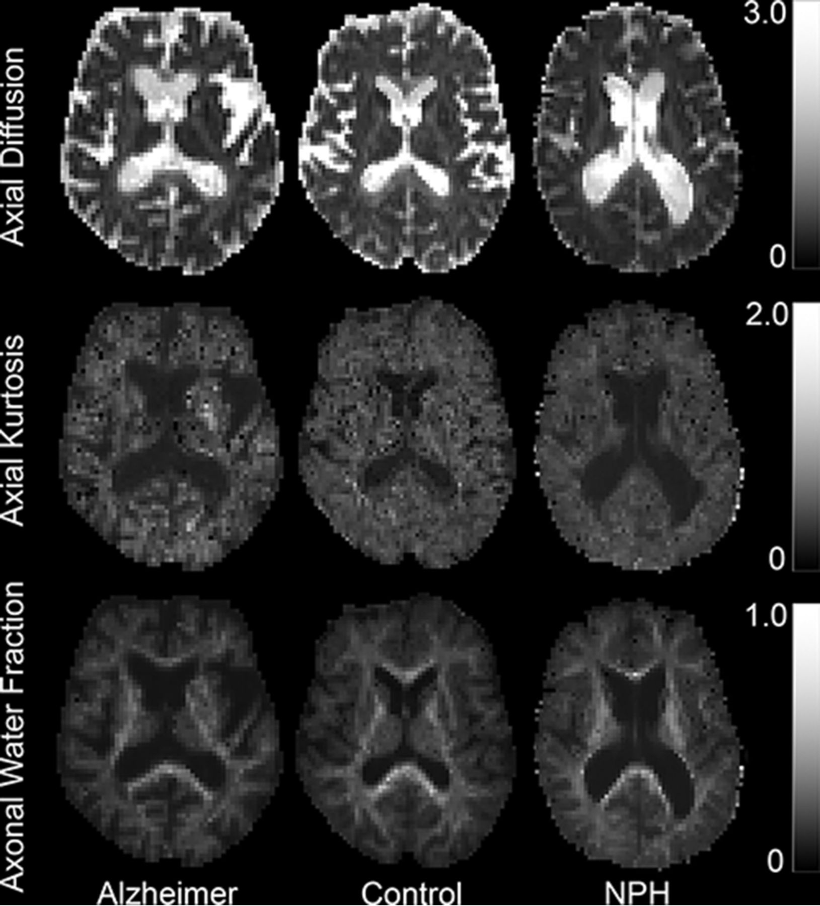
  • Fig 2.
    • Download figure
    • Open in new tab
    • Download powerpoint
    Fig 2.

    Sample parametric maps for patients with Alzheimer disease, healthy controls, and those with normal pressure hydrocephalus to qualitatively demonstrate values that are being mapped to the CST. From upper to lower, the maps show values for axial diffusivity, axial kurtosis, and axonal water fraction.
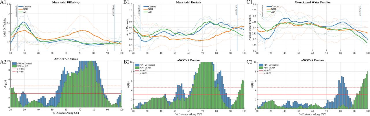
  • Fig 3.
    • Download figure
    • Open in new tab
    • Download powerpoint
    Fig 3.

    A: Along-tract group differences in axial diffusivity among patient groups. A1: Axial diffusion at each point along the CST in the right hemisphere. A2: ANCOVA F-test results show a significant difference between the NPH group and the Alzheimer and control groups at distances between 56% and 88%. B: Along-tract group differences in axial kurtosis among patient groups. B1: Axial kurtosis at each point along the CST in the right hemisphere. B2: ANCOVA F-test results show a significant difference between the NPH group and the Alzheimer and control groups at distances between 50% and 90%. C: Along-tract group differences in the axonal water fraction among patient groups. C1: Axonal water fraction at each point along the CST in the right hemisphere. C2: ANCOVA F-test results show a significant difference between the NPH group and the Alzheimer and control groups at distances between 67% and 86%.

  • Fig 4.
    • Download figure
    • Open in new tab
    • Download powerpoint
    Fig 4.

    Leukoaraiosis heat maps. Yellow areas show regions where patients are most likely to have leukoaraiosis. Probability maps are shown on top of the 1-mm brain of the Montreal Neurological Institute 152 space.

Tables

  • Figures
    • View popup
    Table 1:

    Key demographic information

    No. of SubjectsAge (Range) (Mean) (yr)Male/Female Ratio
    NPH2358–87 (76.9)13:10
    Alzheimer disease1056–81 (74.9)5:5
    Controls1160–87 (75)6:5
    • View popup
    Table 2:

    Values measured in the corona radiata, located at 70%–75% of the distance along the CSTa

    NC (n = 11) (Mean)Alzheimer Disease (n = 10) (Mean)NPH (n = 23) (Mean)NPH vs Alzheimer DiseaseNPH vs NCAlzheimer Disease vs NC
    P ValueAUCP ValueAUCP ValueAUC
    FA0.44 ± 0.070.39 ± 0.050.51 ± 0.100.01b0.880.040.760.120.76
    MD0.88 ± 0.050.89 ± 0.061.30 ± 0.370.02b0.87<0.01b0.940.800.79
    RD0.67 ± 0.050.69 ± 0.070.93 ± 0.370.180.650.04b0.760.530.84
    AD1.30 ± 0.081.26 ± 0.072.04 ± 0.39<0.01c0.96<0.01c1.00.600.51
    MK1.08 ± 0.091.04 ± 0.090.99 ± 0.190.940.520.350.730.660.82
    RK1.40 ± 0.141.19 ± 0.221.54 ± 0.420.030.820.340.620.100.82
    AK0.86 ± 0.070.97 ± 0.060.63 ± 0.11<0.01c0.97<0.01c0.980.110.67
    AWF0.42 ± 0.020.39 ± 0.030.38 ± 0.070.880.660.220.740.120.86
    • Note:—MD indicates mean diffusivity; RD, radial diffusivity; MK, mean kurtosis; RK, radial kurtosis; NC, healthy controls.

    • ↵a Values for the left and right hemispheres did not differ significantly (paired sample t test) and were averaged.

    • ↵b P < .05.

    • ↵c P < .01.

PreviousNext
Back to top

In this issue

American Journal of Neuroradiology: 39 (12)
American Journal of Neuroradiology
Vol. 39, Issue 12
1 Dec 2018
  • Table of Contents
  • Index by author
  • Complete Issue (PDF)
Advertisement
Print
Download PDF
Email Article

Thank you for your interest in spreading the word on American Journal of Neuroradiology.

NOTE: We only request your email address so that the person you are recommending the page to knows that you wanted them to see it, and that it is not junk mail. We do not capture any email address.

Enter multiple addresses on separate lines or separate them with commas.
Diffusional Kurtosis along the Corticospinal Tract in Adult Normal Pressure Hydrocephalus
(Your Name) has sent you a message from American Journal of Neuroradiology
(Your Name) thought you would like to see the American Journal of Neuroradiology web site.
CAPTCHA
This question is for testing whether or not you are a human visitor and to prevent automated spam submissions.
Cite this article
B. Ades-Aron, S. Yeager, N. Miskin, E. Fieremans, A. George, J. Golomb
Diffusional Kurtosis along the Corticospinal Tract in Adult Normal Pressure Hydrocephalus
American Journal of Neuroradiology Dec 2018, 39 (12) 2218-2223; DOI: 10.3174/ajnr.A5845

Citation Manager Formats

  • BibTeX
  • Bookends
  • EasyBib
  • EndNote (tagged)
  • EndNote 8 (xml)
  • Medlars
  • Mendeley
  • Papers
  • RefWorks Tagged
  • Ref Manager
  • RIS
  • Zotero
0 Responses
Respond to this article
Share
Bookmark this article
Diffusional Kurtosis along the Corticospinal Tract in Adult Normal Pressure Hydrocephalus
B. Ades-Aron, S. Yeager, N. Miskin, E. Fieremans, A. George, J. Golomb
American Journal of Neuroradiology Dec 2018, 39 (12) 2218-2223; DOI: 10.3174/ajnr.A5845
del.icio.us logo Twitter logo Facebook logo Mendeley logo
  • Tweet Widget
  • Facebook Like
  • Google Plus One
Purchase

Jump to section

  • Article
    • Abstract
    • ABBREVIATIONS:
    • Materials and Methods
    • Results
    • Discussion
    • Conclusions
    • Acknowledgments
    • Footnotes
    • References
  • Figures & Data
  • Supplemental
  • Info & Metrics
  • Responses
  • References
  • PDF

Related Articles

  • PubMed
  • Google Scholar

Cited By...

  • No citing articles found.
  • Crossref (5)
  • Google Scholar

This article has been cited by the following articles in journals that are participating in Crossref Cited-by Linking.

  • The role of diffusion tensor imaging in idiopathic normal pressure hydrocephalus: A literature review
    Irene Grazzini, Duccio Venezia, Gian Luca Cuneo
    The Neuroradiology Journal 2021 34 2
  • Corticospinal tract abnormalities and ventricular dilatation: A transdiagnostic comparative tractography study
    Alessia Sarica, Andrea Quattrone, Alessandro Mechelli, Maria Grazia Vaccaro, Maurizio Morelli, Aldo Quattrone
    NeuroImage: Clinical 2021 32
  • Progression in Neuroimaging of Normal Pressure Hydrocephalus
    Rui Yin, Junxian Wen, Junji Wei
    Frontiers in Neurology 2021 12
  • Diffusion Spectrum Imaging of Corticospinal Tracts in Idiopathic Normal Pressure Hydrocephalus
    Hong Zhang, Wen-Jie He, Li-Hong Liang, Han-Wen Zhang, Xie-Jun Zhang, Liang Zeng, Si-Ping Luo, Fan Lin, Yi Lei
    Frontiers in Neurology 2021 12
  • Diffusion spectrum imaging in patients with idiopathic normal pressure hydrocephalus: correlation with ventricular enlargement
    Qian Wu, Wenjie He, Chenyuan Liu, Xiaolin Yang, Jiakuan Chen, Boyan Xu, Xi Zhou, Guodong Huang, Jun Xia
    BMC Neurology 2024 24 1

More in this TOC Section

Adult Brain

  • Diagnostic Neuroradiology of Monoclonal Antibodies
  • Clinical Outcomes After Chiari I Decompression
  • Segmentation of Brain Metastases with BLAST
Show more Adult Brain

Functional

  • Kurtosis and Epileptogenic Tubers: A Pilot Study
  • Glutaric Aciduria Type 1: DK vs. Conventional MRI
  • Brain Iron in Niemann-Pick Type C: 7T Study
Show more Functional

Similar Articles

Advertisement

Indexed Content

  • Current Issue
  • Accepted Manuscripts
  • Article Preview
  • Past Issues
  • Editorials
  • Editor's Choice
  • Fellows' Journal Club
  • Letters to the Editor
  • Video Articles

Cases

  • Case Collection
  • Archive - Case of the Week
  • Archive - Case of the Month
  • Archive - Classic Case

More from AJNR

  • Trainee Corner
  • Imaging Protocols
  • MRI Safety Corner
  • Book Reviews

Multimedia

  • AJNR Podcasts
  • AJNR Scantastics

Resources

  • Turnaround Time
  • Submit a Manuscript
  • Submit a Video Article
  • Submit an eLetter to the Editor/Response
  • Manuscript Submission Guidelines
  • Statistical Tips
  • Fast Publishing of Accepted Manuscripts
  • Graphical Abstract Preparation
  • Imaging Protocol Submission
  • Evidence-Based Medicine Level Guide
  • Publishing Checklists
  • Author Policies
  • Become a Reviewer/Academy of Reviewers
  • News and Updates

About Us

  • About AJNR
  • Editorial Board
  • Editorial Board Alumni
  • Alerts
  • Permissions
  • Not an AJNR Subscriber? Join Now
  • Advertise with Us
  • Librarian Resources
  • Feedback
  • Terms and Conditions
  • AJNR Editorial Board Alumni

American Society of Neuroradiology

  • Not an ASNR Member? Join Now

© 2025 by the American Society of Neuroradiology All rights, including for text and data mining, AI training, and similar technologies, are reserved.
Print ISSN: 0195-6108 Online ISSN: 1936-959X

Powered by HighWire