
Article Figures & Data
Figures
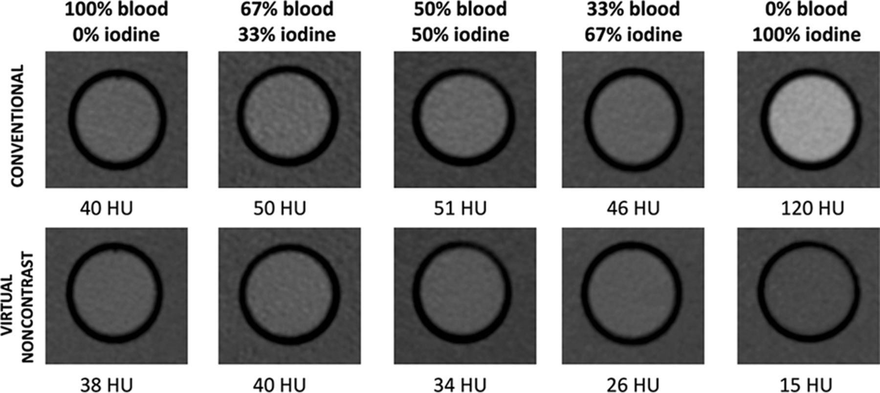
- Fig 1.
Comparison of the attenuation of diluted blood, iodine, and blood-iodine mixtures on conventional and virtual noncontrast images. There is an incremental decrease of VNC attenuation values with decreasing blood content, compared with conventional CT attenuation values.
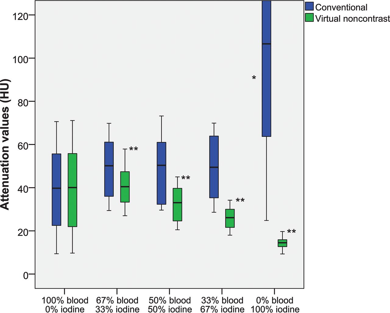
- Fig 2.
Attenuation values (HU) of diluted blood, blood-iodine mixtures, and diluted iodine on spectral detector CT conventional and virtual noncontrast images. There is a significant incremental decrease of VNC attenuation values with decreasing blood content. The asterisk indicates a significant difference of conventional CT attenuation compared with other compositions (P < .01); double asterisks, significant differences of VNC attenuation among these compositions (P < .01).
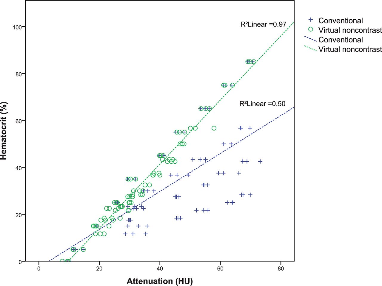
- Fig 3.
Correlation between the hematocrit in our dilutions and the attenuation in the conventional and VNC images.
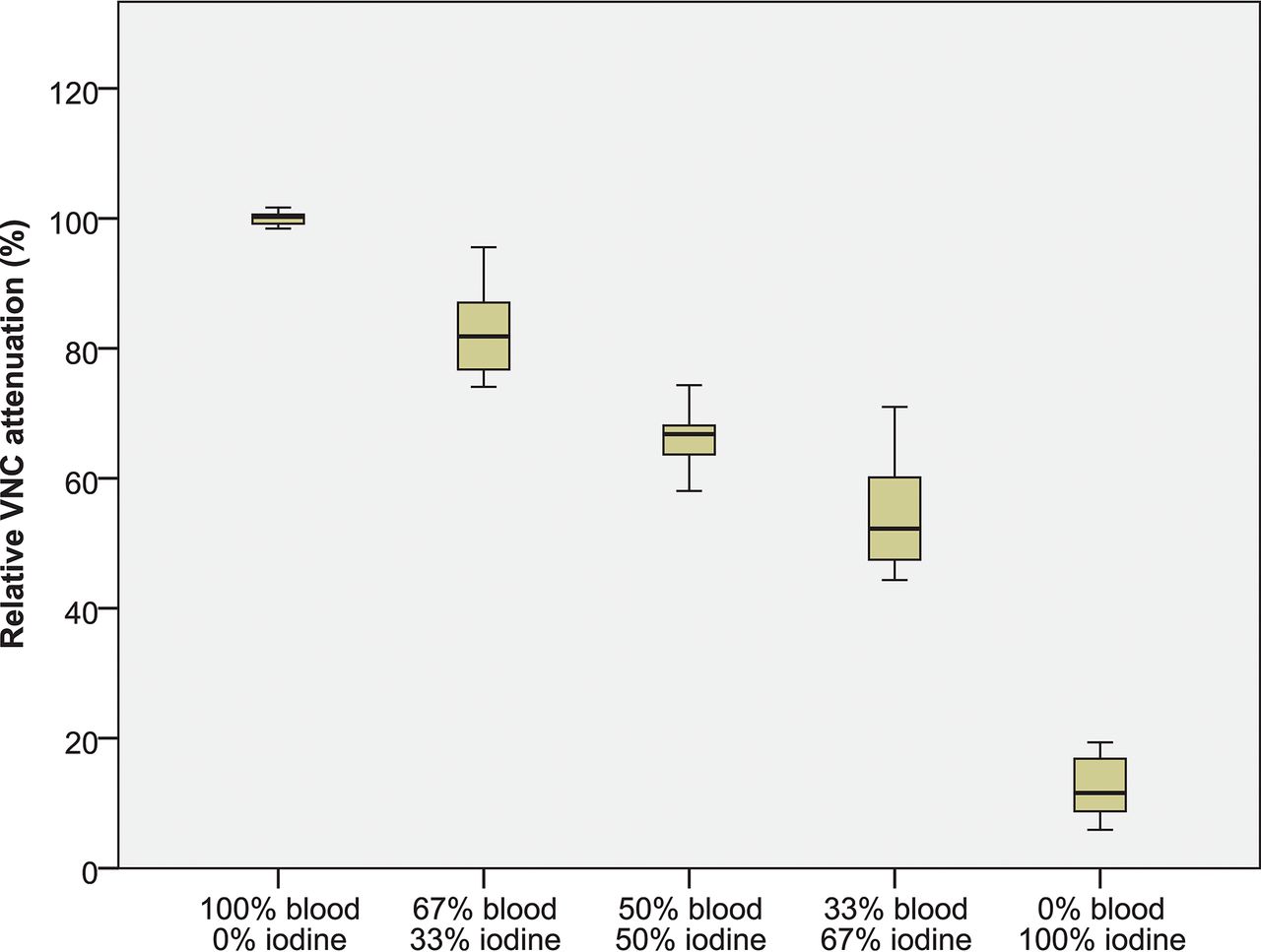
- Fig 4.
Relative VNC attenuation (%), by comparing VNC attenuation with the attenuation on conventional CT for all investigated categories (diluted blood, blood-iodine mixtures, and diluted iodine). R-VNC values among all compositions were significantly different (P < .01).
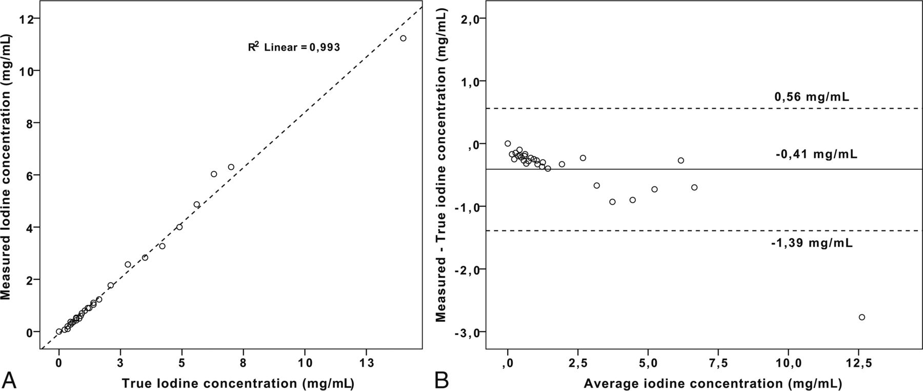
- Fig 5.
Results of the iodine quantification measurements by comparison of measured-to-true iodine concentrations (A) and errors in iodine quantification measurements by a Bland-Altman plot (B). A, Correlation between measured and true iodine concentrations is excellent (R2 > 0.99, P < .01). B, Mean iodine quantification error (± 95% CI) was −0.41 ± 0.31–0.50 mg/mL).
- Fig 6.
Performance of conventional CT attenuation, virtual noncontrast attenuation, and relative VNC attenuation for the detection of blood. A, Receiver operating characteristic curve analysis shows the highest area under the curve for VNC (0.97 ± 0.94–0.99), followed by R-VNC attenuation (0.87 ± 0.77–0.97) and attenuation in the conventional CT images (0.29 ± 0.16–0.41), and the area under the curve was lowest for iodine quantification (0.16 ± 0.06–0.25). B, When we combined a ≥40% R-VNC (dashed line) and ≥10 HU VNC cutoff, there is 100% differentiation between blood-containing (diluted blood and blood-iodine mixtures) and diluted iodine samples.


Tables
Attenuation values (HU) of diluted blood, blood-iodine mixtures, and diluted iodine on SDCT conventional and VNC imagesa
Conventional (HU) VNC (HU) R-VNC (%) Mean 95% CI Mean 95% CI Mean 95% CI 100% blood–0% iodine 39.1 31.5–46.7 39.1 31.5–46.7 99.6 98.8–100.4 67% blood–33% iodine 49.5 42.8–56.2 40.4 35.9–44.9 82.8 79.5–86.1 50% blood–50% iodine 49.2 41.7–56.7 32.6 28.6–36.6 67.6 64.4–70.9 33% blood–67% iodine 49.4 42.2–56.6 26.0 23.3–28.6 54.3 50.6–58.0 0% blood –100% iodine 129.5 99.4–159.6 14.5 13.4–15.6 15.7 12.4–19.1 ↵a R-VNC attenuation (%) was calculated by comparing VNC attenuation with the attenuation on conventional CT for all investigated categories.


Jump to section
Related Articles
Cited By...
- Determinants and Clinical Relevance of Iodine Contrast Extravasation after Endovascular Thrombectomy: A Dual-Energy CT Study
- Utilizing dual energy CT to distinguish blood from contrast leakage following middle meningeal artery embolization for chronic subdural hematomas
- Prediction of Hemorrhage after Successful Recanalization in Patients with Acute Ischemic Stroke: Improved Risk Stratification Using Dual-Energy CT Parenchymal Iodine Concentration Ratio Relative to the Superior Sagittal Sinus
- Virtual Monoenergetic Images from Spectral Detector CT Enable Radiation Dose Reduction in Unenhanced Cranial CT