
Article Figures & Data
Figures
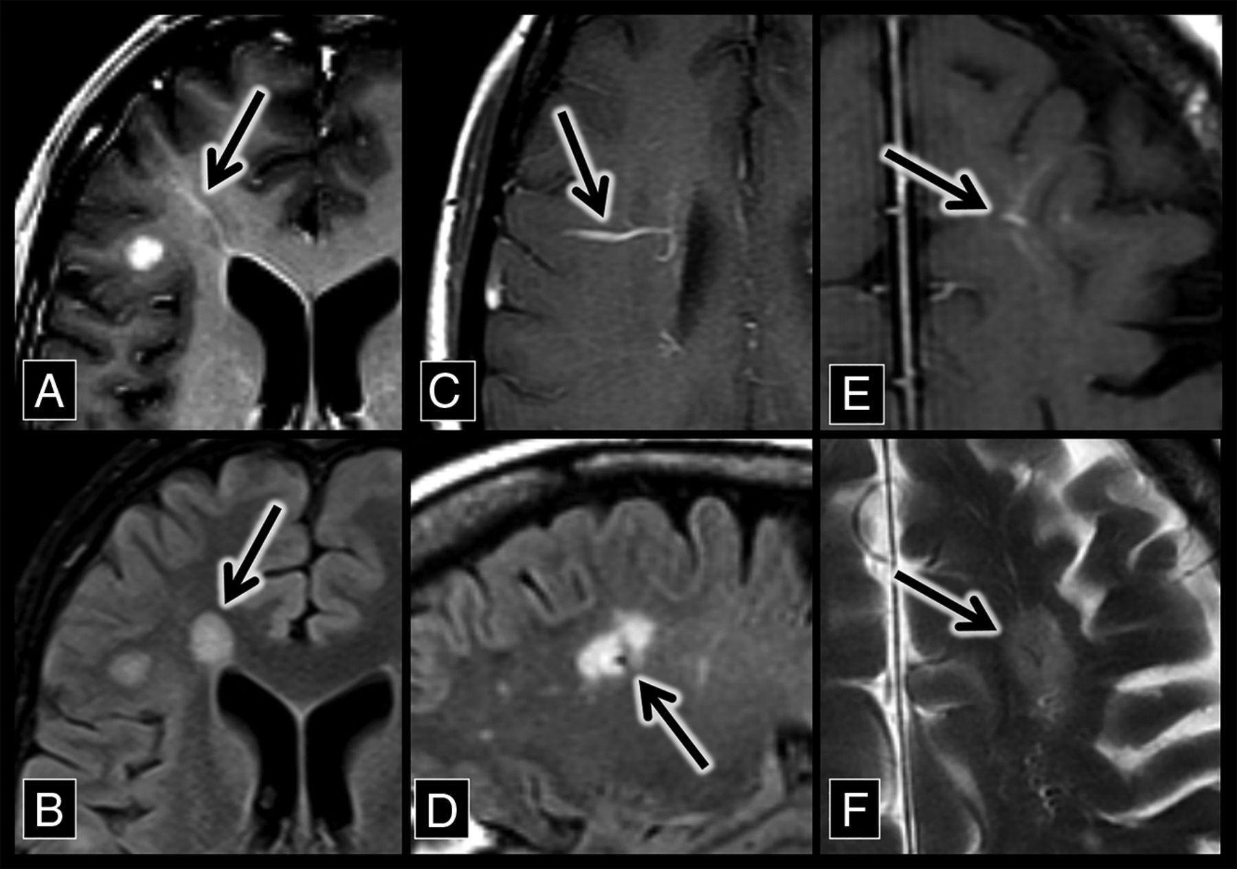
- Fig 1.
Developmental venous anomaly–associated lesions in patients with MS. A, An axial contrast-enhanced T1 sequence shows a right frontal lobe DVA (arrow) with surrounding T1 hypointensity. B, An axial FLAIR sequence shows hyperintensity (arrow) that corresponds to the DVA and associated T1 hypointensity in A. C, An axial contrast-enhanced T1 sequence shows a right frontal lobe DVA (arrow). D, A sagittal FLAIR sequence shows a flow void with adjacent hyperintensity (the central vein sign, arrow), which corresponds to the DVA in C. E, An axial contrast-enhanced T1 sequence shows a left frontal lobe DVA (arrow). F, An axial T2 sequence shows a flow void with adjacent hyperintensity (the central vein sign, arrow), which corresponds to the DVA in E.
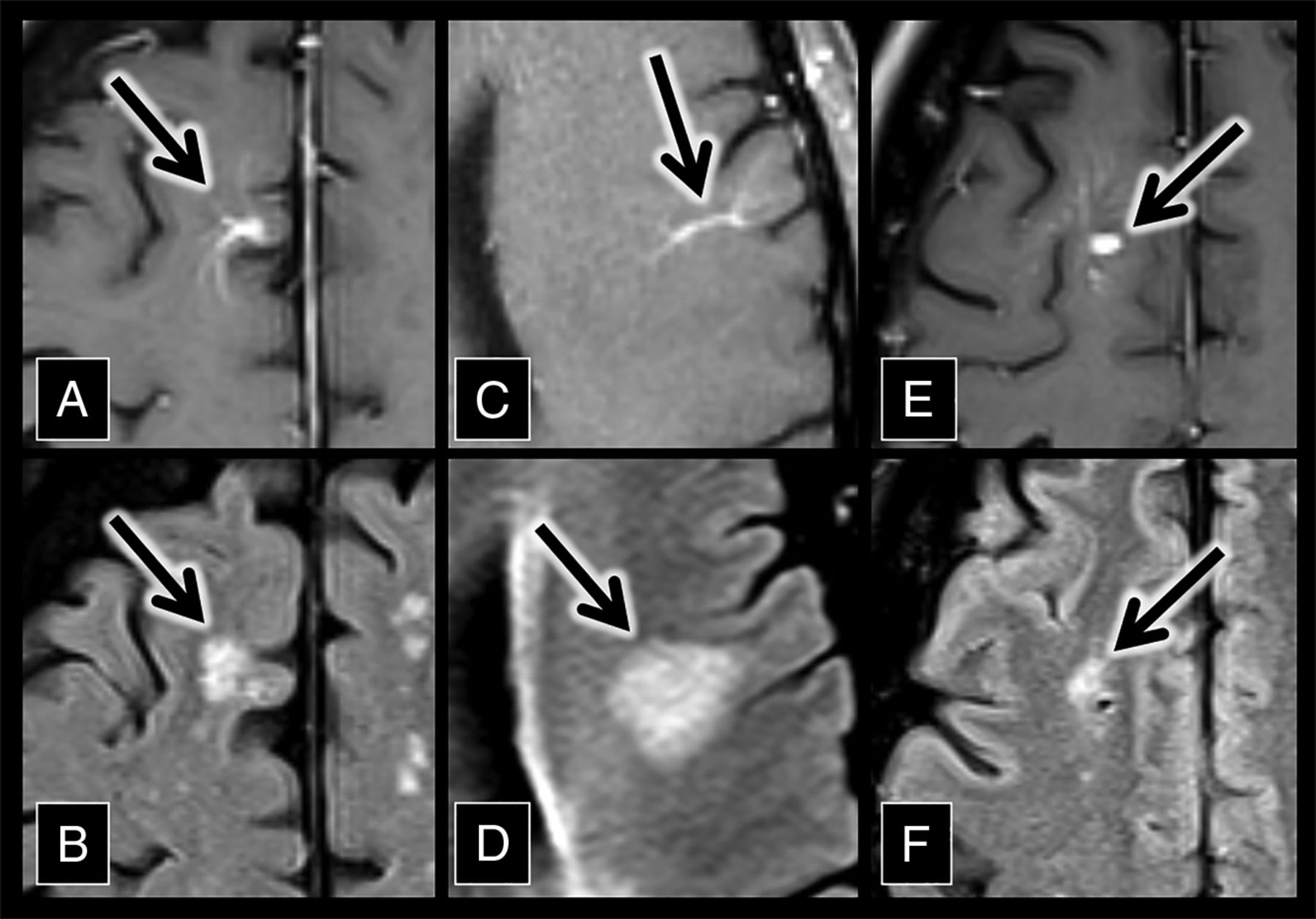
- Fig 2.
Developmental venous anomaly–associated lesions in the control group. A, An axial contrast-enhanced T1 sequence shows a right frontal lobe DVA (arrow). B, An axial FLAIR sequence shows hyperintensity (arrow) adjacent to the DVA in A. C, An axial contrast-enhanced T1 sequence shows a left frontal lobe DVA (arrow). D, An axial FLAIR sequence shows hyperintensity (arrow) adjacent to the DVA in C. D, An axial contrast-enhanced T1 sequence shows a right frontal lobe DVA (arrow). F, An axial FLAIR sequence shows hyperintensity (arrow) adjacent to the DVA in E.
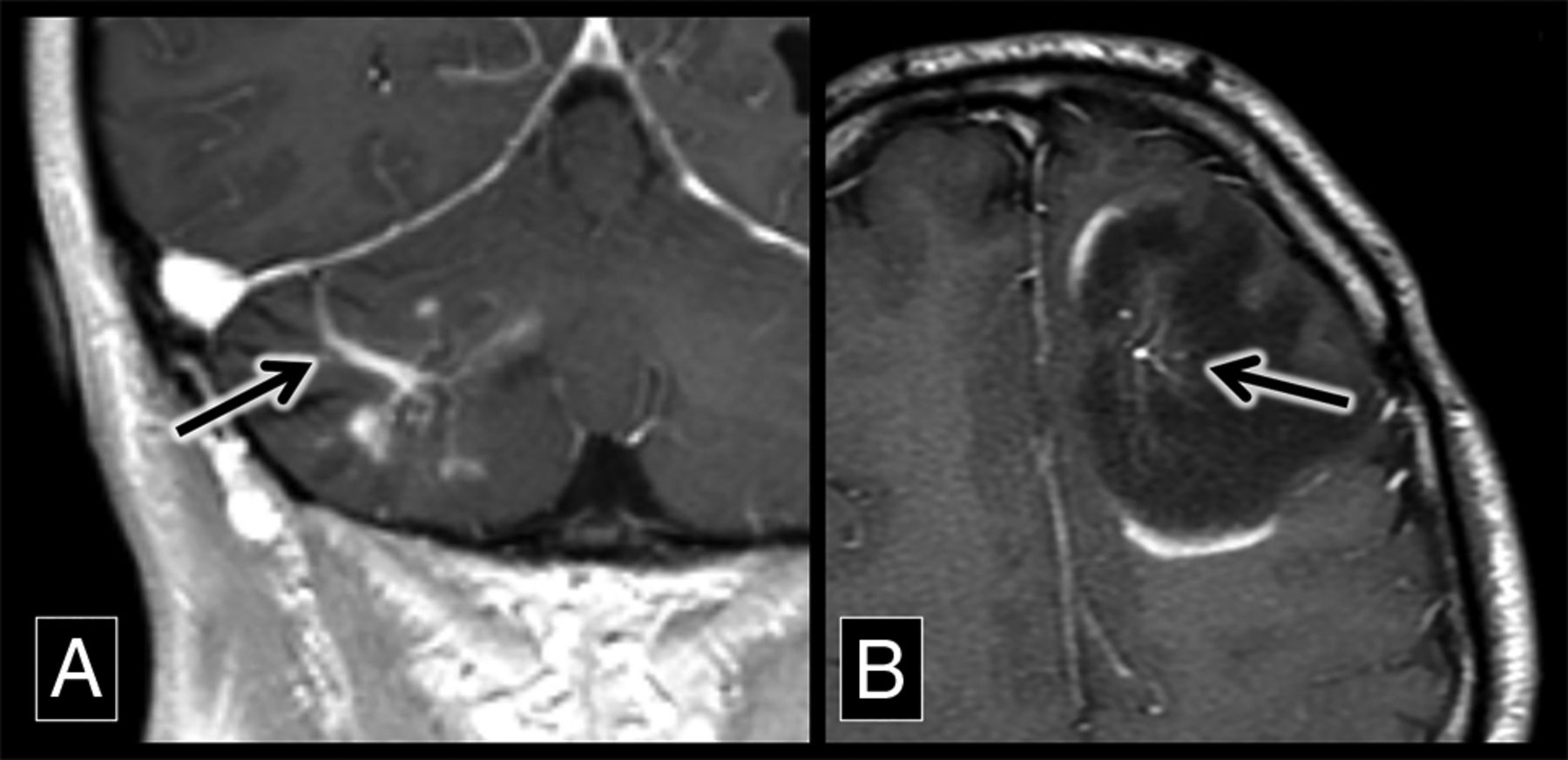
- Fig 3.
Demyelinating lesions around developmental venous anomalies with enhancement. A, A coronal contrast-enhanced T1 sequence shows a superficially draining right cerebellar DVA (arrow) associated with enhancing parenchymal lesions. This was biopsy-proved demyelination. B, An axial contrast-enhanced T1 sequence shows a left frontal lobe DVA (arrow) with surrounding T1 hypointensity and discontinuous peripheral enhancement, typical of demyelination in a patient with MS.


Tables
Characteristic Multiple Sclerosis (n = 74) Controls (n = 74) P Value Mean age (SD) (yr) 45.1 (13.5) 50.7 (18.4) .036 Sex: female 49 (66.2%) 37 (50.0%) .045 MRI field strength 3T 18 (24.3%) 11 (14.9%) MRI field strength 1.5T 56 (75.7%) 63 (85.1%) DVA location Lobar 49 (66.2%) 51 (68.9%) Basal ganglia 6 (8.1%) 5 (6.7%) Cerebellum 18 (24.3%) 15 (20.3%) Brain stem 1 (1.4%) 3 (4.1%) DVA drainage Superficial 50 (67.5%) 46 (62.1%) Deep 21 (28.4%) 25 (33.8%) Both 3 (4.1%) 3 (4.1%) Hypertension 13 (17.6%) 15 (20.3%) Diabetes 3 (4.1%) 6 (8.1%) Migraines 10 (13.5%) 10 (13.5%) Intracranial vasculitis 1 (1.4%) 1 (1.4%) Variable Multiple Sclerosis (n = 74) Controls (n = 74) P Value Positive FLAIR hyperintensity with a central vein 35 (47.3%) 10 (13.5%) <.001 FLAIR hyperintensity depth Juxtacortical 13 (37.1%) 5 (50.0%) Subcortical 12 (37.5%) 3 (30.0%) Periventricular 10 (28.6%) 2 (20.0%) Mean FLAIR hyperintensity width (SD) (mm) 8.5 (7.3) 8.0 (4.5) Enhancing lesions 4 (11.4%) 0 (0%) Location of DVA with associated FLAIR hyperintensity Lobar 29 (82.9%) 10 (100%) Basal ganglia 2 (5.7%) 0 (0%) Cerebellum 4 (11.4%) 0 (0%) Brain stem 0 (0%) 0 (0%) Drainage of DVA with associated FLAIR hyperintensity Superficial 23 (65.7%) 6 (60%) Deep 12 (34.3%) 2 (20%) Both 0 (0%) 2 (20%)


Jump to section
Related Articles
Cited By...
- No citing articles found.