Advertisement

Main menu

  • Home
  • Content
    • Current Issue
    • Accepted Manuscripts
    • Article Preview
    • Past Issue Archive
    • Video Articles
    • AJNR Case Collection
    • Case of the Week Archive
    • Case of the Month Archive
    • Classic Case Archive
  • Special Collections
    • AJNR Awards
    • Low-Field MRI
    • Alzheimer Disease
    • ASNR Foundation Special Collection
    • Photon-Counting CT
    • View All
  • Multimedia
    • AJNR Podcasts
    • AJNR SCANtastic
    • Trainee Corner
    • MRI Safety Corner
    • Imaging Protocols
  • For Authors
    • Submit a Manuscript
    • Submit a Video Article
    • Submit an eLetter to the Editor/Response
    • Manuscript Submission Guidelines
    • Statistical Tips
    • Fast Publishing of Accepted Manuscripts
    • Graphical Abstract Preparation
    • Imaging Protocol Submission
    • Author Policies
  • About Us
    • About AJNR
    • Editorial Board
    • Editorial Board Alumni
  • More
    • Become a Reviewer/Academy of Reviewers
    • Subscribers
    • Permissions
    • Alerts
    • Feedback
    • Advertisers
    • ASNR Home

User menu

  • Alerts
  • Log in

Search

  • Advanced search
American Journal of Neuroradiology

American Journal of Neuroradiology

ASHNR American Society of Functional Neuroradiology ASHNR American Society of Pediatric Neuroradiology ASSR
  • Alerts
  • Log in

Advanced Search

  • Home
  • Content
    • Current Issue
    • Accepted Manuscripts
    • Article Preview
    • Past Issue Archive
    • Video Articles
    • AJNR Case Collection
    • Case of the Week Archive
    • Case of the Month Archive
    • Classic Case Archive
  • Special Collections
    • AJNR Awards
    • Low-Field MRI
    • Alzheimer Disease
    • ASNR Foundation Special Collection
    • Photon-Counting CT
    • View All
  • Multimedia
    • AJNR Podcasts
    • AJNR SCANtastic
    • Trainee Corner
    • MRI Safety Corner
    • Imaging Protocols
  • For Authors
    • Submit a Manuscript
    • Submit a Video Article
    • Submit an eLetter to the Editor/Response
    • Manuscript Submission Guidelines
    • Statistical Tips
    • Fast Publishing of Accepted Manuscripts
    • Graphical Abstract Preparation
    • Imaging Protocol Submission
    • Author Policies
  • About Us
    • About AJNR
    • Editorial Board
    • Editorial Board Alumni
  • More
    • Become a Reviewer/Academy of Reviewers
    • Subscribers
    • Permissions
    • Alerts
    • Feedback
    • Advertisers
    • ASNR Home
  • Follow AJNR on Twitter
  • Visit AJNR on Facebook
  • Follow AJNR on Instagram
  • Join AJNR on LinkedIn
  • RSS Feeds

AJNR Awards, New Junior Editors, and more. Read the latest AJNR updates

Editorial

The Woven EndoBridge Finally Coming Home across the Atlantic: What to Expect?

W.J. van Rooij, S.B.T. van Rooij, H.G. Kortman and J.P. Peluso
American Journal of Neuroradiology November 2018, 39 (11) 1964-1966; DOI: https://doi.org/10.3174/ajnr.A5798
W.J. van Rooij
aFrom the Department of Radiology (W.J.v.R., H.G.K., J.P.P.), Elisabeth Tweesteden Ziekenhuis, Tilburg, The Netherlands
  • Find this author on Google Scholar
  • Find this author on PubMed
  • Search for this author on this site
  • ORCID record for W.J. van Rooij
S.B.T. van Rooij
bDepartment of Radiology (S.B.T.v.R.), Noordwest Ziekenhuisgroep, Alkmaar, The Netherlands.
  • Find this author on Google Scholar
  • Find this author on PubMed
  • Search for this author on this site
  • ORCID record for S.B.T. van Rooij
H.G. Kortman
aFrom the Department of Radiology (W.J.v.R., H.G.K., J.P.P.), Elisabeth Tweesteden Ziekenhuis, Tilburg, The Netherlands
  • Find this author on Google Scholar
  • Find this author on PubMed
  • Search for this author on this site
  • ORCID record for H.G. Kortman
J.P. Peluso
aFrom the Department of Radiology (W.J.v.R., H.G.K., J.P.P.), Elisabeth Tweesteden Ziekenhuis, Tilburg, The Netherlands
  • Find this author on Google Scholar
  • Find this author on PubMed
  • Search for this author on this site
  • ORCID record for J.P. Peluso
  • Article
  • Figures & Data
  • Supplemental
  • Info & Metrics
  • Responses
  • References
  • PDF
Loading

The Woven EndoBridge (WEB; Sequent Medical, Aliso Viejo, California) is an intrasaccular flow diverter primarily developed for wide-neck bifurcation aneurysms without the need for supporting stents and concomitant dual-antiplatelet therapy. The WEB consists of a self-expanding, oblong or spheric braid of platinum-cored nitinol wires intended to implant inside the aneurysmal sac, and it can be electrothermally detached. Since its introduction in 2010, the WEB has evolved from a high-profile dual-layer to a low-profile single-layer construction that can be delivered through a 0.017-inch microcatheter in sizes up to 7 mm.1 To date, more than 6000 WEBs have been implanted worldwide, most of these in Europe.

In the United States, FDA approval is expected soon, and our American colleagues will then have an exciting new tool available for endovascular treatment of intracranial aneurysms. The extensive European experience with the WEB provides for a flying start in the United States.

In this article, we share our personal experience with the WEB after having treated almost 300 aneurysms with the device.

How Did We Start?

In 2014, we evaluated our results of stent-assisted coiling and found that stent placement in 45 patients with acutely ruptured aneurysms resulted in permanent neurologic deficit or death in 10 (22%), in 5 due to thromboembolic complications and in 5 due to early rebleed in the postoperative period despite the aneurysms being adequately coiled.2 In February 2015, we considered an alternative for stent-assisted coiling in the new-generation WEB with a single layer, suitable for a 0.021-inch microcatheter. We started to use the WEB in wide-neck unruptured aneurysms without supporting stents or balloons, and our first results were encouraging. To further master the technique of the WEB, we gradually expanded the indication to all aneurysms suitable for the WEB; in general, small aneurysms from 3 to 10 mm, regardless of neck size or location.3⇓–5

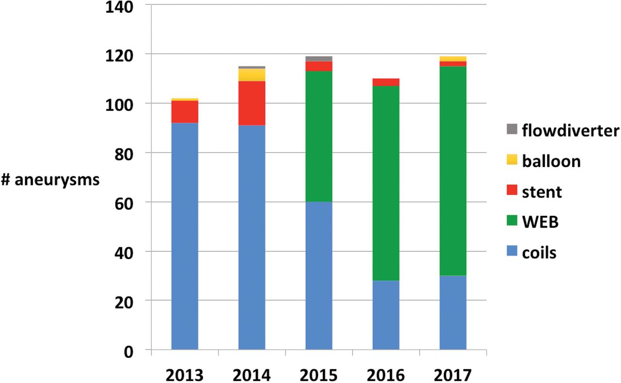
The Figure is a graphic display of the use of the WEB, coils, additional supporting devices, and flow diverters for endovascular treatment of intracranial aneurysms in our practice during 2013–2017.

FIGURE.
  • Download figure
  • Open in new tab
  • Download powerpoint
FIGURE.

Evolution of devices used for endovascular treatment of intracranial aneurysms between 2013 and 2017.

Access and Periprocedural Anticoagulation

We use a biaxial approach with a short (11 cm) 6F sheath and a Benchmark (Penumbra, Alameda, California) guiding catheter positioned in the petrous carotid artery or V3 segment of the vertebral artery. Only with very tortuous vessels do we use a 90-cm-long sheath (Neuron Max; Penumbra) with a 6F or 5F distal-access guiding catheter.

In our hospital (St Elisabeth Hospital Tilburg, The Netherlands), all admitted patients receive subcutaneous heparin, 2500 U twice daily, as a thrombotic prophylaxis. In patients with ruptured aneurysms, we use no other anticoagulation than heparin in the pressure bags for flushing (5000 U/L). In patients with unruptured aneurysms, heparin, a 2500–5000 U bolus, is given at the beginning of the procedure. Antiplatelet medication is not routinely administered.

In case of a thromboembolic complication, we try to remove the thrombus with thrombosuction or a stent retriever, and for peripheral emboli, tirofiban is used. When antiplatelet medication is considered necessary, aspirin, 500 mg, is given intravenously, later followed by prasugrel or clopidogrel (Plavix).

WEB Sizing and Technique of Implantation

The aneurysm is measured from 3D rotational images. Oversizing the WEB is imperative to assure anchoring the device to the aneurysm walls. In general, the WEB is oversized by 1–2 mm from the mean width of the aneurysm in 2 perpendicular planes. Because compression in the diameter results in increased height, the height is chosen 1–2 mm less than the actual aneurysm height. In regularly shaped aneurysms, WEB sizing is straightforward. In irregularly shaped aneurysms, some spatial awareness is helpful. Throughout our experience with WEB sizing, our sizing failure rate is about 25%, which has not improved with time.

In the sizes of 4–7 mm, there is overlap between the WEB 21 and 17 systems. In general, the WEB 17 performs better in tortuous and difficult anatomy and the WEB 21 is more stable. Our personal preference is WEB 17 in the smallest sizes, 3–4 mm, and WEB 21 in the larger sizes, 5–8 mm.

The VIA 17 and 21 Microcatheters (Sequent Medical) are specifically developed for the WEB. These catheters can be steam-shaped when necessary (about 1 in 10 in our practice) and are stable during WEB delivery. The technique of WEB implantation is as follows: The tip of the microcatheter is positioned in the dome of the aneurysm. Then, the first part of the WEB is slowly unsheathed by microcatheter withdrawal until half of the WEB is deployed. At that point, the WEB has become soft, and puncture of the aneurysm wall with the distal marker has become highly unlikely. Next, the microcatheter is slightly pushed forward to ensure that the WEB is in the fundus of the aneurysm. At this point, the WEB can be completely deployed by further retracting the microcatheter while keeping the WEB in place.

The position of the WEB is checked with an angiographic run. Sometimes a rotational angiographic run may be helpful to evaluate the WEB position in relation to the aneurysm neck and parent vessels. We never use VasoCT (Philips Healthcare, Best, the Netherlands) for this purpose because this is time-consuming and we are somewhat afraid to recapture the WEB after several minutes have passed; possible thrombus formed inside the WEB may be fragmented and dispersed.

When a good WEB position is confirmed, the device is detached and the procedure is terminated, despite the possible presence of some flow inside the WEB and aneurysm.

The Appendix contains several links to videos of WEB treatments of intracranial aneurysms.

Use of Additional Devices

Additional coils are only rarely necessary to occlude the aneurysm or part of it. In most irregularly shaped aneurysms, the WEB can be used to cork the neck. Parts of the aneurysm not covered with the WEB will then thrombose anyway.

We never use a supporting balloon during WEB placement. In 2 cases with previously treated aneurysms, a stent was placed as a bailout procedure to stabilize the WEB after it tilted on detachment.

Imaging Follow-Up after WEB Treatment

Because many aneurysms still show some filling immediately after WEB placement, initial occlusion rates, as with coiling, cannot be given. We perform angiography after 3 months to confirm the absence of residual filling inside the WEB. Further follow-up is scheduled with 3T MRA.6 Because the WEB acts like a Faraday cage, possible residual flow may remain undetected. Fortunately, residual flow at 3 months is extremely rare; we encountered only a few cases in larger aneurysms.

Additional Applications of the WEB

The WEB can also be applied for parent vessel occlusion of both large and small arteries.7 For ICA occlusion, 2 WEBs are sufficient to occlude the artery. As opposed to detachable balloons, the WEB can be placed exactly in the desired position in the parent artery without fear of premature detachment. We maximally oversize the WEB in larger arteries such as the ICA or V4. The occlusion of small vessels such as the PICA (distal flow aneurysm) or the distal posterior cerebral artery (dissecting aneurysm) can be performed with the smallest WEB 17 devices.

The WEB can also be used in previously treated aneurysms.8 In wide-neck remnants, stents can be avoided. In large and giant partially thrombosed aneurysms, results with the WEB are as poor as with other techniques; the WEB will migrate into intraluminal thrombus just like coil meshes do.

Clinical and Imaging Results and Implications for Practice

Our published results with the WEB, both for ruptured and unruptured aneurysms, are good with a reopening/retreatment rate of 7%, about 95% adequate occlusion, and no bleeds or rebleeds during follow-up. We encountered only 1 procedural rupture without clinical sequelae. Most thromboembolic complications were due to catheterization and not to the WEB.1,3⇓–5,7

Clinical results in our practice have improved since the WEB replaced stent-assisted coiling, especially in ruptured aneurysms.2,4 The simple periprocedural anticoagulation protocol without antiplatelet medication is a great advantage.

The WEB procedure is a single-catheter treatment and is usually straightforward and quick. After gradually gaining confidence with the WEB, we now treat most aneurysms (about 70%) with this device (Figure). Only large aneurysms and shallow aneurysms are treated otherwise. In our practice, stents and flow diverters are never used in ruptured aneurysms and only rarely in unruptured aneurysms (Figure).

Although the use of the WEB has some technical issues, these can readily be learned by anyone with some experience with endovascular aneurysm treatment. The procedure is similar to simple coiling. Proper training on phantoms and proctoring of first cases can essentially avoid a learning curve. We recommend that first-time users start with simple aneurysms rather than with complex wide-neck aneurysms. Simple aneurysms can be wide-neck as long as there is sufficient height to accommodate oversizing in width. With growing confidence, more challenging aneurysms can be treated, sometimes surprisingly easily.

Improvements and Perspectives

The development of the WEB is ongoing. More shallow sizes are being developed, and experiments are being performed with changes in the WEB shape; for example, a hybrid version between Single-Layer and Single-Layer Sphere. The larger WEB sizes, 9–11 mm, may become available in lower profiles suitable for a 0.027-inch instead of a 0.033-inch microcatheter.

To reduce the proportion of incorrect sizing of about 1 in 4, an automated 3D software program would be a valuable tool.

The WEB is now marketed as an expensive device, comparable with the price of stent-assisted coiling. With growing popularity, prices are likely to drop.

In summary, in our practice, the WEB has rapidly gained popularity and largely replaced the use of coils, stents, and flow diverters. The avoidance of dual antiplatelet medication is a great advantage over stents and flow diverters, resulting in fewer thromboembolic complications and fewer early rebleeds. The easy, quick, and safe technique has a convincing advantage over balloon- or stent-assisted coil treatments.

Footnotes

  • Disclosures: Willem Jan van Rooij—RELATED: Consulting Fee or Honorarium: Terumo, MicroVention; Other: Terumo, MicroVention, Comments: Proctoring WEB cases.* Sanne van Rooij—RELATED: Grants: Noordwest Ziekenhuisgroep Alkmaar, and unrestricted grant from MicroVention; Comments: 20% of resident salary during 2 years.* Jo P. Peluso—RELATED: Consultancy: Proctoring WEB cases.* *Money paid to individual.

References

  1. 1.↵
    1. van Rooij SB,
    2. Peluso JP,
    3. Sluzewski M, et al
    . The new low-profile WEB 17 system for treatment of intracranial aneurysms: first clinical experiences. AJNR Am J Neuroradiol 2018;39:859–63 doi:10.3174/ajnr.A5608 pmid:29567650
    Abstract/FREE Full Text
  2. 2.↵
    1. Bechan RS,
    2. Sprengers ME,
    3. Majoie CB, et al
    . Stent-assisted coil embolization of intracranial aneurysms: complication in acutely ruptured versus unruptured aneurysms. AJNR Am J Neuroradiol 2016;37:502–07 doi:10.3174/ajnr.A4542 pmid:26405089
    Abstract/FREE Full Text
  3. 3.↵
    1. van Rooij WJ,
    2. Peluso JP,
    3. Bechan RS, et al
    . WEB treatment of ruptured intracranial aneurysms. AJNR Am J Neuroradiol 2016;37:1679–83 doi:10.3174/ajnr.A4811 pmid:27102307
    Abstract/FREE Full Text
  4. 4.↵
    1. van Rooij SB,
    2. van Rooij WJ,
    3. Peluso JP, et al
    . WEB treatment of ruptured intracranial aneurysms: a single center cohort of 100 patients. AJNR Am J Neuroradiol 2017;38:2282–87 doi:10.3174/ajnr.A5371 pmid:28882858
    Abstract/FREE Full Text
  5. 5.↵
    1. van Rooij SB,
    2. van Rooij WJ,
    3. Peluso JP, et al
    . The Woven EndoBridge (WEB) as primary treatment for unruptured intracranial aneurysms. Interv Neuroradiol 2018 Jan 1. [Epub ahead of print] doi:10.1177/1591019918772174 pmid:29768963
    CrossRefPubMed
  6. 6.↵
    1. van Rooij S,
    2. Peluso JP,
    3. Sluzewski M, et al
    . Mid-term 3T-MRA follow-up of intracranial aneurysms treated with the Woven EndoBridge (WEB). Interv Neuroradiol 2018 Jan 1. [Epub ahead of print] doi:10.1177/1591019918788346 pmid:30001648
    CrossRefPubMed
  7. 7.↵
    1. van Rooij WJ,
    2. Sluzewski M,
    3. Bechan RS, et al
    . Therapeutic internal carotid or vertebral artery occlusion using the WEB device. Interv Neuroradiol 2016;22:365–67 doi:10.1177/1591019916631146 pmid:26861025
    CrossRefPubMed
  8. 8.↵
    1. van Rooij SB,
    2. van Rooij WJ,
    3. Sluzewski M, et al
    . The Woven EndoBridge (WEB) for recurrent aneurysms: clinical and imaging results. Interv Neuroradiol 2018 Sep 12 [Epub ahead of print]
  • Received June 1, 2018.
  • Accepted after revision June 27, 2018.
  • © 2018 by American Journal of Neuroradiology
PreviousNext
Back to top

In this issue

American Journal of Neuroradiology: 39 (11)
American Journal of Neuroradiology
Vol. 39, Issue 11
1 Nov 2018
  • Table of Contents
  • Index by author
  • Complete Issue (PDF)
Advertisement
Print
Download PDF
Email Article

Thank you for your interest in spreading the word on American Journal of Neuroradiology.

NOTE: We only request your email address so that the person you are recommending the page to knows that you wanted them to see it, and that it is not junk mail. We do not capture any email address.

Enter multiple addresses on separate lines or separate them with commas.
The Woven EndoBridge Finally Coming Home across the Atlantic: What to Expect?
(Your Name) has sent you a message from American Journal of Neuroradiology
(Your Name) thought you would like to see the American Journal of Neuroradiology web site.
CAPTCHA
This question is for testing whether or not you are a human visitor and to prevent automated spam submissions.
Cite this article
W.J. van Rooij, S.B.T. van Rooij, H.G. Kortman, J.P. Peluso
The Woven EndoBridge Finally Coming Home across the Atlantic: What to Expect?
American Journal of Neuroradiology Nov 2018, 39 (11) 1964-1966; DOI: 10.3174/ajnr.A5798

Citation Manager Formats

  • BibTeX
  • Bookends
  • EasyBib
  • EndNote (tagged)
  • EndNote 8 (xml)
  • Medlars
  • Mendeley
  • Papers
  • RefWorks Tagged
  • Ref Manager
  • RIS
  • Zotero
0 Responses
Respond to this article
Share
Bookmark this article
The Woven EndoBridge Finally Coming Home across the Atlantic: What to Expect?
W.J. van Rooij, S.B.T. van Rooij, H.G. Kortman, J.P. Peluso
American Journal of Neuroradiology Nov 2018, 39 (11) 1964-1966; DOI: 10.3174/ajnr.A5798
del.icio.us logo Twitter logo Facebook logo Mendeley logo
  • Tweet Widget
  • Facebook Like
  • Google Plus One
Purchase

Jump to section

  • Article
    • Footnotes
    • References
  • Figures & Data
  • Supplemental
  • Info & Metrics
  • Responses
  • References
  • PDF

Related Articles

  • PubMed
  • Google Scholar

Cited By...

  • Lateral Compression Manipulation: A Simple Approach for Sizing Taller-Than-Wide Intracranial Aneurysms with the Woven EndoBridge Device
  • Crossref (7)
  • Google Scholar

This article has been cited by the following articles in journals that are participating in Crossref Cited-by Linking.

  • Retreatment of Residual and Recurrent Aneurysms After Embolization With the Woven EndoBridge Device: Multicenter Case Series
    Visish M. Srinivasan, Adam A. Dmytriw, Robert W. Regenhardt, Juan Vicenty-Padilla, Naif M. Alotaibi, Elad Levy, Muhammad Waqas, Jacob Cherian, Jeremiah N. Johnson, Pascal Jabbour, Ahmad Sweid, Bradley Gross, Robert M. Starke, Ajit Puri, Francesco Massari, Christoph J. Griessenauer, Joshua S. Catapano, Caleb Rutledge, Omar Tanweer, Parham Yashar, Gustavo M. Cortez, Mohammad A. Aziz-Sultan, Aman B. Patel, Andrew F. Ducruet, Felipe C. Albuquerque, Ricardo A. Hanel, Michael T. Lawton, Peter Kan
    Neurosurgery 2022 90 5
  • Postmarket American Experience With Woven EndoBridge Device: Adjudicated Multicenter Case Series
    Jacob Cherian, Stephen R Chen, Ajit Puri, Kunal Vakharia, Elad Levy, Sheila Eshraghi, Brian M Howard, Frank C Tong, C Michael Cawley, Bradley Gross, Matthew D Alexander, Ramesh Grandhi, Visish M Srinivasan, Jan-Karl Burkhardt, Jeremiah N Johnson, Peter Kan
    Neurosurgery 2021 89 2
  • Bailout Strategies for Abrupt Change in Woven Endobridge 17 Device Orientation After Detachments: Technical Note of 2 Anterior Communicating Artery Aneurysm Cases
    Mohamed M. Salem, Aryan Ali, Howard A. Riina, Jan-Karl Burkhardt
    World Neurosurgery 2022 162
  • Lateral Compression is the Strongest Independent Predictor of Aneurysm Occlusion After Endovascular Treatment of Intracranial Aneurysms With the Woven EndoBridge Device
    Josser E. Delgado Almandoz, Yasha Kayan, Alexander Z. Copelan, Ashley M. Ertelt, Jill M. Scholz
    Neurosurgery Practice 2023 4 3
  • The Woven EndoBridge Embolization Device for the Treatment of Intracranial wide-necked Bifurcation Aneurysms
    Kenneth A Moore, Nitin Goyal, Adam S Arthur
    Future Cardiology 2021 17 6
  • Letter: Food and Drug Administration Approval of the Wove Endobridge Device: What do the Numbers Tell Us?
    Ivo S Muskens, Marike L D Broekman, Geert J Lycklama à Nijeholt, Wouter A Moojen
    Neurosurgery 2019 85 3
  • Lateral Compression Manipulation: A Simple Approach for Sizing Taller-Than-Wide Intracranial Aneurysms with the Woven EndoBridge Device
    Han Seng Chew, Ming Yao Chong, Waleed Butt, Samer Al-Ali, Benjamin Butler, Mohammad Al-Tibi, Swarupsinh Chavda, Saleh Lamin
    American Journal of Neuroradiology 2024 45 6

Similar Articles

Advertisement

Indexed Content

  • Current Issue
  • Accepted Manuscripts
  • Article Preview
  • Past Issues
  • Editorials
  • Editor's Choice
  • Fellows' Journal Club
  • Letters to the Editor
  • Video Articles

Cases

  • Case Collection
  • Archive - Case of the Week
  • Archive - Case of the Month
  • Archive - Classic Case

More from AJNR

  • Trainee Corner
  • Imaging Protocols
  • MRI Safety Corner
  • Book Reviews

Multimedia

  • AJNR Podcasts
  • AJNR Scantastics

Resources

  • Turnaround Time
  • Submit a Manuscript
  • Submit a Video Article
  • Submit an eLetter to the Editor/Response
  • Manuscript Submission Guidelines
  • Statistical Tips
  • Fast Publishing of Accepted Manuscripts
  • Graphical Abstract Preparation
  • Imaging Protocol Submission
  • Evidence-Based Medicine Level Guide
  • Publishing Checklists
  • Author Policies
  • Become a Reviewer/Academy of Reviewers
  • News and Updates

About Us

  • About AJNR
  • Editorial Board
  • Editorial Board Alumni
  • Alerts
  • Permissions
  • Not an AJNR Subscriber? Join Now
  • Advertise with Us
  • Librarian Resources
  • Feedback
  • Terms and Conditions
  • AJNR Editorial Board Alumni

American Society of Neuroradiology

  • Not an ASNR Member? Join Now

© 2025 by the American Society of Neuroradiology All rights, including for text and data mining, AI training, and similar technologies, are reserved.
Print ISSN: 0195-6108 Online ISSN: 1936-959X

Powered by HighWire