
Article Figures & Data
Figures
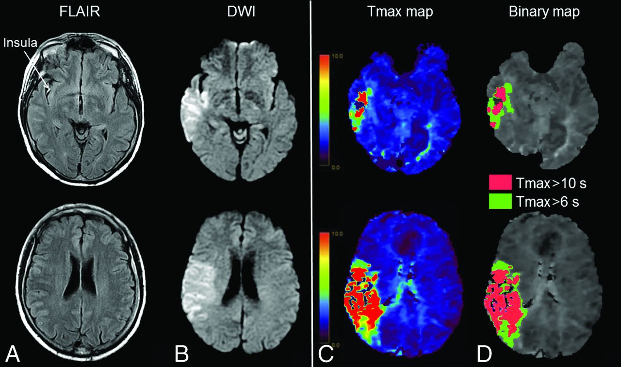
- Fig 1.
Extensive FVHs and low hypoperfusion intensity ratio. MR imaging obtained 110 minutes after stroke onset (NIHSS score = 12) in an 88-year-old woman. A, FVHs in 6 ASPECTS areas (insula, M2–M6), corresponding to a 6-point FVH-score. B, Thirteen-milliliter diffusion-weighted imaging lesion. Tmax map (C) with 85-mL Tmax >6-second lesion and 15-mL Tmax >10-second lesion (D) (HIR = 0.18). After intravenous thrombolysis, the 24-hour NIHSS score was 1, and the DWI lesion was 12 mL (not shown). The 3-month mRS was 1.
- Fig 2.
Few FVHs and high hypoperfusion intensity ratio. MR imaging obtained 180 minutes after stroke onset (NIHSS score = 12) in a 56-year-old man. A, FVHs in the insula, corresponding to a 1-point FVH score. B, Ninety-four milliliter diffusion-weighted imaging lesion. Tmax map (C) with 91-mL Tmax >6-second lesion and 53-mL Tmax >10-second lesion (D) (HIR = 0.58). After intravenous thrombolysis, the 24-hour NIHSS score was 14, and the DWI lesion was 150 mL (not shown). The 3-month mRS was 3.


Tables
Characteristics Low HIR (n = 122) High HIR (n = 122) P Value Age 70 (56–79) 71 (60–79) .446 Male (No.) (%) 55 (45) 83 (68) <.001 Hypertension (No.) (%)b 68 (57) 66 (55) .685 Diabetes mellitus (No.) (%)b 12 (10) 20 (17) .142 Hyperlipidemia (No.) (%)b 31 (26) 40 (33) .251 Current smoking (No.) (%)c 23 (20) 25 (21) .771 Systolic BP (mm Hg) 149 (139–163) 156 (139–173) .141 Diastolic BP (mm Hg) 82 (71–89) 81 (70–93) .990 Serum glucose (mmol/L)d 6.1 (5.4–7.0) 6.5 (5.5–7.9) .038 NIHSSb score 11 (7–17) 16 (11–21) <.001 Baseline MRI Onset-to-MRI time (min) 112 (86–153) 112 (84–156) .838 Proximal occlusion (No.) (%) 81 (66) 80 (66) .893 DWI1 volume (mL) 8 (4–19) 41 (16–96) <.001 Tmax >6-sec volume (mL) 44 (17–85) 115 (56–147) <.001 Tmax >10-sec volume (mL) 9 (2–22) 52 (27–75) <.001 PWI-DWI mismatch (No.) (%) 85 (70) 59 (48) <.001 FVH score (No.) (%) .016e 0–1 6 (5) 13 (11) 2–3 30 (25) 33 (27) 4–5 54 (44) 59 (48) 6–7 32 (26) 17 (14) 24-hr follow-up NIHSS scoref 6 (2–15) 14 (7–20) <.001 ENI (No.) (%)f 51 (42) 27 (22) .001 DWI2 volume (Ml) 18 (9–45) 87 (33–161) <.001 Infarct growth (Ml) 6 (1–26) 29 (10–75) <.001 Complete recanalization (No.) (%) 40 (33) 45 (37) .502 3-mo mRS ≤ 1 (No.) (%)g 49 (45) 25 (21) <.001 3-mo mRS ≤ 2 (No.) (%)g 63 (58) 51 (44) .033 Note:—BP indicates blood pressure; ENI, early neurologic improvement.
↵a Unless specified, numbers are median (interquartile range).
↵b Missing data for 4 patients.
↵c Missing data for 9 patients.
↵d Missing data for 13 patients.
↵e P for Trend.
↵f Missing data for 5 patients.
↵g Missing data for 18 patients.
Characteristics Adjusted OR (95% CI) P Value Male 0.98 (0.42–2.29) .953 Serum glucose, per 1-mmol/L increasea 0.99 (0.83–1.18) .882 Baseline NIHSS score, per 1-point increaseb 1.04 (0.96–1.12) .343 DWI1 volume, per 1-mL increase 1.01 (0.99–1.03) .446 Tmax >10-sec volume, per 1-mL increasec 0.89 (0.85–0.92) <.001 PWI-DWI mismatch 8.95 (2.65–30.22) <.001 FVH-score – .002d 0–1 1 – 2–3 1.17 (0.25–5.50) .844 4–5 2.39 (0.56–10.22) .242 6–7 34.20 (4.35–268.60) .001 FVH Score P Value 0–1 (n = 19) 2–3 (n = 63) 4–5 (n = 113) 6–7 (n = 49) Age (yr) 73 (60–80) 70 (57–79) 71 (56–80) 67 (62–77) .339 Male (No.) (%) 12 (63) 31 (49) 68 (60) 27 (55) .500 Hypertension (No.) (%)b 12 (63) 38 (60) 58 (51) 26 (53) .537 Diabetes mellitus (No.) (%)b 4 (21) 9 (14) 13 (12) 6 (12) .708 Hyperlipidemia (No.) (%)b 9 (47) 18 (29) 43 (38) 18 (37) .477 Current smoking (No.) (%)c 3 (16) 12 (19) 23 (20) 10 (20) .954 Systolic BP (mm Hg) 148 (140–172) 159 (142–173) 151 (139–167) 148 (134–160) .071 Diastolic BP (mm Hg) 87 (76–93) 84 (69–95) 80 (70–89) 80 (72–90) .334 Serum glucose (mmol/L)d 6.6 (5.5–8.1) 6.7 (5.4–7.3) 6.4 (5.5–7.6) 6.0 (5.3–7.0) .189 Baseline NIHSS scoreb 13 (8–20) 13 (7–18) 15 (8–20) 15 (11–21) .321 Onset-to-MRI time (min) 142 (84–196) 115 (89–176) 115 (91–151) 97 (83–130) .007 Proximal occlusion (No.) (%) 6 (31) 31 (49) 81 (72) 43 (88) <.001 DWI1 volume (mL) 24 (12–102) 19 (5–67) 18 (7–49) 12 (6–34) .077 Tmax >10-sec volume (mL) 26 (5–76) 17 (4–43) 24 (11–53) 30 (11–64) .200 Tmax >6-sec volume (mL) 65 (19–133) 47 (19–102) 78 (40–123) 96 (66–134) .002 HIR ≤ 0.35 (No.) (%) 6 (32) 30 (48) 54 (47) 32 (65) .057 HIR 0.44 (0.30–0.54) 0.39 (0.19–0.48) 0.33 (0.19–0.47) 0.32 (0.14–0.44) .049 PWI-DWI mismatch (No.) (%) 6 (32) 32 (51) 71 (63) 35 (51) .011 DWI2 volume (mL) 73 (14–150) 43 (13–147) 37 (13–104) 29 (12–52) .075 24-hr infarct growth (mL) 27 (2–87) 19 (4–57) 14 (1–45) 13 (4–28) .125 Complete recanalization (No.) (%) 7 (37) 19 (30) 38 (34) 21 (43) .551 ENI (No.) (%)e 7 (37) 12 (20) 30 (27) 29 (53) <.001 3-mo mRS ≤ 1 (No.) (%)f 3 (17) 14 (24) 35 (34) 22 (45) .039 3-mo mRS ≤ 2 (No.) (%)f 9 (47) 26 (41) 47 (42) 32 (65) .057


Jump to section
Related Articles
Cited By...
- New insights on the predictive value of hypoperfusion intensity ratio in thrombectomy: an updated systematic review and meta-analysis with multiple cut-offs
- Persistent perfusion abnormalities at day 1 correspond to different clinical trajectories after stroke
- Association of fluid-attenuated inversion recovery vascular hyperintensity with ischaemic events in internal carotid artery or middle cerebral artery occlusion
- FLAIR Vascular Hyperintensities as a Surrogate of Collaterals in Acute Stroke: DWI Matters
- Association between fluid-attenuated inversion recovery vascular hyperintensity and outcome varies with different lesion patterns in patients with intravenous thrombolysis
- Clinical prognosis of FLAIR hyperintense arteries in ischaemic stroke patients: a systematic review and meta-analysis
- The Association between FLAIR Vascular Hyperintensity and Stroke Outcome Varies with Time from Onset