Advertisement

Main menu

  • Home
  • Content
    • Current Issue
    • Accepted Manuscripts
    • Article Preview
    • Past Issue Archive
    • Video Articles
    • AJNR Case Collection
    • Case of the Week Archive
    • Case of the Month Archive
    • Classic Case Archive
  • Special Collections
    • AJNR Awards
    • Low-Field MRI
    • Alzheimer Disease
    • ASNR Foundation Special Collection
    • Photon-Counting CT
    • View All
  • Multimedia
    • AJNR Podcasts
    • AJNR SCANtastic
    • Trainee Corner
    • MRI Safety Corner
    • Imaging Protocols
  • For Authors
    • Submit a Manuscript
    • Submit a Video Article
    • Submit an eLetter to the Editor/Response
    • Manuscript Submission Guidelines
    • Statistical Tips
    • Fast Publishing of Accepted Manuscripts
    • Graphical Abstract Preparation
    • Imaging Protocol Submission
    • Author Policies
  • About Us
    • About AJNR
    • Editorial Board
    • Editorial Board Alumni
  • More
    • Become a Reviewer/Academy of Reviewers
    • Subscribers
    • Permissions
    • Alerts
    • Feedback
    • Advertisers
    • ASNR Home

User menu

  • Alerts
  • Log in

Search

  • Advanced search
American Journal of Neuroradiology

American Journal of Neuroradiology

ASHNR American Society of Functional Neuroradiology ASHNR American Society of Pediatric Neuroradiology ASSR
  • Alerts
  • Log in

Advanced Search

  • Home
  • Content
    • Current Issue
    • Accepted Manuscripts
    • Article Preview
    • Past Issue Archive
    • Video Articles
    • AJNR Case Collection
    • Case of the Week Archive
    • Case of the Month Archive
    • Classic Case Archive
  • Special Collections
    • AJNR Awards
    • Low-Field MRI
    • Alzheimer Disease
    • ASNR Foundation Special Collection
    • Photon-Counting CT
    • View All
  • Multimedia
    • AJNR Podcasts
    • AJNR SCANtastic
    • Trainee Corner
    • MRI Safety Corner
    • Imaging Protocols
  • For Authors
    • Submit a Manuscript
    • Submit a Video Article
    • Submit an eLetter to the Editor/Response
    • Manuscript Submission Guidelines
    • Statistical Tips
    • Fast Publishing of Accepted Manuscripts
    • Graphical Abstract Preparation
    • Imaging Protocol Submission
    • Author Policies
  • About Us
    • About AJNR
    • Editorial Board
    • Editorial Board Alumni
  • More
    • Become a Reviewer/Academy of Reviewers
    • Subscribers
    • Permissions
    • Alerts
    • Feedback
    • Advertisers
    • ASNR Home
  • Follow AJNR on Twitter
  • Visit AJNR on Facebook
  • Follow AJNR on Instagram
  • Join AJNR on LinkedIn
  • RSS Feeds

AJNR Awards, New Junior Editors, and more. Read the latest AJNR updates

Research ArticleSpine

Analysis of 30 Spinal Angiograms Falsely Reported as Normal in 18 Patients with Subsequently Documented Spinal Vascular Malformations

P. Barreras, D. Heck, B. Greenberg, J.-P. Wolinsky, C.A. Pardo and P. Gailloud
American Journal of Neuroradiology September 2017, 38 (9) 1814-1819; DOI: https://doi.org/10.3174/ajnr.A5275
P. Barreras
aFrom the Transverse Myelitis Center, Department of Neurology (P.B., C.A.P.)
  • Find this author on Google Scholar
  • Find this author on PubMed
  • Search for this author on this site
  • ORCID record for P. Barreras
D. Heck
bDivision of Interventional Neuroradiology (D.H., P.G.)
  • Find this author on Google Scholar
  • Find this author on PubMed
  • Search for this author on this site
  • ORCID record for D. Heck
B. Greenberg
dTransverse Myelitis Center (B.G.), University of Texas Southwestern Medical Center, Dallas, Texas.
  • Find this author on Google Scholar
  • Find this author on PubMed
  • Search for this author on this site
  • ORCID record for B. Greenberg
J.-P. Wolinsky
cDepartment of Neurosurgery (J.-P.W.), The Johns Hopkins Hospital, Baltimore, Maryland
  • Find this author on Google Scholar
  • Find this author on PubMed
  • Search for this author on this site
  • ORCID record for J.-P. Wolinsky
C.A. Pardo
aFrom the Transverse Myelitis Center, Department of Neurology (P.B., C.A.P.)
  • Find this author on Google Scholar
  • Find this author on PubMed
  • Search for this author on this site
  • ORCID record for C.A. Pardo
P. Gailloud
bDivision of Interventional Neuroradiology (D.H., P.G.)
  • Find this author on Google Scholar
  • Find this author on PubMed
  • Search for this author on this site
  • ORCID record for P. Gailloud
  • Article
  • Figures & Data
  • Info & Metrics
  • Responses
  • References
  • PDF
Loading

Article Figures & Data

Figures

  • Tables
  • Fig 1.
    • Download figure
    • Open in new tab
    • Download powerpoint
    Fig 1.

    Grading of flow voids on sagittal T2-weighted images (−, +, ++, +++). Left, 50-year-old man (patient 2) with a left L5 SDAVF. Center left, 73-year-old man (patient 8) with a left L4 SEAVF. Center right, 75-year-old man (patient 10) with a right T12 SDAVF. Right, 49-year-old man (patient 12) with a right S2 SDAVF.

  • Fig 2.
    • Download figure
    • Open in new tab
    • Download powerpoint
    Fig 2.

    66-year-old woman (patient 15) with a right T10 SDAVF and 1 prior negative spinal angiogram. A, DSA, nonselective injection at the T9 level, posteroanterior view, arterial phase (first study). The right T10 intersegmental artery supplies an arteriovenous shunt with early opacification of perimedullary venous structures (arrow). The anomaly was not detected. B, DSA, right T10 injection, posteroanterior view, arterial phase (second study). Selective angiography confirms the presence of an SDAVF fed by the right T10 radicular artery (arrowhead) and documents its extensive perimedullary drainage (arrow).
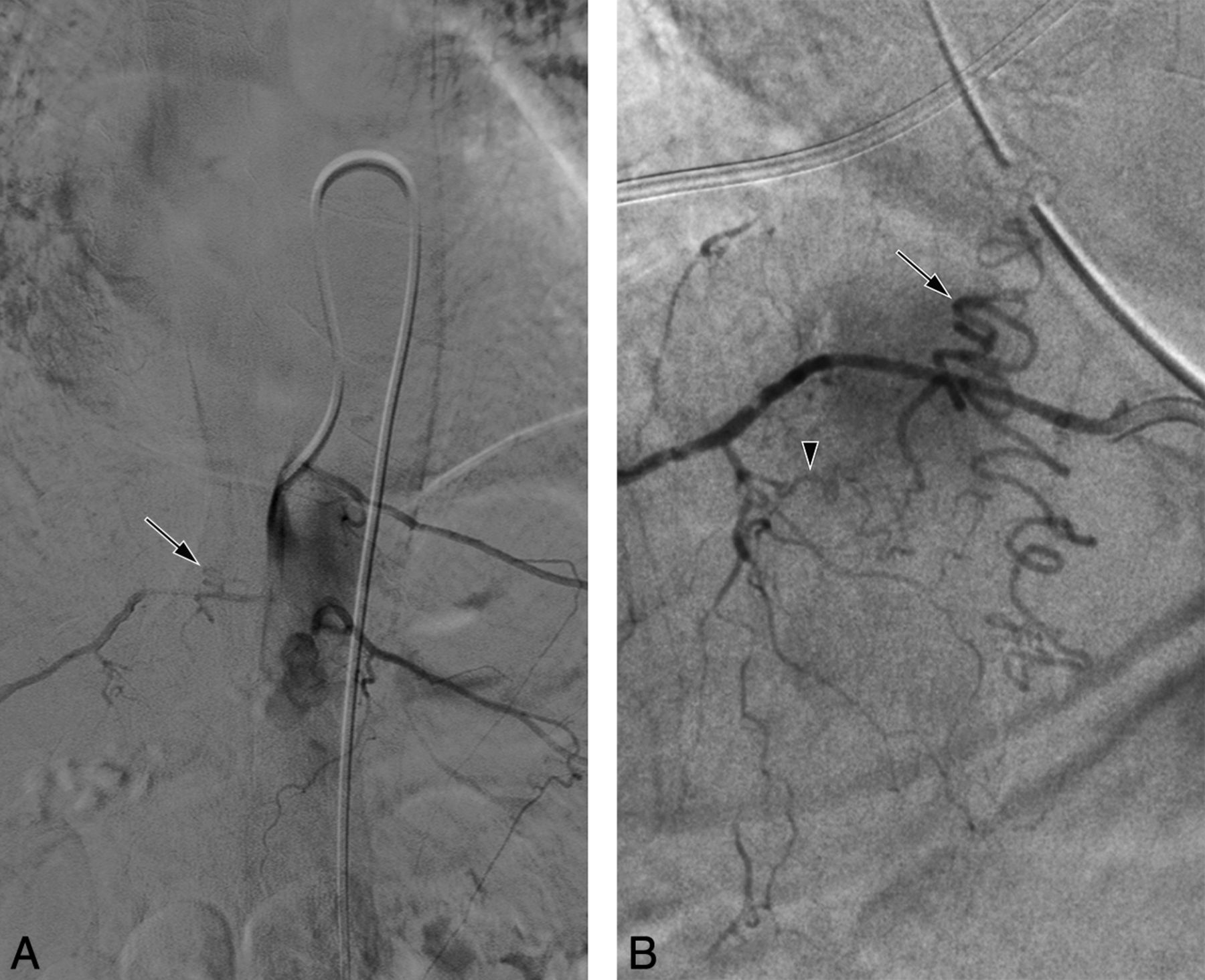
  • Fig 3.
    • Download figure
    • Open in new tab
    • Download powerpoint
    Fig 3.

    60-year-old man (patient 5) with a right T4 SDAVF and 1 prior negative angiogram. The patient consulted for a second opinion after substantial clinical pejoration during intravenous steroid therapy. A, DSA, right T4 injection, posteroanterior view, arterial phase (first study). A midline vessel (arrows) was noted but interpreted as being the anterior spinal artery. B, DSA, right T4 injection, posteroanterior view, arterial phase (second study). A right T4 radiculomeningeal branch (white arrowhead) supplies an arteriovenous shunt draining into a posterior radiculomedullary vein (black arrowhead) and the posterior-median spinal vein (black arrows).

  • Fig 4.
    • Download figure
    • Open in new tab
    • Download powerpoint
    Fig 4.

    75-year-old man (patient 11) with a right L3 spinal dural arteriovenous fistula (SDAVF) and 3 prior negative angiograms. A, DSA, nonselective injection at the level of L3, posteroanterior view, arterial phase (first study), showing no evidence of vascular malformation. In 1 of the other negative angiograms, L3 was not investigated at all, and the third study was not available for review. B, DSA, right L3 injection, posteroanterior view, arterial phase (second study), an arteriovenous shunt supplied by a right L3 radiculomeningeal branch and draining into a right L3 radiculomedullary vein (arrowheads). C, DSA, right L3 injection, posteroanterior view, arterial phase (second study). The congested perimedullary venous system surrounding the lumbosacral spinal cord is documented by this acquisition with a larger field of view.

Tables

  • Figures
    • View popup
    Table 1:

    Aminoff-Logue Disability Scale

    Disturbances of Gait
        1. Leg weakness, abnormal stance, or gait without restriction of locomotor activity
        2. Restricted exercise tolerance
        3. Requires stick or some support for walking
        4. Requires crutches or 2 sticks for walking
        5. Unable to stand, confined to bed or wheelchair
    Micturition
        1. Mild: hesitancy, urgency, or frequency
        2. Moderate: occasional incontinence or retention
        3. Severe: total incontinence or persistent retention
    Bowel
        1. Mild: constipation
        2. Moderate: occasional incontinence or severe constipation
        3. Persistent fecal incontinence
    • View popup
    Table 2:

    Imaging and treatment characteristics

    CaseAge/SexGadFlow VoidsInitial DiagnosisNegative DSAFinal DiagnosisLocaTreatAminoff-Logue Score
    PrePost
    140/M+−TM2SEAVFT9Surg86
    250/M+−TM2SDAVFL5Endo88
    373/M++TM1SDAVFL4Endo85
    480/M+++TM2SEAVFL5Endo97
    560/M+TM1SDAVFT4Endo98
    6b56/F++TM1SEAVFsL2Endo108
    L4Endo
    789/M−Tumor1SEAVFS1Endo31
    873/F+−TM1SEAVFL4Endo105
    958/M++TM1PmAVFT10Surg1010
    1075/M+++TM1SDAVFT12Endo107
    1175/M+−TM3SDAVFL3Surg89
    1249/M+++TM6SDAVFS2Endo64
    1369/M−TM2SEAVFL5Endo1111
    1456/M+++Syrinx1PmAVFT8Surg51
    1566/F+−TM1SDAVFT10Endo117
    1671/M+TM2SEAVFS1None10None
    1761/M+−NMO1SDAVFT4Surg96
    1825/M+++TM1SEAVFT12Surg114
    • Note:—Endo indicates endovascular; Gad, spinal cord enhancement after gadolinium; Loc, lesion location; NMO, neuromyelitis optica; Pre, pretreatment; Post, posttreatment; Surg, surgery; TM, transverse myelitis; Treat, treatment.

    • ↵a For the 2 PmAVFs, the indicated level corresponds to the feeding artery.

    • ↵b Patient 6 had 2 separate SEAVFs treated in separate endovascular procedures.

PreviousNext
Back to top

In this issue

American Journal of Neuroradiology: 38 (9)
American Journal of Neuroradiology
Vol. 38, Issue 9
1 Sep 2017
  • Table of Contents
  • Index by author
  • Complete Issue (PDF)
Advertisement
Print
Download PDF
Email Article

Thank you for your interest in spreading the word on American Journal of Neuroradiology.

NOTE: We only request your email address so that the person you are recommending the page to knows that you wanted them to see it, and that it is not junk mail. We do not capture any email address.

Enter multiple addresses on separate lines or separate them with commas.
Analysis of 30 Spinal Angiograms Falsely Reported as Normal in 18 Patients with Subsequently Documented Spinal Vascular Malformations
(Your Name) has sent you a message from American Journal of Neuroradiology
(Your Name) thought you would like to see the American Journal of Neuroradiology web site.
CAPTCHA
This question is for testing whether or not you are a human visitor and to prevent automated spam submissions.
Cite this article
P. Barreras, D. Heck, B. Greenberg, J.-P. Wolinsky, C.A. Pardo, P. Gailloud
Analysis of 30 Spinal Angiograms Falsely Reported as Normal in 18 Patients with Subsequently Documented Spinal Vascular Malformations
American Journal of Neuroradiology Sep 2017, 38 (9) 1814-1819; DOI: 10.3174/ajnr.A5275

Citation Manager Formats

  • BibTeX
  • Bookends
  • EasyBib
  • EndNote (tagged)
  • EndNote 8 (xml)
  • Medlars
  • Mendeley
  • Papers
  • RefWorks Tagged
  • Ref Manager
  • RIS
  • Zotero
0 Responses
Respond to this article
Share
Bookmark this article
Analysis of 30 Spinal Angiograms Falsely Reported as Normal in 18 Patients with Subsequently Documented Spinal Vascular Malformations
P. Barreras, D. Heck, B. Greenberg, J.-P. Wolinsky, C.A. Pardo, P. Gailloud
American Journal of Neuroradiology Sep 2017, 38 (9) 1814-1819; DOI: 10.3174/ajnr.A5275
del.icio.us logo Twitter logo Facebook logo Mendeley logo
  • Tweet Widget
  • Facebook Like
  • Google Plus One
Purchase

Jump to section

  • Article
    • Abstract
    • ABBREVIATIONS:
    • Materials and Methods
    • Results
    • Discussion
    • Conclusions
    • Footnotes
    • References
  • Figures & Data
  • Info & Metrics
  • Responses
  • References
  • PDF

Related Articles

  • No related articles found.
  • PubMed
  • Google Scholar

Cited By...

  • Application of Spinal Subtraction and Bone Background Fusion CTA in the Accurate Diagnosis and Evaluation of Spinal Vascular Malformations
  • Arterioectatic Spinal Angiopathy of Childhood: Clinical, Imaging, Laboratory, Histologic, and Genetic Description of a Novel CNS Vascular Pathology
  • Volumetric T2-weighted MRI improves the diagnostic accuracy of spinal vascular malformations: comparative analysis with a conventional MR study
  • Comparative Analysis of Volumetric High-Resolution Heavily T2-Weighted MRI and Time-Resolved Contrast-Enhanced MRA in the Evaluation of Spinal Vascular Malformations
  • Spinal Angiogram: A Treacherous Criterion Standard...
  • Crossref (28)
  • Google Scholar

This article has been cited by the following articles in journals that are participating in Crossref Cited-by Linking.

  • Clinical characteristics, misdiagnosis and outcomes of patients with low-flow spinal arteriovenous fistulas
    Olwen C. Murphy, Abderrahmane Hedjoudje, Andrea Salazar-Camelo, Carlos A. Pardo, Philippe Gailloud
    Journal of the Neurological Sciences 2020 413
  • Evaluation and Management of Acute Myelopathy
    Elia Sechi, Eoin P. Flanagan
    Seminars in Neurology 2021 41 05
  • Spinal vascular lesions: anatomy, imaging techniques and treatment
    Valerio Da Ros, Eliseo Picchi, Valentina Ferrazzoli, Tommaso Schirinzi, Federico Sabuzi, Piergiorgio Grillo, Massimo Muto, Francesco Garaci, Mario Muto, Francesca Di Giuliano
    European Journal of Radiology Open 2021 8
  • Clinical and neuroimaging findings of spinal dural arteriovenous fistulas: How to avoid misdiagnosis of this disease
    Keisuke Takai, Makoto Taniguchi
    Journal of Orthopaedic Science 2019 24 6
  • Identification of specific causes of myelopathy in a large cohort of patients initially diagnosed with transverse myelitis
    Olwen C. Murphy, Paula Barreras, Andres Villabona-Rueda, Maureen Mealy, Carlos A. Pardo
    Journal of the Neurological Sciences 2022 442
  • Acute idiopathic spinal subdural hematoma: What to do in an emergency?
    C. Joubert, S. Gazzola, A. Sellier, A. Dagain
    Neurochirurgie 2019 65 2-3
  • Introduction to Diagnostic and Therapeutic Spinal Angiography
    Philippe Gailloud
    Neuroimaging Clinics of North America 2019 29 4
  • Volumetric T2-weighted MRI improves the diagnostic accuracy of spinal vascular malformations: comparative analysis with a conventional MR study
    Santhosh Kumar Kannath, Adhithyan Rajendran, Bejoy Thomas, Jayadevan Enakshy Rajan
    Journal of NeuroInterventional Surgery 2019 11 10
  • Spinal dural arteriovenous fistula: A rare but treatable disease that should not be missed by orthopedic surgeons
    Baohui Yang, Teng Lu, Xijing He, Haopeng Li
    Frontiers in Neurology 2022 13
  • Interventional Neuroradiology
    Philippe Gailloud
    2021 176

More in this TOC Section

Spine

  • Bern Score Validity for SIH
  • MP2RAGE 7T in MS Lesions of the Cervical Spine
  • Resisted Inspiration for CSF-Venous Fistula
Show more Spine

Interventional

  • CT-Guided C2 Dorsal Root Ganglion Radiofrequency Ablation for the Treatment of Cervicogenic Headache: Case Series and Clinical Outcomes
  • Safety, Efficacy, and Durability of Stent-Assisted Coiling Treatment of M2 (Insular) Segment MCA Aneurysms
  • Endovascular Management of Intracranial Dural AVFs: Transvenous Approach
Show more Interventional

Similar Articles

Advertisement

Indexed Content

  • Current Issue
  • Accepted Manuscripts
  • Article Preview
  • Past Issues
  • Editorials
  • Editor's Choice
  • Fellows' Journal Club
  • Letters to the Editor
  • Video Articles

Cases

  • Case Collection
  • Archive - Case of the Week
  • Archive - Case of the Month
  • Archive - Classic Case

More from AJNR

  • Trainee Corner
  • Imaging Protocols
  • MRI Safety Corner
  • Book Reviews

Multimedia

  • AJNR Podcasts
  • AJNR Scantastics

Resources

  • Turnaround Time
  • Submit a Manuscript
  • Submit a Video Article
  • Submit an eLetter to the Editor/Response
  • Manuscript Submission Guidelines
  • Statistical Tips
  • Fast Publishing of Accepted Manuscripts
  • Graphical Abstract Preparation
  • Imaging Protocol Submission
  • Evidence-Based Medicine Level Guide
  • Publishing Checklists
  • Author Policies
  • Become a Reviewer/Academy of Reviewers
  • News and Updates

About Us

  • About AJNR
  • Editorial Board
  • Editorial Board Alumni
  • Alerts
  • Permissions
  • Not an AJNR Subscriber? Join Now
  • Advertise with Us
  • Librarian Resources
  • Feedback
  • Terms and Conditions
  • AJNR Editorial Board Alumni

American Society of Neuroradiology

  • Not an ASNR Member? Join Now

© 2025 by the American Society of Neuroradiology All rights, including for text and data mining, AI training, and similar technologies, are reserved.
Print ISSN: 0195-6108 Online ISSN: 1936-959X

Powered by HighWire