
Article Figures & Data
Figures
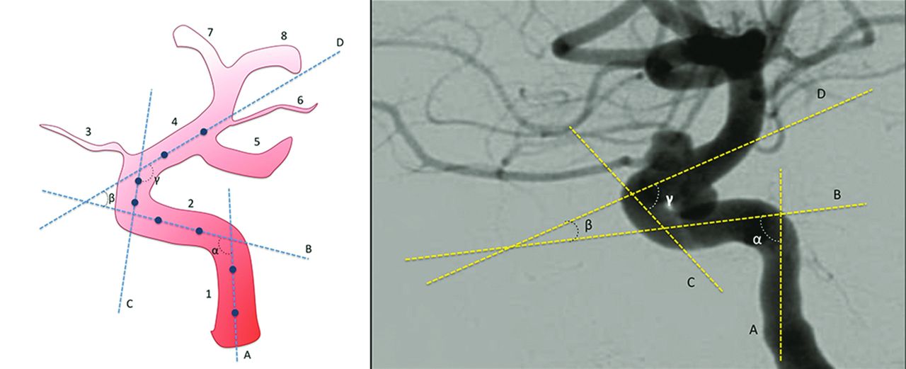
- Fig 1.
Technique for measurement of carotid siphon angles. On the left, an illustration of a carotid siphon with lines traced to cross the midpoints of the diameters of the straight segments of the siphon. On the right, an example of an actual measurement process. A, Line through the vertical petrous segment. B, Line through the horizontal cavernous segment. C, Line through the vertical cavernous segment. D, Line through the supraclinoid segment. α, Posterior bend angle. β, Anterior bend angle. γ, Anterosuperior bend angle. 1, Ascending petrous segment. 2, Intracavernous segment. 3, Ophthalmic artery. 4, Supraclinoid segment. 5, Posterior communicating artery. 6, Anterior choroidal artery. 7, Anterior cerebral artery. 8, Middle cerebral artery.

Tables
Variables Total No. of IAs (%) 692 (100) Mean age (yr) (54.75 ± 13.13) Sex Women with IAs (No.) (%) 457 (66.04) Mean age (yr) (54.66 ± 13.13) Men with IAs (No.) (%) 235 (33.96) Mean age (yr) (54.70 ± 13.16) Rupture (No.) (%) Ruptured IAs 225 (32.51) Unruptured IAs 467 (67.49) IA location (No.) (%) Siphon 218 (31.50) Cavernous segment 45 (6.50) Supraclinoid segment 173 (25.00) Ophthalmic 67 (9.68) PcomA 96 (13.87) Ant. chor 10 (1.45) Postsiphon (No.) (%) 474 (68.50) Bifurcation 35 (5.06) MCA 237 (34.25) AcomA 170 (24.57) ACA/pericallosal 32 (4.62) IA size (No.) (%) ≤5 mm 334 (48.27) 6–10 mm 233 (33.67) 11–25 mm 97 (14.02) >25 mm 28 (4.05) Note:—IA indicates intracranial aneurysm; PcomA, posterior communicating segment; Ant. chor, anterior choroidal segment; ACA, anterior cerebral artery; AcomA, anterior communicating artery.
- Table 2:
Distribution of the study variables and association with aneurysm rupture based on crude and adjusted prevalence ratios according to the Poisson regression model with robust variance and their respective 95% CIs
Variables Crude PR Adjusted PR PR (95% CI) P Value PR (95% CI) P Value Sex .428 .938 Female 1 – 1 – Male 1.09 (0.88–1.37) .428 1.01 (0.81–1.25) .938 Age .413 .640 55 yr or younger 1.09 (0.88–1.36) .413 1.05 (0.83–1.31) .640 Older than 55 yr 1 – 1 – Aneurysm location .009 .022 Postsiphon 1.18 (0.91–1.52) .208 1.11 (0.86–1.44) .416 Supraclinoid segment 1 – 1 – Aneurysm size .134 .445 ≤5 mm 3.92 (1.04–14.81) .044 2.18 (0.57–8.27) .252 6–10 mm 3.49 (0.92–13.27) .066 1.96 (0.51–7.52) .326 11–25 mm 3.20 (0.83–12.42) .092 1.91 (0.49–7.40) .350 >25 mm 1 – 1 – Anterior knee angle .001 .005 ≤15.40° 1 – 1 – >15.40° 1.45 (1.16–1.80) .001 1.36 (1.09–1.69) .005 Posterior knee angle .685 .773 ≤88.15° 1 – 1 – >88.15° 1.05 (0.84–1.30) .685 1.03 (0.84–1.27) .773 Note:—PR indicates prevalence ratio.
- Table 3:
Analysis of rupture odds ratio by location subgroups according to anterior angles above or below the median and their respective 95% CIs
Location/Angle Unruptured (%) Ruptured (%) P Value OR (95% CI) Supraclinoid .091 1.75 (0.91–3.36) ≤15.40° 71 (74.74) 24 (25.26) >15.40° 49 (62.82) 29 (37.18) MCA .354 1.32 (0.73–2.40) ≤15.40° 83 (77.57) 24 (22.43) >15.40° 94 (72.31) 36 (27.69) AcomA .049 1.84 (1.00–3.38) ≤15.40° 45 (55.56) 36 (59.55) >15.40° 36 (40.45) 53 (59.55) Bifurcation .470 1.90 (0.43–8.48) ≤15.40° 14 (77.78) 4 (22.22) >15.40° 11 (64.71) 6 (35.29) Note:—Supraclinoid indicates supraclinoid segments, including the ophthalmic, posterior communicating, and anterior choroidal segments; Bifurcation, internal carotid artery bifurcation; AcomA, anterior communicating artery.
- Table 4:
Distribution of the study variables and association with aneurysm location based on crude and adjusted odds ratios according to the generalized logistic regression model and their respective 95% CIs
Variables Crude OR (Postsiphon/Siphon) Adjusted OR (Cavernous/Supraclinoid) OR (95% CI) P Value OR (95% CI) P Value Sex <.001 .427 Male 2.23 (1.48–3.37) <.001 0.69 (0.27–1.74) .427 Female 1 – 1 – Age .184 .684 55 yr or younger 1.28 (0.89–1.84) .184 1.16 (0.57–2.38) .684 Older than 55 yr 1 – 1 – Rupture .423 No 1 – Yes 1.17 (0.80–1.72) .423 Aneurysm size ≤5 mm 2.38 (0.69–8.20) .169 0.08 (0.02–0.28) <.001 6–10 mm 2.54 (0.73–8.85) .143 0.09 (0.03–0.35) <.001 11–25 mm 1.34 (0.37–4.81) .652 0.12 (0.03–0.45) .002 >25 mm 1 – 1 – Anterior knee angle .034 .066 ≤15.40° 1 – 1 – >15.40° 1.48 (1.03–2.13) .034 0.48 (0.22–1.05) .066 Posterior knee angle .122 .093 ≤88.15° 1 – 1 – >88.15° 1.33 (0.93–1.91) .122 1.84 (0.90–3.73) .093 Variables Associated with Larger Aneurysms P Value 95% CI Aneurysm location Postsiphon/supraclinoid .016 0.901–0.989 Cavernous/supraclinoid <.001 1.133–1.350 Sex Male/female .103 0.993–1.081 Rupture Yes/no .026 0.913–0.994 Anterior knee angle .015 1.000–1.002 Posterior knee angle .804 1.000–1.000 Age .003 1.000–1.003


Jump to section
Related Articles
Cited By...
- No citing articles found.