Advertisement

Main menu

  • Home
  • Content
    • Current Issue
    • Accepted Manuscripts
    • Article Preview
    • Past Issue Archive
    • Video Articles
    • AJNR Case Collection
    • Case of the Week Archive
    • Case of the Month Archive
    • Classic Case Archive
  • Special Collections
    • AJNR Awards
    • Low-Field MRI
    • Alzheimer Disease
    • ASNR Foundation Special Collection
    • Photon-Counting CT
    • View All
  • Multimedia
    • AJNR Podcasts
    • AJNR SCANtastic
    • Trainee Corner
    • MRI Safety Corner
    • Imaging Protocols
  • For Authors
    • Submit a Manuscript
    • Submit a Video Article
    • Submit an eLetter to the Editor/Response
    • Manuscript Submission Guidelines
    • Statistical Tips
    • Fast Publishing of Accepted Manuscripts
    • Graphical Abstract Preparation
    • Imaging Protocol Submission
    • Author Policies
  • About Us
    • About AJNR
    • Editorial Board
    • Editorial Board Alumni
  • More
    • Become a Reviewer/Academy of Reviewers
    • Subscribers
    • Permissions
    • Alerts
    • Feedback
    • Advertisers
    • ASNR Home

User menu

  • Alerts
  • Log in

Search

  • Advanced search
American Journal of Neuroradiology

American Journal of Neuroradiology

ASHNR American Society of Functional Neuroradiology ASHNR American Society of Pediatric Neuroradiology ASSR
  • Alerts
  • Log in

Advanced Search

  • Home
  • Content
    • Current Issue
    • Accepted Manuscripts
    • Article Preview
    • Past Issue Archive
    • Video Articles
    • AJNR Case Collection
    • Case of the Week Archive
    • Case of the Month Archive
    • Classic Case Archive
  • Special Collections
    • AJNR Awards
    • Low-Field MRI
    • Alzheimer Disease
    • ASNR Foundation Special Collection
    • Photon-Counting CT
    • View All
  • Multimedia
    • AJNR Podcasts
    • AJNR SCANtastic
    • Trainee Corner
    • MRI Safety Corner
    • Imaging Protocols
  • For Authors
    • Submit a Manuscript
    • Submit a Video Article
    • Submit an eLetter to the Editor/Response
    • Manuscript Submission Guidelines
    • Statistical Tips
    • Fast Publishing of Accepted Manuscripts
    • Graphical Abstract Preparation
    • Imaging Protocol Submission
    • Author Policies
  • About Us
    • About AJNR
    • Editorial Board
    • Editorial Board Alumni
  • More
    • Become a Reviewer/Academy of Reviewers
    • Subscribers
    • Permissions
    • Alerts
    • Feedback
    • Advertisers
    • ASNR Home
  • Follow AJNR on Twitter
  • Visit AJNR on Facebook
  • Follow AJNR on Instagram
  • Join AJNR on LinkedIn
  • RSS Feeds

AJNR Awards, New Junior Editors, and more. Read the latest AJNR updates

Research ArticleAdult Brain
Open Access

Identification and Quantitative Assessment of Different Components of Intracranial Atherosclerotic Plaque by Ex Vivo 3T High-Resolution Multicontrast MRI

Y. Jiang, W. Peng, B. Tian, C. Zhu, L. Chen, X. Wang, Q. Liu, Y. Wang, Z. Xiang, A.J. Degnan, Z. Teng, D. Saloner and J. Lu
American Journal of Neuroradiology September 2017, 38 (9) 1716-1722; DOI: https://doi.org/10.3174/ajnr.A5266
Y. Jiang
aFrom the Department of Radiology (Y.J.), Wuhan General Hospital of the People's Liberation Army, Wuhan, China
bDepartments of Radiology (Y.J., W.P., B.T., L.C., X.W., Q.L., J.L.)
  • Find this author on Google Scholar
  • Find this author on PubMed
  • Search for this author on this site
  • ORCID record for Y. Jiang
W. Peng
bDepartments of Radiology (Y.J., W.P., B.T., L.C., X.W., Q.L., J.L.)
  • Find this author on Google Scholar
  • Find this author on PubMed
  • Search for this author on this site
  • ORCID record for W. Peng
B. Tian
bDepartments of Radiology (Y.J., W.P., B.T., L.C., X.W., Q.L., J.L.)
  • Find this author on Google Scholar
  • Find this author on PubMed
  • Search for this author on this site
  • ORCID record for B. Tian
C. Zhu
dDepartment of Radiology and Biomedical Imaging (C.Z., D.S.), University of California, San Francisco, San Francisco, California
  • Find this author on Google Scholar
  • Find this author on PubMed
  • Search for this author on this site
  • ORCID record for C. Zhu
L. Chen
bDepartments of Radiology (Y.J., W.P., B.T., L.C., X.W., Q.L., J.L.)
  • Find this author on Google Scholar
  • Find this author on PubMed
  • Search for this author on this site
  • ORCID record for L. Chen
X. Wang
bDepartments of Radiology (Y.J., W.P., B.T., L.C., X.W., Q.L., J.L.)
  • Find this author on Google Scholar
  • Find this author on PubMed
  • Search for this author on this site
  • ORCID record for X. Wang
Q. Liu
bDepartments of Radiology (Y.J., W.P., B.T., L.C., X.W., Q.L., J.L.)
  • Find this author on Google Scholar
  • Find this author on PubMed
  • Search for this author on this site
  • ORCID record for Q. Liu
Y. Wang
cPathology (Y.W., Z.X.), Changhai Hospital, Shanghai, China
  • Find this author on Google Scholar
  • Find this author on PubMed
  • Search for this author on this site
  • ORCID record for Y. Wang
Z. Xiang
cPathology (Y.W., Z.X.), Changhai Hospital, Shanghai, China
  • Find this author on Google Scholar
  • Find this author on PubMed
  • Search for this author on this site
  • ORCID record for Z. Xiang
A.J. Degnan
eDepartment of Radiology (A.J.D.), University of Pittsburgh Medical Center, Pittsburgh, Pennsylvania
  • Find this author on Google Scholar
  • Find this author on PubMed
  • Search for this author on this site
  • ORCID record for A.J. Degnan
Z. Teng
fDepartment of Radiology (Z.T.), University of Cambridge, Cambridge, UK.
  • Find this author on Google Scholar
  • Find this author on PubMed
  • Search for this author on this site
  • ORCID record for Z. Teng
D. Saloner
dDepartment of Radiology and Biomedical Imaging (C.Z., D.S.), University of California, San Francisco, San Francisco, California
  • Find this author on Google Scholar
  • Find this author on PubMed
  • Search for this author on this site
  • ORCID record for D. Saloner
J. Lu
bDepartments of Radiology (Y.J., W.P., B.T., L.C., X.W., Q.L., J.L.)
  • Find this author on Google Scholar
  • Find this author on PubMed
  • Search for this author on this site
  • ORCID record for J. Lu
  • Article
  • Figures & Data
  • Supplemental
  • Info & Metrics
  • Responses
  • References
  • PDF
Loading

Article Figures & Data

Figures

  • Tables
  • Fig 1.
    • Download figure
    • Open in new tab
    • Download powerpoint
    Fig 1.

    A basilar artery plaque with a thick fibrous cap and a large lipid core (type IV–V) was identified on multicontrast MR imaging and matched histology (20× magnification). Contours have been drawn for the lumen (red), vessel wall (green), and lipid core (yellow) on a T2-weighted image and hematoxylin-eosin-stained section. The lumen, total vessel, and lipid core areas were 4.34, 13.43, and 2.35 mm2 and 4.66, 10.60, and 1.42 mm2 for MR imaging and histology. The vessel wall areas calculated by total vessel area minus lumen area were 9.09 and 6.00 mm2. The fibrous component areas calculated by vessel wall area minus lipid core area were 6.70 and 4.56 mm2. The percentages of plaque components (Specific Component Area / Vessel Wall Area × 100%) were 25.85% versus 23.63% for the lipid core and 74.15% versus 76.37% for fibrous tissue. Plaque burden (Vessel Wall Area / Total Vessel Area × 100%) was 67.68% versus 56.24%.
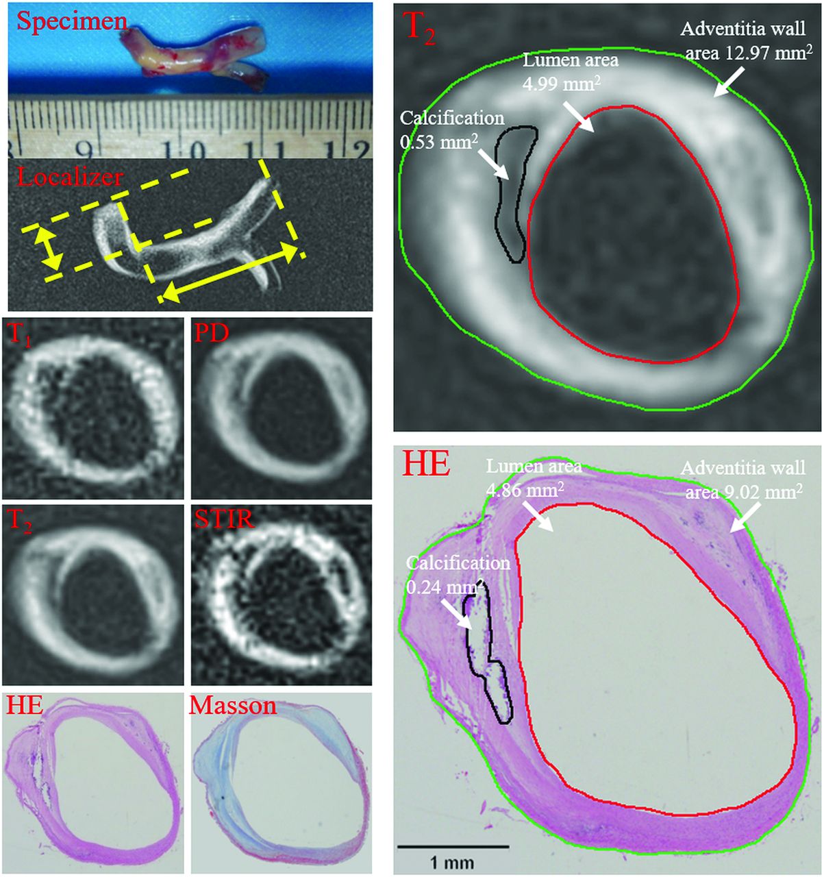
  • Fig 2.
    • Download figure
    • Open in new tab
    • Download powerpoint
    Fig 2.

    Vertebral artery plaque with fibrous tissue and calcification was identified on multicontrast MR imaging and matched histology (20× magnification). Contours have been drawn for lumen (red), outer vessel wall (green), and calcification (black) on T2-weighted imaging and a hematoxylin-eosin-stained section. The lumen, total vessel, and calcification areas were 4.99, 12.97, and 0.53 mm2 on MR imaging and 4.86, 9.02, and 0.24 mm2 on histology. The vessel wall areas calculated by total vessel area minus lumen area were 7.98 and 4.16 mm2. The fibrous component area calculated by vessel wall area minus the calcification area was 7.45 and 3.93 mm2. The percentages of components (Specific Component Area / Vessel Wall Area × 100%) were 6.64% versus 5.73% for calcification and 93.36% versus 94.27% for fibrous tissue. Plaque burden (Vessel Wall Area / Total Vessel Area × 100%) was 61.52% versus 46.09%.

Tables

  • Figures
    • View popup
    Table 1:

    The criteria for plaque component identification by multicontrast MRI

    Lipid CoreFibrous CapCalcification
    T1Isointense/hyperintenseIsointenseHypointense
    T2Isointense/hypointenseIsointense/hyperintenseHypointense
    PDIsointense/hypointenseIsointense/hyperintenseHypointense
    STIRHypointenseHyperintenseHypointense
    • View popup
    Table 2:

    Comparison of the percentages of areas and thicknesses and overall plaque burden between MRI and histologya

    MRIHistologyPrICC (95% CI)
    Fibrous component area (%)81.86 ± 10.5981.87 ± 11.59.9970.9010.898 (0.832–0.938)
    Lipid core area (%)19.51 ± 10.7619.86 ± 11.56.6600.8880.885 (0.804–0.934)
    Calcification area (%)9.68 ± 5.218.83 ± 5.67.0300.9330.930 (0.745–0.982)
    Fibrous cap thickness (%)31.10 ± 11.2830.83 ± 8.51.8900.4380.421 (0.155–0.630)
    Plaque burden area (%)65.18 ± 9.0152.71 ± 14.58<.0010.8620.771 (0.640–0.858)
    • Note:—r indicates the Pearson correlation coefficient.

    • ↵a Results are expressed as mean ± SD with P values derived from a linear mixed-effects model.

PreviousNext
Back to top

In this issue

American Journal of Neuroradiology: 38 (9)
American Journal of Neuroradiology
Vol. 38, Issue 9
1 Sep 2017
  • Table of Contents
  • Index by author
  • Complete Issue (PDF)
Advertisement
Print
Download PDF
Email Article

Thank you for your interest in spreading the word on American Journal of Neuroradiology.

NOTE: We only request your email address so that the person you are recommending the page to knows that you wanted them to see it, and that it is not junk mail. We do not capture any email address.

Enter multiple addresses on separate lines or separate them with commas.
Identification and Quantitative Assessment of Different Components of Intracranial Atherosclerotic Plaque by Ex Vivo 3T High-Resolution Multicontrast MRI
(Your Name) has sent you a message from American Journal of Neuroradiology
(Your Name) thought you would like to see the American Journal of Neuroradiology web site.
CAPTCHA
This question is for testing whether or not you are a human visitor and to prevent automated spam submissions.
Cite this article
Y. Jiang, W. Peng, B. Tian, C. Zhu, L. Chen, X. Wang, Q. Liu, Y. Wang, Z. Xiang, A.J. Degnan, Z. Teng, D. Saloner, J. Lu
Identification and Quantitative Assessment of Different Components of Intracranial Atherosclerotic Plaque by Ex Vivo 3T High-Resolution Multicontrast MRI
American Journal of Neuroradiology Sep 2017, 38 (9) 1716-1722; DOI: 10.3174/ajnr.A5266

Citation Manager Formats

  • BibTeX
  • Bookends
  • EasyBib
  • EndNote (tagged)
  • EndNote 8 (xml)
  • Medlars
  • Mendeley
  • Papers
  • RefWorks Tagged
  • Ref Manager
  • RIS
  • Zotero
0 Responses
Respond to this article
Share
Bookmark this article
Identification and Quantitative Assessment of Different Components of Intracranial Atherosclerotic Plaque by Ex Vivo 3T High-Resolution Multicontrast MRI
Y. Jiang, W. Peng, B. Tian, C. Zhu, L. Chen, X. Wang, Q. Liu, Y. Wang, Z. Xiang, A.J. Degnan, Z. Teng, D. Saloner, J. Lu
American Journal of Neuroradiology Sep 2017, 38 (9) 1716-1722; DOI: 10.3174/ajnr.A5266
del.icio.us logo Twitter logo Facebook logo Mendeley logo
  • Tweet Widget
  • Facebook Like
  • Google Plus One
Purchase

Jump to section

  • Article
    • Abstract
    • ABBREVIATIONS:
    • Materials and Methods
    • Results
    • Discussion
    • Conclusions
    • Footnotes
    • References
  • Figures & Data
  • Supplemental
  • Info & Metrics
  • Responses
  • References
  • PDF

Related Articles

  • PubMed
  • Google Scholar

Cited By...

  • Plaque characteristics associated with failure of primary balloon angioplasty for intracranial atherosclerotic stenosis: a retrospective study
  • Crossref (5)
  • Google Scholar

This article has been cited by the following articles in journals that are participating in Crossref Cited-by Linking.

  • Inter-rater and scan–rescan reproducibility of the detection of intracranial atherosclerosis on contrast-enhanced 3D vessel wall MRI
    Mahmud Mossa-Basha, Hiroko Watase, Jie Sun, Dean K. Shibata, Daniel S. Hippe, Niranjan Balu, Thomas Hatsukami, Chun Yuan
    The British Journal of Radiology 2019 92 1097
  • Histology-Verified Intracranial Artery Calcification and Its Clinical Relevance With Cerebrovascular Disease
    Heng Du, Wenjie Yang, Xiangyan Chen
    Frontiers in Neurology 2022 12
  • Why is middle cerebral artery plaque augmented by contrast media? A phantom study using middle cerebral artery stenotic silicon model
    Sol-Ki Kim, Hyo Sung Kwak, Gyung Ho Chung, Seung-Bae Hwang
    Neuroradiology 2019 61 10
  • Plaque characteristics associated with failure of primary balloon angioplasty for intracranial atherosclerotic stenosis: a retrospective study
    Yuexin Mei, Shan Yu, Zhuhao Li, Hongbing Chen, Jian Zhang, Shuangquan Tan, Jinsheng Zeng, Shihui Xing, Xinran Chen
    Journal of NeuroInterventional Surgery 2025 17 1
  • Wall characteristics of atherosclerotic middle cerebral arteries in patients with single or multiple infarcts: A high-resolution MRI Study
    Zelan Ma, Mengjuan Huo, Jiajun Xie, Guoqing Liu, Guoming Li, Qiang Liu, Liting Mao, Weikang Huang, Bo Liu, Xian Liu
    Frontiers in Neurology 2022 13

More in this TOC Section

  • Diagnostic Neuroradiology of Monoclonal Antibodies
  • Clinical Outcomes After Chiari I Decompression
  • Segmentation of Brain Metastases with BLAST
Show more ADULT BRAIN

Similar Articles

Advertisement

Indexed Content

  • Current Issue
  • Accepted Manuscripts
  • Article Preview
  • Past Issues
  • Editorials
  • Editor's Choice
  • Fellows' Journal Club
  • Letters to the Editor
  • Video Articles

Cases

  • Case Collection
  • Archive - Case of the Week
  • Archive - Case of the Month
  • Archive - Classic Case

More from AJNR

  • Trainee Corner
  • Imaging Protocols
  • MRI Safety Corner
  • Book Reviews

Multimedia

  • AJNR Podcasts
  • AJNR Scantastics

Resources

  • Turnaround Time
  • Submit a Manuscript
  • Submit a Video Article
  • Submit an eLetter to the Editor/Response
  • Manuscript Submission Guidelines
  • Statistical Tips
  • Fast Publishing of Accepted Manuscripts
  • Graphical Abstract Preparation
  • Imaging Protocol Submission
  • Evidence-Based Medicine Level Guide
  • Publishing Checklists
  • Author Policies
  • Become a Reviewer/Academy of Reviewers
  • News and Updates

About Us

  • About AJNR
  • Editorial Board
  • Editorial Board Alumni
  • Alerts
  • Permissions
  • Not an AJNR Subscriber? Join Now
  • Advertise with Us
  • Librarian Resources
  • Feedback
  • Terms and Conditions
  • AJNR Editorial Board Alumni

American Society of Neuroradiology

  • Not an ASNR Member? Join Now

© 2025 by the American Society of Neuroradiology All rights, including for text and data mining, AI training, and similar technologies, are reserved.
Print ISSN: 0195-6108 Online ISSN: 1936-959X

Powered by HighWire