Advertisement

Main menu

  • Home
  • Content
    • Current Issue
    • Accepted Manuscripts
    • Article Preview
    • Past Issue Archive
    • Video Articles
    • AJNR Case Collection
    • Case of the Week Archive
    • Case of the Month Archive
    • Classic Case Archive
  • Special Collections
    • Advancing NeuroMRI with High-Relaxivity Contrast Agents
    • AJNR Awards
    • Low-Field MRI
    • Alzheimer Disease
    • ASNR Foundation Special Collection
    • Photon-Counting CT
    • View All
  • Multimedia
    • AJNR Podcasts
    • AJNR SCANtastic
    • Trainee Corner
    • MRI Safety Corner
    • Imaging Protocols
  • For Authors
    • Submit a Manuscript
    • Submit a Video Article
    • Submit an eLetter to the Editor/Response
    • Manuscript Submission Guidelines
    • Statistical Tips
    • Fast Publishing of Accepted Manuscripts
    • Graphical Abstract Preparation
    • Imaging Protocol Submission
    • Author Policies
  • About Us
    • About AJNR
    • Editorial Board
    • Editorial Board Alumni
  • More
    • Become a Reviewer/Academy of Reviewers
    • Subscribers
    • Permissions
    • Alerts
    • Feedback
    • Advertisers
    • ASNR Home

User menu

  • Alerts
  • Log in

Search

  • Advanced search
American Journal of Neuroradiology

American Journal of Neuroradiology

ASHNR American Society of Functional Neuroradiology ASHNR American Society of Pediatric Neuroradiology ASSR
  • Alerts
  • Log in

Advanced Search

  • Home
  • Content
    • Current Issue
    • Accepted Manuscripts
    • Article Preview
    • Past Issue Archive
    • Video Articles
    • AJNR Case Collection
    • Case of the Week Archive
    • Case of the Month Archive
    • Classic Case Archive
  • Special Collections
    • Advancing NeuroMRI with High-Relaxivity Contrast Agents
    • AJNR Awards
    • Low-Field MRI
    • Alzheimer Disease
    • ASNR Foundation Special Collection
    • Photon-Counting CT
    • View All
  • Multimedia
    • AJNR Podcasts
    • AJNR SCANtastic
    • Trainee Corner
    • MRI Safety Corner
    • Imaging Protocols
  • For Authors
    • Submit a Manuscript
    • Submit a Video Article
    • Submit an eLetter to the Editor/Response
    • Manuscript Submission Guidelines
    • Statistical Tips
    • Fast Publishing of Accepted Manuscripts
    • Graphical Abstract Preparation
    • Imaging Protocol Submission
    • Author Policies
  • About Us
    • About AJNR
    • Editorial Board
    • Editorial Board Alumni
  • More
    • Become a Reviewer/Academy of Reviewers
    • Subscribers
    • Permissions
    • Alerts
    • Feedback
    • Advertisers
    • ASNR Home
  • Follow AJNR on Twitter
  • Visit AJNR on Facebook
  • Follow AJNR on Instagram
  • Join AJNR on LinkedIn
  • RSS Feeds

AJNR Awards, New Junior Editors, and more. Read the latest AJNR updates


Improved Turnaround Times | Median time to first decision: 12 days

Research ArticleAdult Brain

Head-to-Head Visual Comparison between Brain Perfusion SPECT and Arterial Spin-Labeling MRI with Different Postlabeling Delays in Alzheimer Disease

T. Kaneta, O. Katsuse, T. Hirano, M. Ogawa, K. Yoshida, T. Odawara, Y. Hirayasu and T. Inoue
American Journal of Neuroradiology August 2017, 38 (8) 1562-1568; DOI: https://doi.org/10.3174/ajnr.A5238
T. Kaneta
aFrom the Departments of Radiology (T.K., T.H., M.O., K.Y., T.I.)
  • Find this author on Google Scholar
  • Find this author on PubMed
  • Search for this author on this site
  • ORCID record for T. Kaneta
O. Katsuse
bPsychiatry (O.K., T.O., Y.H.), Yokohama City University, Yokohama, Japan.
  • Find this author on Google Scholar
  • Find this author on PubMed
  • Search for this author on this site
  • ORCID record for O. Katsuse
T. Hirano
aFrom the Departments of Radiology (T.K., T.H., M.O., K.Y., T.I.)
  • Find this author on Google Scholar
  • Find this author on PubMed
  • Search for this author on this site
  • ORCID record for T. Hirano
M. Ogawa
aFrom the Departments of Radiology (T.K., T.H., M.O., K.Y., T.I.)
  • Find this author on Google Scholar
  • Find this author on PubMed
  • Search for this author on this site
  • ORCID record for M. Ogawa
K. Yoshida
aFrom the Departments of Radiology (T.K., T.H., M.O., K.Y., T.I.)
  • Find this author on Google Scholar
  • Find this author on PubMed
  • Search for this author on this site
  • ORCID record for K. Yoshida
T. Odawara
bPsychiatry (O.K., T.O., Y.H.), Yokohama City University, Yokohama, Japan.
  • Find this author on Google Scholar
  • Find this author on PubMed
  • Search for this author on this site
  • ORCID record for T. Odawara
Y. Hirayasu
bPsychiatry (O.K., T.O., Y.H.), Yokohama City University, Yokohama, Japan.
  • Find this author on Google Scholar
  • Find this author on PubMed
  • Search for this author on this site
  • ORCID record for Y. Hirayasu
T. Inoue
aFrom the Departments of Radiology (T.K., T.H., M.O., K.Y., T.I.)
  • Find this author on Google Scholar
  • Find this author on PubMed
  • Search for this author on this site
  • ORCID record for T. Inoue
  • Article
  • Figures & Data
  • Info & Metrics
  • Responses
  • References
  • PDF
Loading

Article Figures & Data

Figures

  • Tables
  • Fig 1.
    • Download figure
    • Open in new tab
    • Download powerpoint
    Fig 1.

    The regions used for scoring brain images. We evaluated 22 regions located at the levels of the vertex, lateral ventricle, basal ganglia, and cerebellum. The raters scored each region with a 4-point scale, where 0 is a normal CBF, 1 is a mild decrease, 2 is a moderate decease, and 3 is a severe decrease.
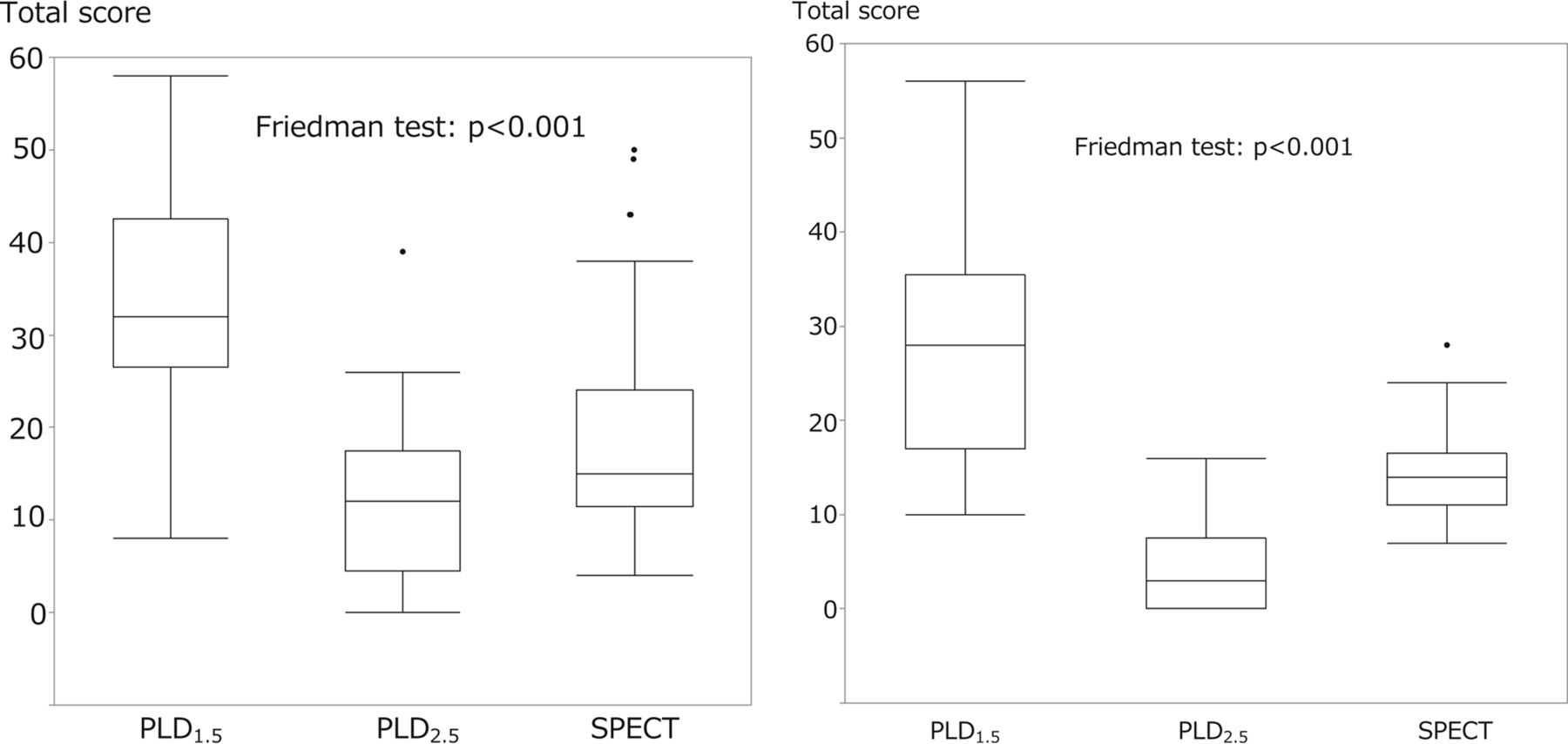
  • Fig 2.
    • Download figure
    • Open in new tab
    • Download powerpoint
    Fig 2.

    The comparisons of the total scores of the 2 raters for PLD1.5, PLD2.5, and SPECT images. The Friedman analysis of variance showed a significant difference among all 3 groups (P < .01), and the Wilcoxon test showed a significant difference between the 2 raters for the PLD1.5, PLD2.5, and SPECT total scores (P < .01).
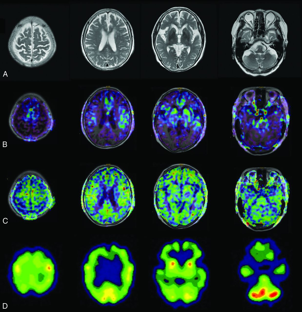
  • Fig 3.
    • Download figure
    • Open in new tab
    • Download powerpoint
    Fig 3.

    An 86-year-old woman with AD. The patient had an MMSE score of 14. Axial images of T2-weighted MR imaging (A), ASL-MR imaging with PLD1.5 in a color-scale fused with T1-weighted MR imaging (B), ASL-MR imaging with PLD2.5 in a color-scale fused with T1-weighted MR imaging (C), and brain perfusion SPECT at the level of the parietal lobe, corona radiata, basal ganglia, and cerebellum (D). Both raters indicated a score of 4 (definitely AD) for PLD1.5 and SPECT and 1 (probably not AD) for PLD2.5.

  • Fig 4.
    • Download figure
    • Open in new tab
    • Download powerpoint
    Fig 4.

    An 83-year-old woman with AD. The patient had an MMSE score of 19. Axial images of T2-weighted MR imaging (A), fused T1-weighted and PLD1.5 ASL-MR imaging (B), fused T1-weighted and PLD2.5 ASL-MR imaging (C), and brain perfusion SPECT (D). Both raters indicated a score of 4 for PLD1.5 and 2 (undetermined) for PLD2.5. One rater indicated a score of 3 (probably AD) and another rater indicated a score of 4 for SPECT.

  • Fig 5.
    • Download figure
    • Open in new tab
    • Download powerpoint
    Fig 5.

    A 73-year-old woman with MCI. The patient had an MMSE score of 23. Axial images of T2-weighted MR imaging (A), fused T1-weighted and PLD1.5 ASL-MR imaging (B), fused T1-weighted and PLD2.5 ASL-MR imaging (C), and brain perfusion SPECT (D). Both raters scored 4 for PLD1.5. However, 1 rater indicated a score of 1 (probably not AD) for PLD2.5 and 2 for SPECT, while another rater indicated a score of 0 (definitely not AD) for PLD2.5, and 1 for SPECT.

Tables

  • Figures
    • View popup
    Table 1:

    Demographic and neuropsychological summary of the participantsa

    ADMCI due to AD
    Age (yr)77.1 (7.7)78.2 (5.6)
    No. of patients1419
    Sex (M/F)3:118:11
    MMSE score16.9 (3.0)b25.2 (2.6)b
    • ↵a Data are presented as mean (±SD).

    • ↵b There was a significant difference in the MMSE score (P < .01) between AD and patients with AD-induced MCI.

    • View popup
    Table 2:

    The results of visual assessments for the diagnosis of AD

    Overall Positive Rate (%)Positive Rate in AD (%)Positive Rate in MCI (%)
    PLD1.5PLD2.5SPECTPLD1.5PLD2.5SPECTPLD1.5PLD2.5SPECT
    Rater 187.99.169.792.914.392.984.228.652.6
    Rater 297.012.190.110028.692.994.7089.5
    Agreement84.83.066.792.97.185.784.2052.6
    • View popup
    Table 3:

    Interrater agreement

    κ Statistic
    5-Point ScoringPositive or Negative for AD Diagnosis
    PLD1.50.0330.3694
    PLD2.50.1410.203
    SPECT0.0100.195
PreviousNext
Back to top

In this issue

American Journal of Neuroradiology: 38 (8)
American Journal of Neuroradiology
Vol. 38, Issue 8
1 Aug 2017
  • Table of Contents
  • Index by author
  • Complete Issue (PDF)
Advertisement
Print
Download PDF
Email Article

Thank you for your interest in spreading the word on American Journal of Neuroradiology.

NOTE: We only request your email address so that the person you are recommending the page to knows that you wanted them to see it, and that it is not junk mail. We do not capture any email address.

Enter multiple addresses on separate lines or separate them with commas.
Head-to-Head Visual Comparison between Brain Perfusion SPECT and Arterial Spin-Labeling MRI with Different Postlabeling Delays in Alzheimer Disease
(Your Name) has sent you a message from American Journal of Neuroradiology
(Your Name) thought you would like to see the American Journal of Neuroradiology web site.
CAPTCHA
This question is for testing whether or not you are a human visitor and to prevent automated spam submissions.
Cite this article
T. Kaneta, O. Katsuse, T. Hirano, M. Ogawa, K. Yoshida, T. Odawara, Y. Hirayasu, T. Inoue
Head-to-Head Visual Comparison between Brain Perfusion SPECT and Arterial Spin-Labeling MRI with Different Postlabeling Delays in Alzheimer Disease
American Journal of Neuroradiology Aug 2017, 38 (8) 1562-1568; DOI: 10.3174/ajnr.A5238

Citation Manager Formats

  • BibTeX
  • Bookends
  • EasyBib
  • EndNote (tagged)
  • EndNote 8 (xml)
  • Medlars
  • Mendeley
  • Papers
  • RefWorks Tagged
  • Ref Manager
  • RIS
  • Zotero
0 Responses
Respond to this article
Share
Bookmark this article
Head-to-Head Visual Comparison between Brain Perfusion SPECT and Arterial Spin-Labeling MRI with Different Postlabeling Delays in Alzheimer Disease
T. Kaneta, O. Katsuse, T. Hirano, M. Ogawa, K. Yoshida, T. Odawara, Y. Hirayasu, T. Inoue
American Journal of Neuroradiology Aug 2017, 38 (8) 1562-1568; DOI: 10.3174/ajnr.A5238
del.icio.us logo Twitter logo Facebook logo Mendeley logo
  • Tweet Widget
  • Facebook Like
  • Google Plus One
Purchase

Jump to section

  • Article
    • Abstract
    • ABBREVIATIONS:
    • Materials and Methods
    • Results
    • Discussion
    • Conclusions
    • Acknowledgments
    • Footnotes
    • REFERENCES
  • Figures & Data
  • Info & Metrics
  • Responses
  • References
  • PDF

Related Articles

  • No related articles found.
  • PubMed
  • Google Scholar

Cited By...

  • No citing articles found.
  • Crossref
  • Google Scholar

This article has not yet been cited by articles in journals that are participating in Crossref Cited-by Linking.

More in this TOC Section

  • Diagnostic Neuroradiology of Monoclonal Antibodies
  • ML for Glioma Molecular Subtype Prediction
  • NCCT vs. MRI for Brain Atrophy in Acute Stroke
Show more Adult Brain

Similar Articles

Advertisement

Indexed Content

  • Current Issue
  • Accepted Manuscripts
  • Article Preview
  • Past Issues
  • Editorials
  • Editor's Choice
  • Fellows' Journal Club
  • Letters to the Editor
  • Video Articles

Cases

  • Case Collection
  • Archive - Case of the Week
  • Archive - Case of the Month
  • Archive - Classic Case

More from AJNR

  • Trainee Corner
  • Imaging Protocols
  • MRI Safety Corner
  • Book Reviews

Multimedia

  • AJNR Podcasts
  • AJNR Scantastics

Resources

  • Turnaround Time
  • Submit a Manuscript
  • Submit a Video Article
  • Submit an eLetter to the Editor/Response
  • Manuscript Submission Guidelines
  • Statistical Tips
  • Fast Publishing of Accepted Manuscripts
  • Graphical Abstract Preparation
  • Imaging Protocol Submission
  • Evidence-Based Medicine Level Guide
  • Publishing Checklists
  • Author Policies
  • Become a Reviewer/Academy of Reviewers
  • News and Updates

About Us

  • About AJNR
  • Editorial Board
  • Editorial Board Alumni
  • Alerts
  • Permissions
  • Not an AJNR Subscriber? Join Now
  • Advertise with Us
  • Librarian Resources
  • Feedback
  • Terms and Conditions
  • AJNR Editorial Board Alumni

American Society of Neuroradiology

  • Not an ASNR Member? Join Now

© 2025 by the American Society of Neuroradiology All rights, including for text and data mining, AI training, and similar technologies, are reserved.
Print ISSN: 0195-6108 Online ISSN: 1936-959X

Powered by HighWire