
Article Figures & Data
Figures
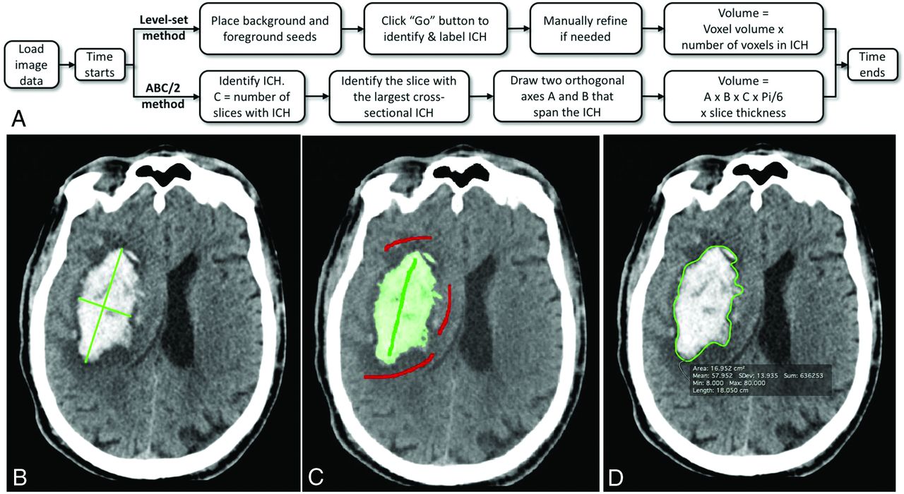
- Fig 1.
Hemorrhage measurement methods. A, A flowchart describes steps in the ABC/2 and level set measurement processes. B, Results from the ABC/2 method. C, Results from the level set method. Green and red strokes were placed by the user. Green indicates desired tissue (foreground), and red indicates undesired tissue (background). The level set region grows in 3D after the user clicks “Go” (Fig 2). D, Manual outlining of the boundary in a single axial section. This process was replicated in each axial section with visible ICH. Each true volume was determined from 4 manual tracings. Two operators each performed 2 repeat manual tracings. A minimum interval of 2 weeks between repeat tracings was required. Any voxel selected in at least 3 of the 4 manual tracings was labeled a “true” lesion voxel and was used to estimate the true lesion volume.
- Fig 2.
The SegTool ICH segmentation process in a CT scan with a 512 × 512 × 36 array. A, The user placed foreground (green) and background (red) seeds in an axial section (Fig 1C). Here the seeds are viewed in 3D. After the user clicks the “Play” button (not shown), the level set evolves to cover the ICH. In this example, a 41-mL ICH is segmented in 3 seconds (B and C). The final segmented region can be viewed in 2D or 3D by rotating the CT scan volume (D).
- Fig 3.
The level set algorithm may leak. An axial (A) and 3D view (B) of 1 segmented ICH lesion (enclosed in a red ellipse) along with level set leakage outside the lesion. Leakage was caused by partial volume effects between hemorrhage and skull (due, in part, to the large section thickness of the clinical imaging examinations). This can be corrected by either increasing the level set surface stiffness or removing the leaked regions with editing tools. However, when the transparency of the segmented (green) region was high (as it was by default), operators found it difficult to detect leaks. Consequently, we asked all operators to perform a blinded review of their level set segmentations. They performed this by interactively reducing the transparency of the green region until it was opaque and then viewing their segmentation in 3D (B). If, for any reason, they were not satisfied with the labeled region, they were asked to re-segment the lesion. In the text, we describe several enhancements to the tool that could be made to help operators detect leaks before saving the final segmented region.
- Fig 4.
The bias (A), precision (B), and time (C) for ICH volume measurement. Measurements made using the ABC/2 method are shown in red. Those made using SegTool are shown purple (initial) or blue (after review). A, Bias: the dashed (purple) line indicates perfect agreement between measured and true volumes. Each true volume was determined from 4 manual tracings placed by experts (details in the text). The solid markers indicate the mean measured volume determined from 8 measurements (4 operators × 2 measurements/operator). The error bars indicate the 95% confidence interval for the mean measured volume. The solid lines show a linear regression fit through all measurements for each method. The shaded zone around each solid line indicates the 95% confidence interval for the slope of the line. Each line is labeled with its equation, the P value from the linear regression, and the coefficient of determination describing goodness of fit (1.0 equals a perfect fit). B, Precision: lower values are better. Changes in ICH volume less than the indicated value can be explained by measurement variability alone, with 95% confidence. The values in the “Between Operator” group are all statistically significantly different (P < .02). The “SegTool (initial)” value in the “Within Operators” group is significantly different than the other values (P < .001), which are not significantly different from each other (P = .8). C, Time: each bar indicates the mean value from 560 measurements (70 ICH lesions × 4 operators × 2 repeated measurements). The SegTool time is based on the initial measurements. The error bars indicate the standard error of the mean. On average, SegTool required an extra 8.9 seconds to measure ICH volume. This difference is statistically significantly different from 0 (P < .0001).


Tables
Summary statistics for our experimentsa
ABC/2 SegTool (Initial) SegTool (after Review) SegTool (Revisions) No. 560 560 560 140 Mean (SD) 35.28 (56.24) 26.02 (40.35) 25.32 (39.76) −2.2 (12.7) 1st Quartile 7.00 4.77 4.84 −1.43 Median 18.48 13.19 13.09 −0.01 3rd Quartile 36.82 28.87 27.47 0.40 ↵a All values are in milliliters, except No. that is dimensionless. The values in the SegTool (revisions) column indicate the change after operators reviewed their initial measurements. Negative values mean that the after-review value was smaller than the initial value.


