Advertisement

Main menu

  • Home
  • Content
    • Current Issue
    • Accepted Manuscripts
    • Article Preview
    • Past Issue Archive
    • Video Articles
    • AJNR Case Collection
    • Case of the Week Archive
    • Case of the Month Archive
    • Classic Case Archive
  • Special Collections
    • Advancing NeuroMRI with High-Relaxivity Contrast Agents
    • AJNR Awards
    • Low-Field MRI
    • Alzheimer Disease
    • ASNR Foundation Special Collection
    • Photon-Counting CT
    • View All
  • Multimedia
    • AJNR Podcasts
    • AJNR SCANtastic
    • Trainee Corner
    • MRI Safety Corner
    • Imaging Protocols
  • For Authors
    • Submit a Manuscript
    • Submit a Video Article
    • Submit an eLetter to the Editor/Response
    • Manuscript Submission Guidelines
    • Statistical Tips
    • Fast Publishing of Accepted Manuscripts
    • Graphical Abstract Preparation
    • Imaging Protocol Submission
    • Author Policies
  • About Us
    • About AJNR
    • Editorial Board
    • Editorial Board Alumni
  • More
    • Become a Reviewer/Academy of Reviewers
    • Subscribers
    • Permissions
    • Alerts
    • Feedback
    • Advertisers
    • ASNR Home

User menu

  • Alerts
  • Log in

Search

  • Advanced search
American Journal of Neuroradiology

American Journal of Neuroradiology

ASHNR American Society of Functional Neuroradiology ASHNR American Society of Pediatric Neuroradiology ASSR
  • Alerts
  • Log in

Advanced Search

  • Home
  • Content
    • Current Issue
    • Accepted Manuscripts
    • Article Preview
    • Past Issue Archive
    • Video Articles
    • AJNR Case Collection
    • Case of the Week Archive
    • Case of the Month Archive
    • Classic Case Archive
  • Special Collections
    • Advancing NeuroMRI with High-Relaxivity Contrast Agents
    • AJNR Awards
    • Low-Field MRI
    • Alzheimer Disease
    • ASNR Foundation Special Collection
    • Photon-Counting CT
    • View All
  • Multimedia
    • AJNR Podcasts
    • AJNR SCANtastic
    • Trainee Corner
    • MRI Safety Corner
    • Imaging Protocols
  • For Authors
    • Submit a Manuscript
    • Submit a Video Article
    • Submit an eLetter to the Editor/Response
    • Manuscript Submission Guidelines
    • Statistical Tips
    • Fast Publishing of Accepted Manuscripts
    • Graphical Abstract Preparation
    • Imaging Protocol Submission
    • Author Policies
  • About Us
    • About AJNR
    • Editorial Board
    • Editorial Board Alumni
  • More
    • Become a Reviewer/Academy of Reviewers
    • Subscribers
    • Permissions
    • Alerts
    • Feedback
    • Advertisers
    • ASNR Home
  • Follow AJNR on Twitter
  • Visit AJNR on Facebook
  • Follow AJNR on Instagram
  • Join AJNR on LinkedIn
  • RSS Feeds

AJNR Awards, New Junior Editors, and more. Read the latest AJNR updates


Improved Turnaround Times | Median time to first decision: 12 days

Research ArticleAdult Brain

Diffusional Kurtosis Imaging of the Corticospinal Tract in Multiple Sclerosis: Association with Neurologic Disability

M.V. Spampinato, M.R. Kocher, J.H. Jensen, J.A. Helpern, H.R. Collins and N.U. Hatch
American Journal of Neuroradiology August 2017, 38 (8) 1494-1500; DOI: https://doi.org/10.3174/ajnr.A5225
M.V. Spampinato
aFrom the Department of Radiology and Radiological Science (M.V.S., M.R.K., J.H.J., J.A.H., H.R.C., N.U.H.)
bCenter for Biomedical Imaging (M.V.S., J.H.J., J.A.H.), Medical University of South Carolina, Charleston, South Carolina.
  • Find this author on Google Scholar
  • Find this author on PubMed
  • Search for this author on this site
  • ORCID record for M.V. Spampinato
M.R. Kocher
aFrom the Department of Radiology and Radiological Science (M.V.S., M.R.K., J.H.J., J.A.H., H.R.C., N.U.H.)
  • Find this author on Google Scholar
  • Find this author on PubMed
  • Search for this author on this site
  • ORCID record for M.R. Kocher
J.H. Jensen
aFrom the Department of Radiology and Radiological Science (M.V.S., M.R.K., J.H.J., J.A.H., H.R.C., N.U.H.)
bCenter for Biomedical Imaging (M.V.S., J.H.J., J.A.H.), Medical University of South Carolina, Charleston, South Carolina.
  • Find this author on Google Scholar
  • Find this author on PubMed
  • Search for this author on this site
  • ORCID record for J.H. Jensen
J.A. Helpern
aFrom the Department of Radiology and Radiological Science (M.V.S., M.R.K., J.H.J., J.A.H., H.R.C., N.U.H.)
bCenter for Biomedical Imaging (M.V.S., J.H.J., J.A.H.), Medical University of South Carolina, Charleston, South Carolina.
  • Find this author on Google Scholar
  • Find this author on PubMed
  • Search for this author on this site
  • ORCID record for J.A. Helpern
H.R. Collins
aFrom the Department of Radiology and Radiological Science (M.V.S., M.R.K., J.H.J., J.A.H., H.R.C., N.U.H.)
  • Find this author on Google Scholar
  • Find this author on PubMed
  • Search for this author on this site
  • ORCID record for H.R. Collins
N.U. Hatch
aFrom the Department of Radiology and Radiological Science (M.V.S., M.R.K., J.H.J., J.A.H., H.R.C., N.U.H.)
  • Find this author on Google Scholar
  • Find this author on PubMed
  • Search for this author on this site
  • ORCID record for N.U. Hatch
  • Article
  • Figures & Data
  • Info & Metrics
  • Responses
  • References
  • PDF
Loading

Article Figures & Data

Figures

  • Tables
  • Fig 1.
    • Download figure
    • Open in new tab
    • Download powerpoint
    Fig 1.

    White matter lesion mask (yellow) of a representative patient with multiple sclerosis overlaid on axial (A) and coronal (B) mean fractional anisotropy images. The mean fractional anisotropy skeleton (green) and the Johns Hopkins University White Matter Tractography Atlas corticospinal tract (red) are overlaid onto the mean axial (C) and coronal (A) fractional anisotropy images. The white matter skeleton was obtained with FSL Tract-Based Spatial Statistics. The corticospinal tract VOIs were obtained from the white matter skeleton with the Johns Hopkins University White Matter Tractography Atlas, after masking out T2 hyperintense areas of individual patients. All images are in the Montreal Neurological Institute standard space.
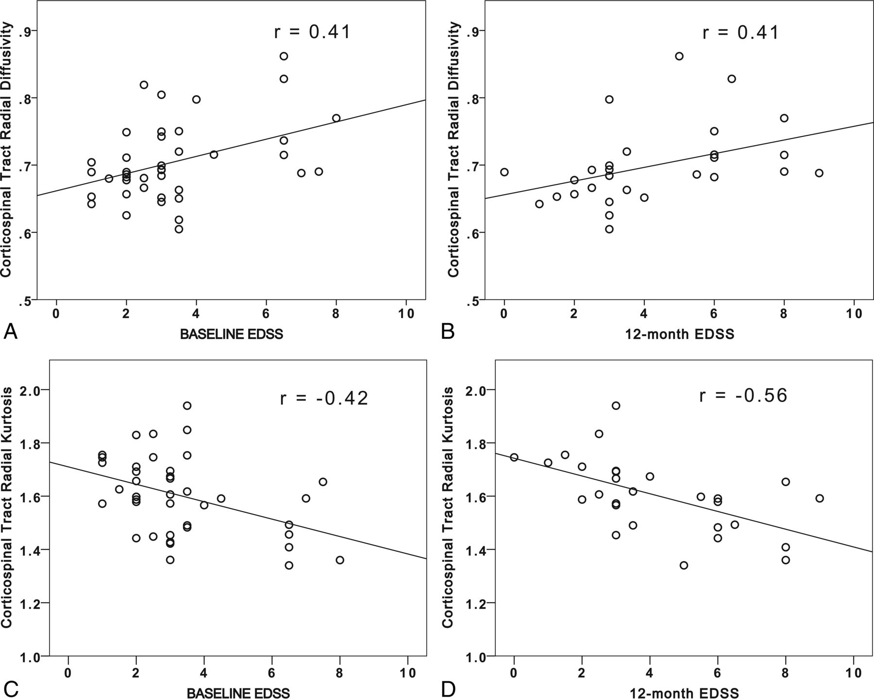
  • Fig 2.
    • Download figure
    • Open in new tab
    • Download powerpoint
    Fig 2.

    Scatterplots for the Expanded Disability Status Scale at the time of the brain MR imaging versus radial diffusivity (A) and radial kurtosis (C) of the corticospinal tracts, and EDSS at the follow-up visit 12 months after the brain MR imaging versus radial diffusivity (B) and radial kurtosis (D) of the corticospinal tracts in patients with MS.

Tables

  • Figures
    • View popup
    Table 1:

    Patient clinical characteristics and imaging metrics

    Entire CohortPatients with 12-Month Follow-Up
    No. of subjects4028
    Sex (F/M)33/724/4
    Age (mean) (yr)42.9 ± 12.542.1 ± 13.1
    EDSS0 (mean)3.4 ± 1.93.6 ± 2
    EDSS12 m (mean)4.2 ± 2.3
    MS type (RR/SP/unknown/PP)32/2/5/123/1/3/1
    Disease duration (mean) (yr)8.0 ± 6.46.9 ± 4.5
    T2 lesion load (mean) (mL)21.3 ± 22.823.3 ± 26.0
    CST FA (mean)0.44 ± 0.030.44 ± 0.03
    CST MD (mean)0.94 ± 0.060.93 ± 0.06
    CST λ⊥ (mean)0.70 ± 0.060.70 ± 0.06
    CST (mean)1.41 ± 0.071.40 ± 0.07
    CST MK (mean)1.17 ± 0.061.17 ± 0.06
    CST K⊥ (mean)1.60 ± 0.151.60 ± 0.14
    CST K‖ (mean)0.96 ± 0.050.97 ± 0.05
    • Note:—RR indicates relapsing-remitting MS; SP, secondary-progressive MS; PP, primary-progressive MS.

    • View popup
    Table 2:

    Correlations between EDSS and age, T2 lesion volume, and diffusion metrics

    Diffusion MetricsEDSS0EDSS12 m
    rP ValuearP Valuea
    Age0.47.0090.38.003
    T2 lesion volume0.38.0180.23.117
    MD0.41.0180.45.018
    λ‖0.34.0230.45.018
    λ⊥0.41.0180.41.023
    FA−0.36.022−0.30.063
    MK−0.29.046−0.42.023
    K‖−0.25.063−0.36.040
    K⊥−0.42.018−0.56.009
    • ↵a P values were corrected for multiple comparisons with the false discovery rate method.

    • View popup
    Table 3:

    Linear regression analyses: coefficients table

    EDSS0EDSS12 m
    βP ValueβP Value
    (Constant).022<.001
    Age.399.007.172.347
    CST MK.074.811.257.432
    CST K‖.055.774.006.979
    CST K⊥−.329.023−.562.002
    T2 lesion volume.153.355−.141.491
PreviousNext
Back to top

In this issue

American Journal of Neuroradiology: 38 (8)
American Journal of Neuroradiology
Vol. 38, Issue 8
1 Aug 2017
  • Table of Contents
  • Index by author
  • Complete Issue (PDF)
Advertisement
Print
Download PDF
Email Article

Thank you for your interest in spreading the word on American Journal of Neuroradiology.

NOTE: We only request your email address so that the person you are recommending the page to knows that you wanted them to see it, and that it is not junk mail. We do not capture any email address.

Enter multiple addresses on separate lines or separate them with commas.
Diffusional Kurtosis Imaging of the Corticospinal Tract in Multiple Sclerosis: Association with Neurologic Disability
(Your Name) has sent you a message from American Journal of Neuroradiology
(Your Name) thought you would like to see the American Journal of Neuroradiology web site.
CAPTCHA
This question is for testing whether or not you are a human visitor and to prevent automated spam submissions.
Cite this article
M.V. Spampinato, M.R. Kocher, J.H. Jensen, J.A. Helpern, H.R. Collins, N.U. Hatch
Diffusional Kurtosis Imaging of the Corticospinal Tract in Multiple Sclerosis: Association with Neurologic Disability
American Journal of Neuroradiology Aug 2017, 38 (8) 1494-1500; DOI: 10.3174/ajnr.A5225

Citation Manager Formats

  • BibTeX
  • Bookends
  • EasyBib
  • EndNote (tagged)
  • EndNote 8 (xml)
  • Medlars
  • Mendeley
  • Papers
  • RefWorks Tagged
  • Ref Manager
  • RIS
  • Zotero
0 Responses
Respond to this article
Share
Bookmark this article
Diffusional Kurtosis Imaging of the Corticospinal Tract in Multiple Sclerosis: Association with Neurologic Disability
M.V. Spampinato, M.R. Kocher, J.H. Jensen, J.A. Helpern, H.R. Collins, N.U. Hatch
American Journal of Neuroradiology Aug 2017, 38 (8) 1494-1500; DOI: 10.3174/ajnr.A5225
del.icio.us logo Twitter logo Facebook logo Mendeley logo
  • Tweet Widget
  • Facebook Like
  • Google Plus One
Purchase

Jump to section

  • Article
    • Abstract
    • ABBREVIATIONS:
    • Materials and Methods
    • Results
    • Discussion
    • Conclusions
    • Acknowledgments
    • Footnotes
    • REFERENCES
  • Figures & Data
  • Info & Metrics
  • Responses
  • References
  • PDF

Related Articles

  • No related articles found.
  • PubMed
  • Google Scholar

Cited By...

  • No citing articles found.
  • Crossref
  • Google Scholar

This article has not yet been cited by articles in journals that are participating in Crossref Cited-by Linking.

More in this TOC Section

  • Diagnostic Neuroradiology of Monoclonal Antibodies
  • Clinical Outcomes After Chiari I Decompression
  • Segmentation of Brain Metastases with BLAST
Show more Adult Brain

Similar Articles

Advertisement

Indexed Content

  • Current Issue
  • Accepted Manuscripts
  • Article Preview
  • Past Issues
  • Editorials
  • Editor's Choice
  • Fellows' Journal Club
  • Letters to the Editor
  • Video Articles

Cases

  • Case Collection
  • Archive - Case of the Week
  • Archive - Case of the Month
  • Archive - Classic Case

More from AJNR

  • Trainee Corner
  • Imaging Protocols
  • MRI Safety Corner
  • Book Reviews

Multimedia

  • AJNR Podcasts
  • AJNR Scantastics

Resources

  • Turnaround Time
  • Submit a Manuscript
  • Submit a Video Article
  • Submit an eLetter to the Editor/Response
  • Manuscript Submission Guidelines
  • Statistical Tips
  • Fast Publishing of Accepted Manuscripts
  • Graphical Abstract Preparation
  • Imaging Protocol Submission
  • Evidence-Based Medicine Level Guide
  • Publishing Checklists
  • Author Policies
  • Become a Reviewer/Academy of Reviewers
  • News and Updates

About Us

  • About AJNR
  • Editorial Board
  • Editorial Board Alumni
  • Alerts
  • Permissions
  • Not an AJNR Subscriber? Join Now
  • Advertise with Us
  • Librarian Resources
  • Feedback
  • Terms and Conditions
  • AJNR Editorial Board Alumni

American Society of Neuroradiology

  • Not an ASNR Member? Join Now

© 2025 by the American Society of Neuroradiology All rights, including for text and data mining, AI training, and similar technologies, are reserved.
Print ISSN: 0195-6108 Online ISSN: 1936-959X

Powered by HighWire