
Article Figures & Data
Figures
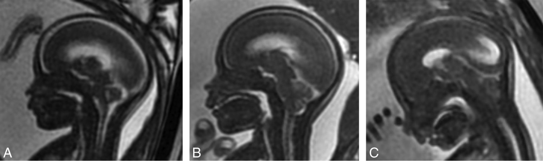
- Fig 1.
Grading of cerebellar ectopia on fetal MR imaging on sagittal FIESTA images of the brain. Grade 1 (A) has no cerebellar ectopia with a patent cisterna magna and fourth ventricle. Grade 2 (B) has cerebellar ectopia with an effaced fourth ventricle but a patent cisterna magna. Grade 3 (C) has cerebellar ectopia with an effaced fourth ventricle and cisterna magna.
- Fig 2.
A and B, Sagittal T2 SSFSE from fetal MR imaging performed at 24 weeks' and 5 days' gestational age (A) demonstrates severe cerebellar ectopia or grade 3 Chiari II malformation (arrow). Note that there is also effacement of the prepontine cistern and extra-axial CSF spaces over the cerebral hemispheres. Sagittal T2 FSE from postnatal MR imaging of the same patient at 2 weeks of age after postnatal repair of OSD shows a persistent grade 3 Chiari II malformation (arrow).
- Fig 3.
Sagittal T2 FSE from postnatal MR imaging in the same patient as in Fig 1A, status post postnatal repair of OSD at 11 days of age again demonstrates no cerebellar ectopia (arrow). Note the presence of other intracranial findings of a Chiari II malformation, including callosal hypogenesis/dysgenesis, a thickened massa intermedia, and tectal beaking.
- Fig 4.
Sagittal T2 SSFSE from fetal MR imaging at 23 weeks' and 3 days' gestational age (A) with severe or grade 3 Chiari II malformation (arrow). Sagittal T2 FSE image from postnatal MR imaging in the same patient at 2 weeks of age after in utero repair of OSD (B) demonstrates resolved cerebellar ectopia (arrow).


Tables
Imaging Findings % of Cohort Cerebellar ectopia 92.2% (94/102) Fetal Chiari grade 1 7.8% (8/102) 2 12.7% (13/102) 3 79.4% (81/102) Effacement of prepontine cistern 92.2% (94/102) Lateral ventricular size Normal 20.6% (21/102) Mild-moderate ventriculomegaly 57.8% (59/102) Severe ventriculomegaly 21.6% (22/102) Third ventriculomegaly 9.8% (10/102) Lateral ventricular rupture 1% (1/102) Extra-axial CSF effacement 92.1% (94/102) Imaging Finding % of Cohort Cerebellar ectopia 63.7% (65/102) Postnatal Chiari grade 1 36.3% (37/102) 2 26.5% (27/102) 3 37.3% (38/102) Effacement of prepontine cistern 14.7% (15/102) Lateral ventricular size Normal 4.9% (5/102) Mild-moderate ventriculomegaly 21.6% (22/102) Severe ventriculomegaly 51% (52/102) Extreme ventriculomegaly 22.5% (23/102) Lateral ventricular rupture 2.9% (3/102) Extra-axial CSF effacement 22.5% (23/102) Prenatal Repair Group (n = 32) Postnatal Repair Group (n = 70) % of fetal grade 3 Chiari II malformation that remained grade 3 postnatally 11.5% (3/26) 65.5% (36/55) % of fetal grade 3 Chiari II malformation that improved or resolved postnatally 88.5% (23/26) 34.5% (19/55) % of fetal grade 2 Chiari II malformation that resolved postnatally (grade 1) 100% (6/6) 28.6% (2/7) % that had a ventricular shunt catheter on postnatal MRI 3.1% (1/32) 47.1% (33/70)


Jump to section
Related Articles
Cited By...
- Chiari II brain malformation is secondary to open spina bifida
- Morphometric Analysis of Spina Bifida after Fetal Repair Shows New Subtypes with Associated Outcomes
- The Perplexity Surrounding Chiari Malformations - Are We Any Wiser Now?
- Fetal Intraventricular Hemorrhage in Open Neural Tube Defects: Prenatal Imaging Evaluation and Perinatal Outcomes
- Reliability of MR Imaging-Based Posterior Fossa and Brain Stem Measurements in Open Spinal Dysraphism in the Era of Fetal Surgery
- Spinal Imaging Findings of Open Spinal Dysraphisms on Fetal and Postnatal MRI