Advertisement

Main menu

  • Home
  • Content
    • Current Issue
    • Accepted Manuscripts
    • Article Preview
    • Past Issue Archive
    • Video Articles
    • AJNR Case Collection
    • Case of the Week Archive
    • Case of the Month Archive
    • Classic Case Archive
  • Special Collections
    • AJNR Awards
    • Low-Field MRI
    • Alzheimer Disease
    • ASNR Foundation Special Collection
    • Photon-Counting CT
    • View All
  • Multimedia
    • AJNR Podcasts
    • AJNR SCANtastic
    • Trainee Corner
    • MRI Safety Corner
    • Imaging Protocols
  • For Authors
    • Submit a Manuscript
    • Submit a Video Article
    • Submit an eLetter to the Editor/Response
    • Manuscript Submission Guidelines
    • Statistical Tips
    • Fast Publishing of Accepted Manuscripts
    • Graphical Abstract Preparation
    • Imaging Protocol Submission
    • Author Policies
  • About Us
    • About AJNR
    • Editorial Board
    • Editorial Board Alumni
  • More
    • Become a Reviewer/Academy of Reviewers
    • Subscribers
    • Permissions
    • Alerts
    • Feedback
    • Advertisers
    • ASNR Home

User menu

  • Alerts
  • Log in

Search

  • Advanced search
American Journal of Neuroradiology

American Journal of Neuroradiology

ASHNR American Society of Functional Neuroradiology ASHNR American Society of Pediatric Neuroradiology ASSR
  • Alerts
  • Log in

Advanced Search

  • Home
  • Content
    • Current Issue
    • Accepted Manuscripts
    • Article Preview
    • Past Issue Archive
    • Video Articles
    • AJNR Case Collection
    • Case of the Week Archive
    • Case of the Month Archive
    • Classic Case Archive
  • Special Collections
    • AJNR Awards
    • Low-Field MRI
    • Alzheimer Disease
    • ASNR Foundation Special Collection
    • Photon-Counting CT
    • View All
  • Multimedia
    • AJNR Podcasts
    • AJNR SCANtastic
    • Trainee Corner
    • MRI Safety Corner
    • Imaging Protocols
  • For Authors
    • Submit a Manuscript
    • Submit a Video Article
    • Submit an eLetter to the Editor/Response
    • Manuscript Submission Guidelines
    • Statistical Tips
    • Fast Publishing of Accepted Manuscripts
    • Graphical Abstract Preparation
    • Imaging Protocol Submission
    • Author Policies
  • About Us
    • About AJNR
    • Editorial Board
    • Editorial Board Alumni
  • More
    • Become a Reviewer/Academy of Reviewers
    • Subscribers
    • Permissions
    • Alerts
    • Feedback
    • Advertisers
    • ASNR Home
  • Follow AJNR on Twitter
  • Visit AJNR on Facebook
  • Follow AJNR on Instagram
  • Join AJNR on LinkedIn
  • RSS Feeds

AJNR Awards, New Junior Editors, and more. Read the latest AJNR updates

Research ArticleHead and Neck Imaging

Benign Miliary Osteoma Cutis of the Face: A Common Incidental CT Finding

D. Kim, G.A. Franco, H. Shigehara, J. Asaumi and P. Hildenbrand
American Journal of Neuroradiology April 2017, 38 (4) 789-794; DOI: https://doi.org/10.3174/ajnr.A5096
D. Kim
aFrom the Department of Radiology (D.K., P.H.), Lahey Hospital and Medical Center, Burlington, Massachusetts
  • Find this author on Google Scholar
  • Find this author on PubMed
  • Search for this author on this site
  • ORCID record for D. Kim
G.A. Franco
bCommonwealth Radiology Associates (G.A.F.), Brockton, Massachusetts
  • Find this author on Google Scholar
  • Find this author on PubMed
  • Search for this author on this site
  • ORCID record for G.A. Franco
H. Shigehara
cDepartment of Oral and Maxillofacial Radiology (H.S., J.A.), Okayama University Graduate School of Medicine, Dentistry, and Pharmaceutical Sciences, Okayama, Japan.
  • Find this author on Google Scholar
  • Find this author on PubMed
  • Search for this author on this site
  • ORCID record for H. Shigehara
J. Asaumi
cDepartment of Oral and Maxillofacial Radiology (H.S., J.A.), Okayama University Graduate School of Medicine, Dentistry, and Pharmaceutical Sciences, Okayama, Japan.
  • Find this author on Google Scholar
  • Find this author on PubMed
  • Search for this author on this site
  • ORCID record for J. Asaumi
P. Hildenbrand
aFrom the Department of Radiology (D.K., P.H.), Lahey Hospital and Medical Center, Burlington, Massachusetts
  • Find this author on Google Scholar
  • Find this author on PubMed
  • Search for this author on this site
  • ORCID record for P. Hildenbrand
  • Article
  • Figures & Data
  • Info & Metrics
  • Responses
  • References
  • PDF
Loading

Article Figures & Data

Figures

  • Tables
  • Fig 1.
    • Download figure
    • Open in new tab
    • Download powerpoint
    Fig 1.

    Axial (A), sagittal (B), and coronal (C) CT images of 3 different representative patients demonstrate multiple millimetric scattered facial and scalp hypodermal calcified nodules with varying degrees of severity. A 3D bone window reconstruction of a patient's sinus CT (D) also demonstrates a relatively large, 4- to 5-mm facial calcified nodule within the right premaxillary skin.
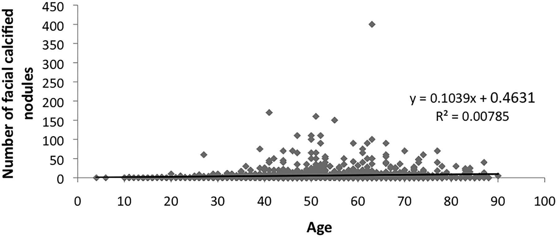
  • Fig 2.
    • Download figure
    • Open in new tab
    • Download powerpoint
    Fig 2.

    A scatterplot and linear regression between age and the number of facial calcified nodules among 553 patients with positive facial calcified nodules. No significant linear quantitative relationship was observed.
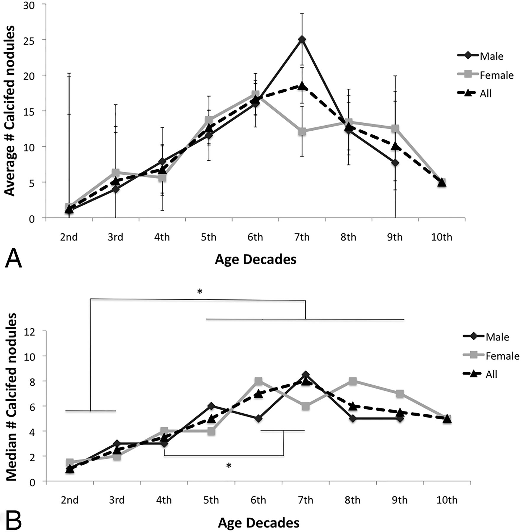
  • Fig 3.
    • Download figure
    • Open in new tab
    • Download powerpoint
    Fig 3.

    A, The average number of facial calcified nodules in each age group by decade. B, The median number of facial calcified nodules in each age group by decade. The Kruskal-Wallis test revealed a statistically significant difference in the median number of nodules among different age-decade groups. Post hoc analysis between each age group in the overall population, including both males and females, revealed a significantly higher (asterisk indicates P < .05) median number of nodules in the fifth–ninth decades compared with the second and third decades. An additional statistically significant difference was observed between the sixth and seventh decades and the fourth decade.

  • Fig 4.
    • Download figure
    • Open in new tab
    • Download powerpoint
    Fig 4.

    Radiographic examination of cadaveric facial skin specimen (A) reveals multiple calcified nodules. A magnified view of inflated buccal skin on an asymptomatic living patient (B) demonstrates multiple calcified nodules with central lucency. Benign breast skin calcification detected in a routine screening mammogram in a middle-aged female subject (C) demonstrates classic imaging findings of central lucency, which resemble the radiographic appearance of facial calcified nodules in (B). Axial (D) and coronal (E) CT images of the buccal area of a patient demonstrate facial calcified nodules. A magnified axial CT image of a relatively large 5-mm maxillary hypodermal calcified nodule (F) demonstrates a subtle central lucency. A and B, Images adapted from Shigehara et al16 and obtained from Dr Shigehara with permission.
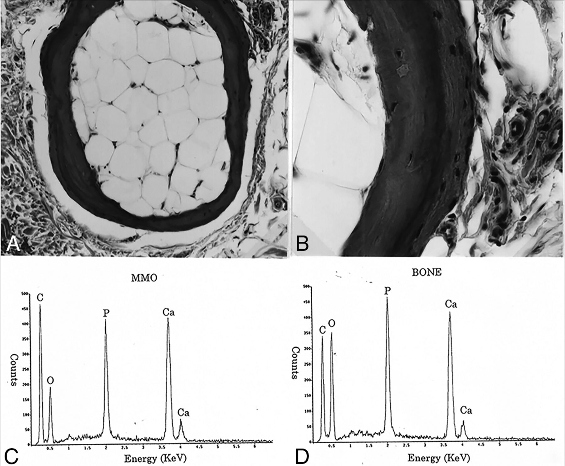
  • Fig 5.
    • Download figure
    • Open in new tab
    • Download powerpoint
    Fig 5.

    A, Pathologic examination of facial calcified nodules from a cadaveric specimen demonstrates mature bone-like characteristics, including a concentric, multilamellated, osteoid cortex and a central adipose medulla. B, A magnified view of a portion of A. C and D, Elemental composition and peak ratios of a cadaveric facial nodule (C) and the clavicle (D) are similar to each other and well-correlated with the composition of hydroxyapatite. Images are adapted from Shigehara et al16 and obtained with permission from Dr Shigehara. MMO indicates multiple miliary osteoma.

Tables

  • Figures
    • View popup
    Table 1:

    Study population and the distribution of CT indicationsa

    IndicationsCases%
    Chronic sinusitis82362.6%
    Upper respiratory infection/rhinitis14210.8%
    Sinus pain/pressure765.8%
    Polyposis/mucocele352.7%
    Fungal infection110.8%
    Allergy251.9%
    Asthma80.6%
    Headache1158.8%
    Obstructive sleep apnea60.5%
    Mass/cyst90.7%
    Head and neck cancer80.6%
    Pituitary mass60.5%
    Wegener granulomatosis40.3%
    Epistaxis120.9%
    CSF leak60.5%
    Trauma30.2%
    Facial infection20.2%
    Facial weakness30.2%
    Anosmia50.4%
    Vertigo/hearing loss80.6%
    Otalgia10.1%
    Acromegaly10.1%
    Unspecified60.5%
    Total1315100%
    • ↵a The first 6 indications relate to sinusitis or upper respiratory symptoms, accounting for the majority of indications (1112 cases, 84.6%; 1315 sinus CT cases; male, 599, 45.6%; female, 716, 54.4%).

    • View popup
    Table 2:

    Distribution of facial calcified nodules by location

    Nodule DistributionAllMaleFemale
    None762347415
    Frontal285126159
    Frontal-maxillary19587108
    Maxillary603129
    Temporal734
    Mandibular/buccal440
    Orbital211
    Subtotal1315599716
PreviousNext
Back to top

In this issue

American Journal of Neuroradiology: 38 (4)
American Journal of Neuroradiology
Vol. 38, Issue 4
1 Apr 2017
  • Table of Contents
  • Index by author
  • Complete Issue (PDF)
Advertisement
Print
Download PDF
Email Article

Thank you for your interest in spreading the word on American Journal of Neuroradiology.

NOTE: We only request your email address so that the person you are recommending the page to knows that you wanted them to see it, and that it is not junk mail. We do not capture any email address.

Enter multiple addresses on separate lines or separate them with commas.
Benign Miliary Osteoma Cutis of the Face: A Common Incidental CT Finding
(Your Name) has sent you a message from American Journal of Neuroradiology
(Your Name) thought you would like to see the American Journal of Neuroradiology web site.
CAPTCHA
This question is for testing whether or not you are a human visitor and to prevent automated spam submissions.
Cite this article
D. Kim, G.A. Franco, H. Shigehara, J. Asaumi, P. Hildenbrand
Benign Miliary Osteoma Cutis of the Face: A Common Incidental CT Finding
American Journal of Neuroradiology Apr 2017, 38 (4) 789-794; DOI: 10.3174/ajnr.A5096

Citation Manager Formats

  • BibTeX
  • Bookends
  • EasyBib
  • EndNote (tagged)
  • EndNote 8 (xml)
  • Medlars
  • Mendeley
  • Papers
  • RefWorks Tagged
  • Ref Manager
  • RIS
  • Zotero
0 Responses
Respond to this article
Share
Bookmark this article
Benign Miliary Osteoma Cutis of the Face: A Common Incidental CT Finding
D. Kim, G.A. Franco, H. Shigehara, J. Asaumi, P. Hildenbrand
American Journal of Neuroradiology Apr 2017, 38 (4) 789-794; DOI: 10.3174/ajnr.A5096
del.icio.us logo Twitter logo Facebook logo Mendeley logo
  • Tweet Widget
  • Facebook Like
  • Google Plus One
Purchase

Jump to section

  • Article
    • Abstract
    • Materials and Methods
    • Results
    • Discussion
    • Conclusions
    • Footnotes
    • REFERENCES
  • Figures & Data
  • Info & Metrics
  • Responses
  • References
  • PDF

Related Articles

  • No related articles found.
  • PubMed
  • Google Scholar

Cited By...

  • No citing articles found.
  • Crossref (19)
  • Google Scholar

This article has been cited by the following articles in journals that are participating in Crossref Cited-by Linking.

  • Multiple miliary osteoma cutis: an overlooked dermatosis?
    B. Duarte, R.R. Pinheiro, J. Cabete
    Journal of the European Academy of Dermatology and Venereology 2018 32 1
  • Two cases of treatment with aromatase inhibitors and development of miliary osteoma cutis—Is there an association?
    Eric D. Schadler, Stephanie L. Mehlis, Thomas L. Cibull, Bernhard Ortel
    JAAD Case Reports 2018 4 7
  • Multiple miliary osteoma cutis treatment response to Q-switched Nd:YAG laser: A case report
    Augustin C. Barolet, Ivan V. Litvinov, Daniel Barolet
    SAGE Open Medical Case Reports 2020 8
  • A Case of Surgical Removal of a Giant and Multiple Acquired Plate-Like Osteoma Cutis After Implantation of a Dilator
    Fangfang Wu, Weilong Qiao, Xiangdong Sun, Chengzhi Li, Huiqing Pan, Renliang He
    Clinical, Cosmetic and Investigational Dermatology 2021 Volume 14
  • High-frequency ultrasonography helps to confirm the diagnosis of multiple miliary osteoma cutis
    Frédéric Jaouen, Matthias Tallegas, Odile Treffandier, Frédéric Patat, Loïc Vaillant, Laurent Machet
    European Journal of Dermatology 2019 29 3
  • Imaging feature of cosmetic fillers in cone-beam computed tomography and its dental consideration
    Chena Lee, Yoon Joo Choi, Kug Jin Jeon, Seong Ho Choi, Sang-Sun Han
    Head & Face Medicine 2022 18 1
  • Multiple miliary osteoma cutis, renal failure and secondary hyperparathyroidism in a geriatric patient
    John K. Brooks, Hokyoung Jang, Arthur S. Harrow, Jeffery B. Price
    Gerodontology 2022 39 2
  • Plewig and Kligman´s Acne and Rosacea
    Gerd Plewig, Bodo Melnik, WenChieh Chen
    2019
  • What do we know about osteoma cutis? Literature review. Clinical cases
    Elena S. Snarskaya, Natalia P. Teplyuk, Ksenia D. Vasileva, E. Yu. Vertieva
    Russian Journal of Skin and Venereal Diseases 2021 24 2
  • A case of miliaris osteoma cutis in a male: Dermoscopic findings and microablation treatment
    Francesco Moro, Francesco Ricci, Ilaria Pennacchia, Cinzia Mazzanti, Damiano Abeni, Luca Fania
    Dermatologic Therapy 2019 32 5

More in this TOC Section

  • ASL Sensitivity for Head and Neck Paraganglioma
  • Post SRS Peritumoral Hyperintense Signal of VSs
  • Contrast Enhanced Pituitary CISS/FIESTA
Show more Head and Neck Imaging

Similar Articles

Advertisement

Indexed Content

  • Current Issue
  • Accepted Manuscripts
  • Article Preview
  • Past Issues
  • Editorials
  • Editor's Choice
  • Fellows' Journal Club
  • Letters to the Editor
  • Video Articles

Cases

  • Case Collection
  • Archive - Case of the Week
  • Archive - Case of the Month
  • Archive - Classic Case

More from AJNR

  • Trainee Corner
  • Imaging Protocols
  • MRI Safety Corner
  • Book Reviews

Multimedia

  • AJNR Podcasts
  • AJNR Scantastics

Resources

  • Turnaround Time
  • Submit a Manuscript
  • Submit a Video Article
  • Submit an eLetter to the Editor/Response
  • Manuscript Submission Guidelines
  • Statistical Tips
  • Fast Publishing of Accepted Manuscripts
  • Graphical Abstract Preparation
  • Imaging Protocol Submission
  • Evidence-Based Medicine Level Guide
  • Publishing Checklists
  • Author Policies
  • Become a Reviewer/Academy of Reviewers
  • News and Updates

About Us

  • About AJNR
  • Editorial Board
  • Editorial Board Alumni
  • Alerts
  • Permissions
  • Not an AJNR Subscriber? Join Now
  • Advertise with Us
  • Librarian Resources
  • Feedback
  • Terms and Conditions
  • AJNR Editorial Board Alumni

American Society of Neuroradiology

  • Not an ASNR Member? Join Now

© 2025 by the American Society of Neuroradiology All rights, including for text and data mining, AI training, and similar technologies, are reserved.
Print ISSN: 0195-6108 Online ISSN: 1936-959X

Powered by HighWire