Advertisement

Main menu

  • Home
  • Content
    • Current Issue
    • Accepted Manuscripts
    • Article Preview
    • Past Issue Archive
    • Video Articles
    • AJNR Case Collection
    • Case of the Week Archive
    • Case of the Month Archive
    • Classic Case Archive
  • Special Collections
    • AJNR Awards
    • Low-Field MRI
    • Alzheimer Disease
    • ASNR Foundation Special Collection
    • Photon-Counting CT
    • View All
  • Multimedia
    • AJNR Podcasts
    • AJNR SCANtastic
    • Trainee Corner
    • MRI Safety Corner
    • Imaging Protocols
  • For Authors
    • Submit a Manuscript
    • Submit a Video Article
    • Submit an eLetter to the Editor/Response
    • Manuscript Submission Guidelines
    • Statistical Tips
    • Fast Publishing of Accepted Manuscripts
    • Graphical Abstract Preparation
    • Imaging Protocol Submission
    • Author Policies
  • About Us
    • About AJNR
    • Editorial Board
    • Editorial Board Alumni
  • More
    • Become a Reviewer/Academy of Reviewers
    • Subscribers
    • Permissions
    • Alerts
    • Feedback
    • Advertisers
    • ASNR Home

User menu

  • Alerts
  • Log in

Search

  • Advanced search
American Journal of Neuroradiology

American Journal of Neuroradiology

ASHNR American Society of Functional Neuroradiology ASHNR American Society of Pediatric Neuroradiology ASSR
  • Alerts
  • Log in

Advanced Search

  • Home
  • Content
    • Current Issue
    • Accepted Manuscripts
    • Article Preview
    • Past Issue Archive
    • Video Articles
    • AJNR Case Collection
    • Case of the Week Archive
    • Case of the Month Archive
    • Classic Case Archive
  • Special Collections
    • AJNR Awards
    • Low-Field MRI
    • Alzheimer Disease
    • ASNR Foundation Special Collection
    • Photon-Counting CT
    • View All
  • Multimedia
    • AJNR Podcasts
    • AJNR SCANtastic
    • Trainee Corner
    • MRI Safety Corner
    • Imaging Protocols
  • For Authors
    • Submit a Manuscript
    • Submit a Video Article
    • Submit an eLetter to the Editor/Response
    • Manuscript Submission Guidelines
    • Statistical Tips
    • Fast Publishing of Accepted Manuscripts
    • Graphical Abstract Preparation
    • Imaging Protocol Submission
    • Author Policies
  • About Us
    • About AJNR
    • Editorial Board
    • Editorial Board Alumni
  • More
    • Become a Reviewer/Academy of Reviewers
    • Subscribers
    • Permissions
    • Alerts
    • Feedback
    • Advertisers
    • ASNR Home
  • Follow AJNR on Twitter
  • Visit AJNR on Facebook
  • Follow AJNR on Instagram
  • Join AJNR on LinkedIn
  • RSS Feeds

AJNR Awards, New Junior Editors, and more. Read the latest AJNR updates

Research ArticleHead & Neck

Optimization of Quantitative Dynamic Postgadolinium MRI Technique Using Normalized Ratios for the Evaluation of Temporomandibular Joint Synovitis in Patients with Juvenile Idiopathic Arthritis

P. Caruso, K. Buch, S. Rincon, R. Hakimelahi, Z.S. Peacock, C.M. Resnick, C. Foster, L. Guidoboni, T. Donahue, R. Macdonald, H. Rothermel, H.D. Curtin and L.B. Kaban
American Journal of Neuroradiology December 2017, 38 (12) 2344-2350; DOI: https://doi.org/10.3174/ajnr.A5424
P. Caruso
aFrom the Division of Neuroradiology, Department of Radiology, Massachusetts General Hospital (P.C., K.B., S.R., R.H., C.F., L.G., T.D., R.M.)
  • Find this author on Google Scholar
  • Find this author on PubMed
  • Search for this author on this site
  • ORCID record for P. Caruso
K. Buch
aFrom the Division of Neuroradiology, Department of Radiology, Massachusetts General Hospital (P.C., K.B., S.R., R.H., C.F., L.G., T.D., R.M.)
  • Find this author on Google Scholar
  • Find this author on PubMed
  • Search for this author on this site
  • ORCID record for K. Buch
S. Rincon
aFrom the Division of Neuroradiology, Department of Radiology, Massachusetts General Hospital (P.C., K.B., S.R., R.H., C.F., L.G., T.D., R.M.)
  • Find this author on Google Scholar
  • Find this author on PubMed
  • Search for this author on this site
  • ORCID record for S. Rincon
R. Hakimelahi
aFrom the Division of Neuroradiology, Department of Radiology, Massachusetts General Hospital (P.C., K.B., S.R., R.H., C.F., L.G., T.D., R.M.)
  • Find this author on Google Scholar
  • Find this author on PubMed
  • Search for this author on this site
  • ORCID record for R. Hakimelahi
Z.S. Peacock
cDepartment of Oral & Maxillofacial Surgery, Massachusetts General Hospital and Mass General Hospital for Children (Z.S.P., L.B.K.)
  • Find this author on Google Scholar
  • Find this author on PubMed
  • Search for this author on this site
  • ORCID record for Z.S. Peacock
C.M. Resnick
dDepartment of Plastic and Oral Surgery, Boston Children's Hospital (C.M.R.)
  • Find this author on Google Scholar
  • Find this author on PubMed
  • Search for this author on this site
  • ORCID record for C.M. Resnick
C. Foster
aFrom the Division of Neuroradiology, Department of Radiology, Massachusetts General Hospital (P.C., K.B., S.R., R.H., C.F., L.G., T.D., R.M.)
  • Find this author on Google Scholar
  • Find this author on PubMed
  • Search for this author on this site
  • ORCID record for C. Foster
L. Guidoboni
aFrom the Division of Neuroradiology, Department of Radiology, Massachusetts General Hospital (P.C., K.B., S.R., R.H., C.F., L.G., T.D., R.M.)
  • Find this author on Google Scholar
  • Find this author on PubMed
  • Search for this author on this site
  • ORCID record for L. Guidoboni
T. Donahue
aFrom the Division of Neuroradiology, Department of Radiology, Massachusetts General Hospital (P.C., K.B., S.R., R.H., C.F., L.G., T.D., R.M.)
  • Find this author on Google Scholar
  • Find this author on PubMed
  • Search for this author on this site
  • ORCID record for T. Donahue
R. Macdonald
aFrom the Division of Neuroradiology, Department of Radiology, Massachusetts General Hospital (P.C., K.B., S.R., R.H., C.F., L.G., T.D., R.M.)
  • Find this author on Google Scholar
  • Find this author on PubMed
  • Search for this author on this site
  • ORCID record for R. Macdonald
H. Rothermel
bDepartment of Pediatrics, Mass General Hospital for Children (H.R.)
  • Find this author on Google Scholar
  • Find this author on PubMed
  • Search for this author on this site
  • ORCID record for H. Rothermel
H.D. Curtin
eDepartment of Radiology, Massachusetts Eye and Ear Infirmary (H.D.C.), Harvard Medical School and Harvard School of Dental Medicine, Boston, Massachusetts.
  • Find this author on Google Scholar
  • Find this author on PubMed
  • Search for this author on this site
  • ORCID record for H.D. Curtin
L.B. Kaban
cDepartment of Oral & Maxillofacial Surgery, Massachusetts General Hospital and Mass General Hospital for Children (Z.S.P., L.B.K.)
  • Find this author on Google Scholar
  • Find this author on PubMed
  • Search for this author on this site
  • ORCID record for L.B. Kaban
  • Article
  • Figures & Data
  • Supplemental
  • Info & Metrics
  • Responses
  • References
  • PDF
Loading

Article Figures & Data

Figures

  • Tables
  • Fig 1.
    • Download figure
    • Open in new tab
    • Download powerpoint
    Fig 1.

    A and B, Coronal scanning plane and ROI-based measurements of the TMJ synovia. An oblique coronal plane was prescribed off the sagittal localizer to include the entire right and left TMJs and the longus capitis in the same coronal FOV. The coronal plane was angled superoanterior to posteroinferior to include both the TMJs from anterior to posterior and the longus capitis muscle; in controls, the oblique coronal plane included both TMJs, the longus capitis, and the sella turcica. C, An example of a single ROI at the superomedial synovial edge of the right TMJ (0.5 mm2) on a precontrast MR imaging and a single ROI placed along the longus capitis muscle (50 mm2), which was used for normalization. D, Example of the 10 ROIs placed on a postgadolinium MR imaging. These ROIs are placed along the superior and inferior synovial compartments of the left TMJ with the following designations: SL indicates superior compartment, lateral edge; SML, superior compartment, midlateral edge; SC, superior compartment, central; SMM, superior compartment, midmedial edge; SM, superior compartment, medial edge; IL, inferior compartment, lateral edge; IML, inferior compartment, midlateral edge, IC, inferior compartment, central; IMM, inferior compartment, midmedial edge; IM, inferior compartment, medial edge. Although attempts were made to measure the signal along the area closest to the disk edge in the expected location of the synovium, these measurements undoubtedly reflect a composite of signal produced by the synovium, cartilage, and perhaps the periosteum. Thus, the notion of “synovial signal” used in this report reflects this currently unavoidable composite measurement.
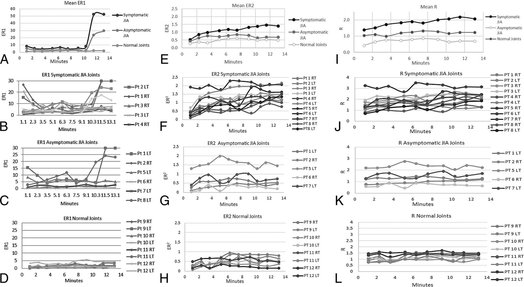
  • Fig 2.
    • Download figure
    • Open in new tab
    • Download powerpoint
    Fig 2.

    A, Mean ER1 for the symptomatic JIA, asymptomatic JIA, and control joints. ER1 shows a monopeak profile with an upward slope that starts at 10 minutes 30 seconds at the end of run 8 and clear separation of the curves between 10 minutes 30 seconds and 13 minutes 10 seconds during runs 9 and 10. B, Individual patient profiles for ER1 for the symptomatic JIA joints. C, Individual patient profiles for ER1 for the asymptomatic JIA joints. D, Individual patient profiles for ER1 for the control TMJ joints. These joints exhibit relatively constant enhancement ratios between 0.73 and 5 at all time points. E, Mean ER2 for the symptomatic JIA, asymptomatic JIA, and control joints. Note the clear separation among the 3 groups at all sampling time points with the mean profile for the symptomatic JIA group showing a monopeak profile. Also, the mean profile of the asymptomatic JIA group is higher at all time points than the mean profile for the control group. F, Individual patient profiles for ER2 for symptomatic JIA joints. All but one of the symptomatic joints, patient 8 RT TMJ, pass above the threshold value of normal (0.95) at multiple time points that thus results in correct classification of 10/11 symptomatic joints. G, Individual patient profiles for ER2 for asymptomatic JIA joints. Most profiles stay below the threshold normal value (0.94) while several pass above. H, Individual patient profiles for ER2 for control TMJ joints. The initial ER2 at 1 minute 10 seconds ranges from 0.15 to 0.5, and the end ER2 for this group at 13 minutes 10 seconds ranges from 0.14 to 0.63. The maximum ratio (peak) reached by any single patient at any time point is 0.94 that serves as a threshold value for normal for ER2. I, Mean R for the symptomatic JIA, asymptomatic JIA, and control joints. Note the clear separation among the 3 groups at all sampling time points. The mean profile for the symptomatic JIA group shows a monopeak profile. J, Individual patient profiles for R for the symptomatic JIA joints. Two joints, patient 2 L TMJ and patient 8 R TMJ, never pass above the 1.55 threshold and thus would be misclassified by R as asymptomatic joints. A third joint, patient 8 L TMJ, passes above the threshold only twice that may result in an indeterminate or borderline classification. K, Individual patient profiles for R for the asymptomatic JIA joints. Two of these joints, patient 5 L TMJ and patient 7 L TMJ pass above the normal threshold. L, Individual patient profiles for R for the control TMJ joints. While most of these joints stay below the 1.55 threshold, one joint, patient 12 L TMJ passes above the threshold at 4 time points, that may lead to misclassification of this joint.

  • Fig 3.
    • Download figure
    • Open in new tab
    • Download powerpoint
    Fig 3.

    Raw T1 hyperintensity curves of the longus capitis muscle as measured on the precontrast image and each of the 10 postgadolinium dynamic images. The raw signal of the longus capitis is similar at all sampling time points in both the JIA and control groups: no statistically significant difference (P = .99) was seen at any time point, suggesting that the longus capitis may serve as a normalization standard for both normal and symptomatic groups.

Tables

  • Figures
    • View popup
    Table 1:

    Dynamic gadolinium MRI technique for imaging of the TMJ

    ParametersDetails
    SequenceCoronal T1 postgadolinium, 70-sec, 10-sec prescan
    TR/TE (ms)400/15
    Frequency × phase300 × 200
    NEX1
    Section thickness3 mm, 0-mm gap
    FOV14–16 cm
    Timing of the serial runsRuns: run 0, pregadolinium run, followed by 10 postgadolinium runs with the following timing intervals: run 1: 0 -1 min 10 sec, run 2: 1 min 20 sec to 2 min 30 sec; run 3: 2 min 40 sec to 3 min 50 sec; run 4: 4 min 00 sec to 5 min 10 sec; run 5: 5 min 20 sec to 6 min 30 sec; run 6: 6 min 40 sec to 7 min 50 sec; run 7: 8 min 00 sec to 9 min 10 sec; run 8: 9 min 20 sec to 10 min 30 sec; run 9: 10 min 40 sec to 11 min 50 sec; run 10: 12 min 00 sec to 13 min 10 sec
    Injection techniqueStandard supine position
    22-ga angiocatheter in right antecubital fossa, attached to 20-cm length of standard tubing connected to a power injector
    Contrast agent and dosingGadoterate meglumine (Dotarem)a maintained at room temperature was drawn up at a dose of 0.2 mL/kg (0.1 mmol/kg) of body weight
    Simultaneous with the start of the first coronal T1 dynamic sequence, run 1, gadoterate was administered by power injector as a single intravenous bolus injection at a flow rate of 2.5 mL/s followed by a 20-mL saline chaser at the same injection rate
    • ↵a Guerbet, Aulnay-sous-Bois, France.

    • View popup
    Table 2:

    Measurements of signal intensity over the synovium and longus capitis muscles and calculation of the normalized ratios ER1, ER2, and R

    Calculations
    Synovial signal intensityPre = (SL+SML+SC+SMM+SM+IL+IML+IC+IMM+IM)/10, where, Pre = average precontrast synovial T1 signal intensity measured at 10 points along the synovia
    Postn = (SL+SML+SC+SMM+SM+IL+IML+IC+IMM+IM)n /10, where, Postn = average postgadolinium synovial signal, for run n (n = 1–10) measured at 10 points along the synovia
    Synovial enhancementn = Postn–Pre, for each run n (n = 1–10)
    Longus capitis intensityLCPRE = precontrast T1 signal in the longus capitis
    LCPOSTn = postcontrast T1 signal in the longus capitis, for run n (n = 1–10)
    Longus capitis enhancementn = LCPOSTn–LCPRE, for each run n (n = 1–10)
    Normalized ratiosER1 = (postgadolinium T1 signal of the TMJ synovium − the pregadolinium T1 signal of the TMJ synovium) divided by the (postgadolinium T1 signal of the longus capitis − the pregadolinium T1 signal of the longus capitis) where ER1 thus represents the ratio of the synovial enhancementn to the longus capitis enhancementn for each run n
    ER2 = (postgadolinium T1 signal of the TMJ synovium − the pregadolinium T1 signal of the TMJ synovium) divided by the postgadolinium T1 signal of the longus capitis where ER2 thus represents the ratio of the synovial enhancementn to the LCPOSTn for each run n
    R = the postgadolinium T1 signal of the TMJ synovium divided by the postgadolinium T1 signal of the longus capitis and thus represents the ratio of Postn to LCPOSTn
    • Note:—n indicates each run; SL, superior compartment, lateral edge; SML, superior compartment, midlateral edge; SC, superior compartment, central; SMM, superior compartment, midmedial edge; SM, superior compartment, medial edge; IL, inferior compartment, lateral edge; IML, inferior compartment, midlateral edge; IC, inferior compartment, central; IMM, inferior compartment, midmedial edge, IM, inferior compartment medial edge; LCPRE, the measurement of the T1 signal in the longus capitis muscle on the coronal pregadolinium T1 sequence; LCPOSTn, the measurement of the T1 signal in the longus capitis on the postgadolinium coronal T1 sequence for runs 1–10.

PreviousNext
Back to top

In this issue

American Journal of Neuroradiology: 38 (12)
American Journal of Neuroradiology
Vol. 38, Issue 12
1 Dec 2017
  • Table of Contents
  • Index by author
  • Complete Issue (PDF)
Advertisement
Print
Download PDF
Email Article

Thank you for your interest in spreading the word on American Journal of Neuroradiology.

NOTE: We only request your email address so that the person you are recommending the page to knows that you wanted them to see it, and that it is not junk mail. We do not capture any email address.

Enter multiple addresses on separate lines or separate them with commas.
Optimization of Quantitative Dynamic Postgadolinium MRI Technique Using Normalized Ratios for the Evaluation of Temporomandibular Joint Synovitis in Patients with Juvenile Idiopathic Arthritis
(Your Name) has sent you a message from American Journal of Neuroradiology
(Your Name) thought you would like to see the American Journal of Neuroradiology web site.
CAPTCHA
This question is for testing whether or not you are a human visitor and to prevent automated spam submissions.
Cite this article
P. Caruso, K. Buch, S. Rincon, R. Hakimelahi, Z.S. Peacock, C.M. Resnick, C. Foster, L. Guidoboni, T. Donahue, R. Macdonald, H. Rothermel, H.D. Curtin, L.B. Kaban
Optimization of Quantitative Dynamic Postgadolinium MRI Technique Using Normalized Ratios for the Evaluation of Temporomandibular Joint Synovitis in Patients with Juvenile Idiopathic Arthritis
American Journal of Neuroradiology Dec 2017, 38 (12) 2344-2350; DOI: 10.3174/ajnr.A5424

Citation Manager Formats

  • BibTeX
  • Bookends
  • EasyBib
  • EndNote (tagged)
  • EndNote 8 (xml)
  • Medlars
  • Mendeley
  • Papers
  • RefWorks Tagged
  • Ref Manager
  • RIS
  • Zotero
0 Responses
Respond to this article
Share
Bookmark this article
Optimization of Quantitative Dynamic Postgadolinium MRI Technique Using Normalized Ratios for the Evaluation of Temporomandibular Joint Synovitis in Patients with Juvenile Idiopathic Arthritis
P. Caruso, K. Buch, S. Rincon, R. Hakimelahi, Z.S. Peacock, C.M. Resnick, C. Foster, L. Guidoboni, T. Donahue, R. Macdonald, H. Rothermel, H.D. Curtin, L.B. Kaban
American Journal of Neuroradiology Dec 2017, 38 (12) 2344-2350; DOI: 10.3174/ajnr.A5424
del.icio.us logo Twitter logo Facebook logo Mendeley logo
  • Tweet Widget
  • Facebook Like
  • Google Plus One
Purchase

Jump to section

  • Article
    • Abstract
    • ABBREVIATIONS:
    • Materials and Methods
    • Results
    • Discussion
    • Conclusions
    • Footnotes
    • References
  • Figures & Data
  • Supplemental
  • Info & Metrics
  • Responses
  • References
  • PDF

Related Articles

  • PubMed
  • Google Scholar

Cited By...

  • No citing articles found.
  • Crossref (10)
  • Google Scholar

This article has been cited by the following articles in journals that are participating in Crossref Cited-by Linking.

  • Temporomandibular joint arthritis in juvenile idiopathic arthritis, now what?
    Matthew L. Stoll, Chung H. Kau, Peter D. Waite, Randy Q. Cron
    Pediatric Rheumatology 2018 16 1
  • An Algorithm for Management of Dentofacial Deformity Resulting From Juvenile Idiopathic Arthritis: Results of a Multinational Consensus Conference
    Cory M. Resnick, Paula Frid, Sven Erik Norholt, Peter Stoustrup, Zachary S. Peacock, Leonard B. Kaban, Thomas Klit Pedersen, Shelly Abramowicz
    Journal of Oral and Maxillofacial Surgery 2019 77 6
  • Current status of MR imaging of juvenile idiopathic arthritis
    Clara Malattia, Mirkamal Tolend, Marta Mazzoni, Jyoti Panwar, Margalit Zlotnik, Tarimobo Otobo, Logi Vidarsson, Andrea S. Doria
    Best Practice & Research Clinical Rheumatology 2020 34 6
  • Assessing the Reliability of the OMERACT Juvenile Idiopathic Arthritis Magnetic Resonance Scoring System for Temporomandibular Joints (JAMRIS-TMJ)
    Mirkamal Tolend, Andrea S. Doria, Arthur B. Meyers, Tore A. Larheim, Shelly Abramowicz, Julien Aguet, Simone Appenzeller, Linda Z. Arvidsson, Lauren W. Averill, Brian M. Feldman, Saurabh Guleria, Emilio J. Inarejos Clemente, Jacob L. Jaremko, Thitiporn Junhasavasdikul, Thekla von Kalle, Eva Kirkhus, Bernd Koos, Elka Miller, Rahim Moineddin, Jyoti Panwar, Zachary S. Peacock, Cory M. Resnick, Marion A. van Rossum, Jennifer Stimec, George Tomlinson, Nikolay Tzaribachev, Christian J. Kellenberger
    Journal of Clinical Medicine 2021 10 18
  • MRI of the temporomandibular joint in children with juvenile idiopathic arthritis: protocol and findings
    Emilio J. Inarejos Clemente, Mirkamal Tolend, Maria Navallas, Andrea S. Doria, Arthur B. Meyers
    Pediatric Radiology 2023 53 8
  • Imaging of the Temporomandibular Joint in Juvenile Idiopathic Arthritis: How Does Quantitative Compare to Semiquantitative MRI Scoring?
    Grace M.Y. Ma, Carly E. Calabrese, Timothy Donohue, Zachary S. Peacock, Paul Caruso, Leonard B. Kaban, Cory M. Resnick
    Journal of Oral and Maxillofacial Surgery 2019 77 5
  • Regional differences in temporomandibular joint inflammation in patients with juvenile idiopathic arthritis: a dynamic post-contrast magnetic resonance imaging study
    K. Buch, Z.S. Peacock, C.M. Resnick, H. Rothermel, L.B. Kaban, P. Caruso
    International Journal of Oral and Maxillofacial Surgery 2020 49 9
  • MRI of the Temporomandibular Joint
    Tiziana Robba, Paolo Tosco, Simone Parisi, Guglielmo Ramieri, Enrico Fusaro, Riccardo Faletti, Giulia Tanteri
    2020
  • Dynamic contrast-enhanced magnetic resonance imaging of masticatory muscles in patients with idiopathic condylar resorption
    Yu-Chen Wang, Hsiao-Chiao Yeh, Yunn-Jy Chen, Tiffany Ting-Fang Shih
    Journal of Dental Sciences 2025
  • Peterson’s Principles of Oral and Maxillofacial Surgery
    Cory M. Resnick, Peter Waite
    2022

More in this TOC Section

  • Chondrosarcoma vs Synovial Chondromatosis: Imaging
  • WHO Classification Update: Nasal&Skull Base Tumors
  • Peritumoral Signal in Vestibular Schwannomas
Show more Head & Neck

Similar Articles

Advertisement

Indexed Content

  • Current Issue
  • Accepted Manuscripts
  • Article Preview
  • Past Issues
  • Editorials
  • Editor's Choice
  • Fellows' Journal Club
  • Letters to the Editor
  • Video Articles

Cases

  • Case Collection
  • Archive - Case of the Week
  • Archive - Case of the Month
  • Archive - Classic Case

More from AJNR

  • Trainee Corner
  • Imaging Protocols
  • MRI Safety Corner
  • Book Reviews

Multimedia

  • AJNR Podcasts
  • AJNR Scantastics

Resources

  • Turnaround Time
  • Submit a Manuscript
  • Submit a Video Article
  • Submit an eLetter to the Editor/Response
  • Manuscript Submission Guidelines
  • Statistical Tips
  • Fast Publishing of Accepted Manuscripts
  • Graphical Abstract Preparation
  • Imaging Protocol Submission
  • Evidence-Based Medicine Level Guide
  • Publishing Checklists
  • Author Policies
  • Become a Reviewer/Academy of Reviewers
  • News and Updates

About Us

  • About AJNR
  • Editorial Board
  • Editorial Board Alumni
  • Alerts
  • Permissions
  • Not an AJNR Subscriber? Join Now
  • Advertise with Us
  • Librarian Resources
  • Feedback
  • Terms and Conditions
  • AJNR Editorial Board Alumni

American Society of Neuroradiology

  • Not an ASNR Member? Join Now

© 2025 by the American Society of Neuroradiology All rights, including for text and data mining, AI training, and similar technologies, are reserved.
Print ISSN: 0195-6108 Online ISSN: 1936-959X

Powered by HighWire