
Article Figures & Data
Figures
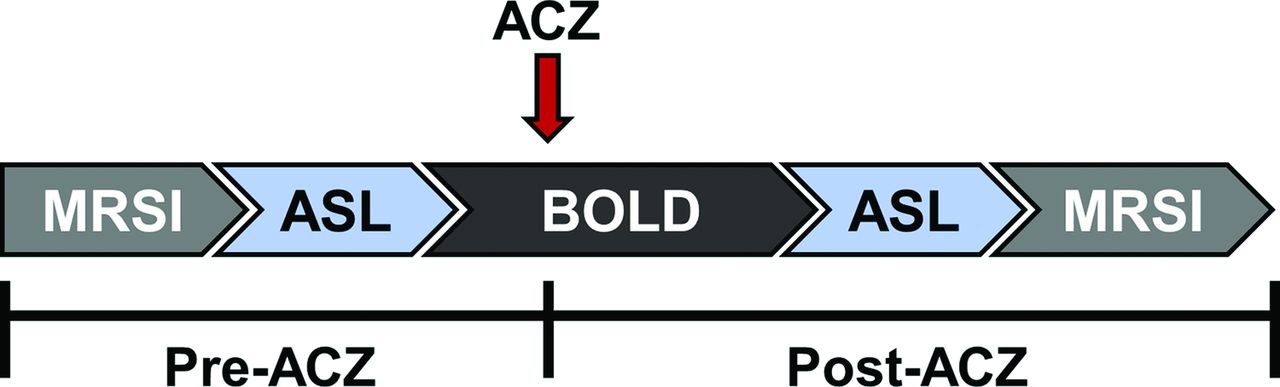
- Fig 1.
Schematic of MR imaging scan protocol. ACZ was injected 5 minutes after the start of the BOLD sequence and data were acquired for ∼10 minutes post-injection. ASL and MRSI were acquired pre- and post-ACZ.
- Fig 2.
Representative CVR percentage augmentation maps calculated with BOLD and ASL, along with a BTR map overlaid on a T1-weighted image. Images are all from the same subject (a 32-year-old woman) with unilateral left MCA stenosis without infarction. The white grid overlay represents the MR thermometry grid derived from multivoxel spectroscopy analysis using the water-NAA chemical shift difference. Diffusion-weighted imaging demonstrates no evidence for acute infarction. Images are displayed in the radiologic convention. Impaired CVR in the left hemisphere is present in both BOLD and ASL, with a greater severity of impairment in ASL likely related to delay sensitivity and tag decay (see text). The BTR map demonstrates an asymmetric thermal response, with less brain cooling following vasodilatory stimulus in the diseased left hemisphere, indicated by lower and more positive BTR values and corresponding primarily to the areas of greatest impairment in the anterior and posterior MCA borderzone territories.
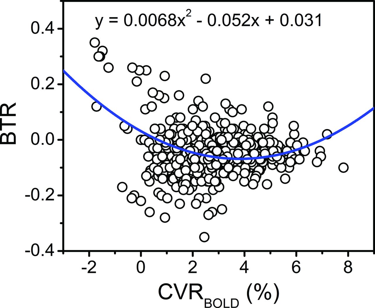
- Fig 3.
Voxelwise relationship of the BTR versus BOLD MR imaging signal augmentation following ACZ administration. The blue line and corresponding equation represent the fit of the raw data (circles) calculated with a mixed-effects model. A significant quadratic relationship between BTR and CVRBOLD was identified for all voxels, wherein an initially negative slope is observed at low CVRBOLD but a positive trend is observed for CVRBOLD augmentation upward of approximately 4% (see “Results” and “Discussion”; F, x2 = 57.5, P < .001; F, x = 74.1, P < .001).


Tables
Patient No. Age (years) Sex Presentation and Diagnosis 1 41 F Left-sided-predominate supraclinoid ICA stenosis 2 61 M Left cervical ICA stenosis; aphasia 3 39 F Right-sided-predominate ICA stenosis; recurrent minor stroke 4 32 F Left M1 stenosis; left monocular vision loss and recurrent right hemiparesis 5 56 M Intracranial ICA stenosis; recurrent TIA 6 68 F Left ICA stenosis; recurrent left hemispheric TIA and borderzone ischemia 7 33 F Right-sided-predominate supraclinoid ICA stenosis; Moyamoya disease following STA-MCA bypass and recurrent TIA 8 36 F Left M1 stenosis; recurrent left deep borderzone ischemia 9 38 F Right-sided-predominate intracranial stenosis; recurrent MCA infarctions 10 51 F Left ICA stenosis; recurrent TIA Note:—STA indicates superficial temporal artery.
- Table 2:
Parameter estimates calculated with a mixed-effects model of BTR and baseline brain temperature as a function of CVR
CVR CVR2 Intercept Coefficient F dfa P Value Coefficient F dfa P Value Coefficient F dfa P Valueb BTR CVRBOLD −0.052 74.1 1, 513 <.001 0.0068 57.5 1, 619 <.001 0.031 6.0 1, 35 .02 CVRASL 0.0018 1.2 1, 8.0 .30 – – – – −0.069 205 1, 3.8 <.001 CVRBOLD < 70c −0.096 59.7 1, 97 <.001 0.024 25.0 1, 128 <.001 0.031 2.8 1, 11 .12 CVRBOLD ≥ 70c 0.0070 5.5 1, 200 .02 – – – – −0.081 46.0 1, 51 <.001 CVRASL < 30d 0.0018 1.1 1, 7.4 .33 – – – – −0.068 183.2 1, 69 <.001 CVRASL ≥ 30d 0.0014 12.1 1, 47 .001 – – – – −0.10 33.5 1, 44 <.001 Tpree (°C) CVRBOLD 0.59 7.0 1, 598 .008 −0.13 14.8 1, 630 <.001 37.5 4871 1, 27 <.001 CVRASL −0.039 0.9 1, 7.3 .39 – – – – 38.4 8655 1, 6.0 <.001 CVRBOLD < 70c 2.1 19.5 1, 139 <.001 −0.65 12.9 1, 150 <.001 37.1 1810 1, 17 <.001 CVRBOLD ≥ 70c −0.38 10.7 1, 429 .001 – – – – 39.0 4575 1, 28 <.001 CVRASL < 30d −0.049 1.1 1, 6.8 .34 – – – – 38.5 12,953 1, 6.5 <.001 CVRASL ≥ 30d 0.0026 0.01 1, 198 .91 – – – – 37.5 1094 1, 27 <.001 ↵a df calculated with the Satterthwaite approximation are reported as numerator, denominator.
↵b P value represents the significance of a nonzero intercept.
↵c CVRBOLD values thresholded to 70% of the subject-wise mean contralateral value.
↵d CVRASL values thresholded to 30% augmentation.
↵e Tpre is the baseline brain temperature.


