
Article Figures & Data
Figures
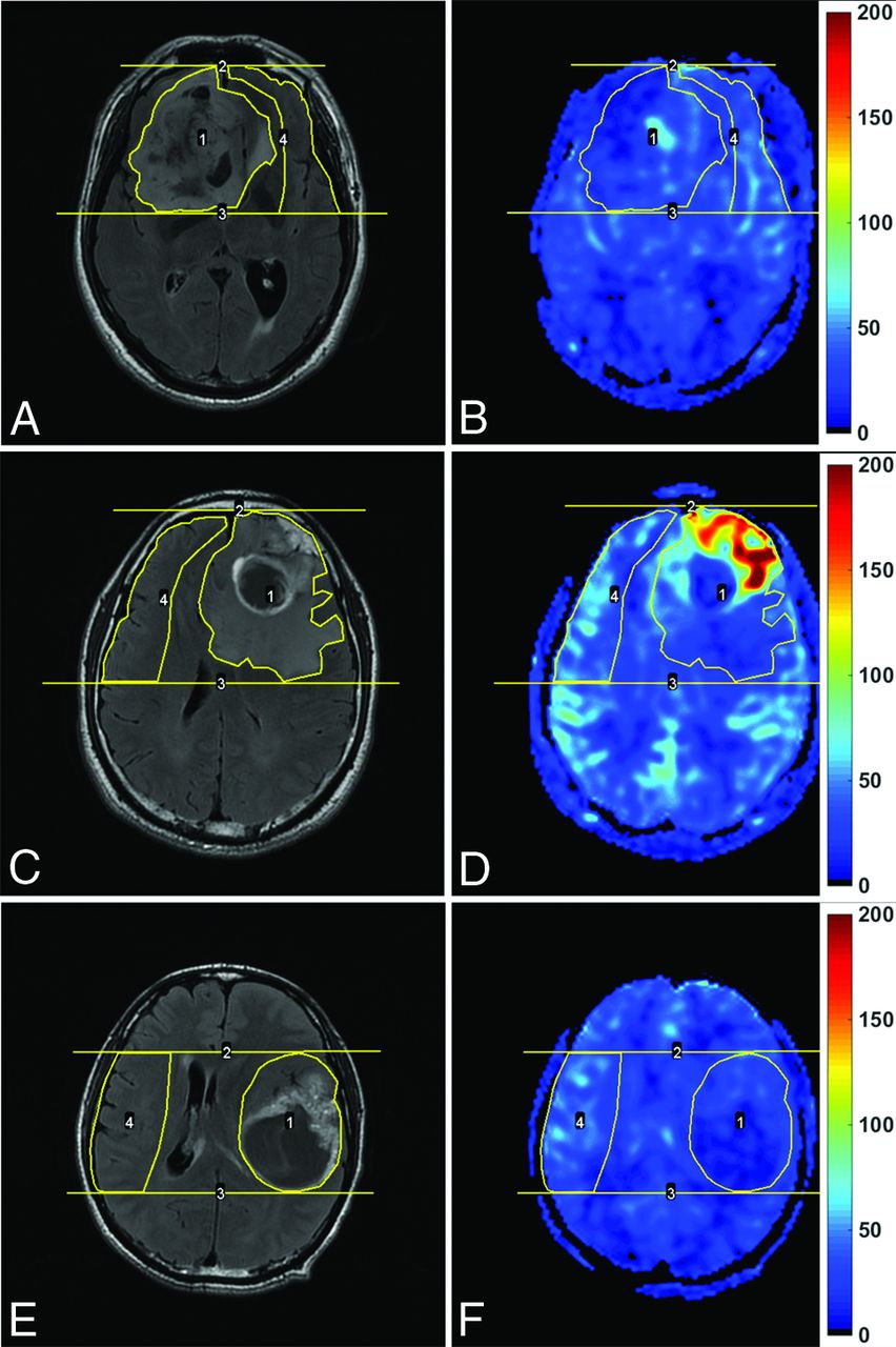
- Fig 1.
Enhanced T2-FLAIR images (A, C, and E) and CBF maps (B, D, and F) of a 69-year-old man with oligoastrocytoma (WHO grade II; Ki-67 index, 10%), a 42-year-old man with glioblastoma (WHO grade IV; Ki-67 index, 20%), and a 43-year-old man with glioblastoma (WHO grade IV; Ki-67 index, 60%), respectively. Note that blood flow is significantly elevated in the glioblastoma with a relatively low Ki-67 index, while it is not elevated in the glioblastoma with a very high Ki-67 index. The unit for CBF maps is milliliters/100 g/min.
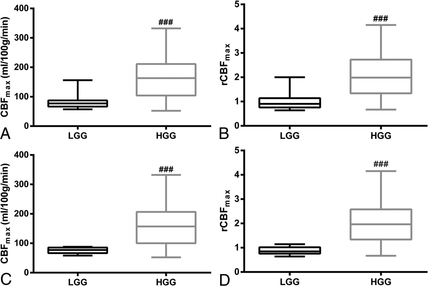
- Fig 2.
Boxplots of CBFmax (A) and rCBFmax (B) in low-grade gliomas and high-grade gliomas for all subjects. The boxplots of CBFmax (C) and rCBFmax (D) in LGGs and HGGs after the oligodendrogliomas and anaplastic oligodendrogliomas were excluded. ### indicates P < .001, compared with LGG.
- Fig 3.
Receiver operating characteristic curves of CBFmax (A) and rCBFmax (B) in distinguishing high- from low-grade gliomas, without (black line) or with (red line) oligodendrogliomas and anaplastic oligodendrogliomas excluded.
- Fig 4.
The linear regression of CBFmax (A–C) and rCBFmax (D–F) with the Ki-67 index in all subjects (A and D), in the low and high Ki-67 groups (B and E), and in glioblastomas (C and F). The low Ki-67 group included patients with a Ki-67 index of <30%, and the high Ki-67 group included patients with a Ki-67 index of ≥30%.


Tables
Grade/Histology No. CBFmax (mL/100 g/min) rCBFmax Tumor Contralateral Grade II (n = 13) Astrocytoma 5 75.4 ± 10.2a,b 83.2 ± 3.90 0.91 ± 0.18c,d Oligoastrocytoma 4 74.8 ± 12.1a,b 93.2 ± 28.8 0.83 ± 0.16c,d Oligodendroglioma 4 109.8 ± 47.8e 80.0 ± 9.6 1.38 ± 0.59e,f Grade III (n = 17) Anaplastic astrocytoma 6 127.2 ± 84.8e 78.0 ± 23.6 1.51 ± 0.68e,f Anaplastic oligoastrocytoma 7 125.1 ± 71.9e 82.4 ± 8.9 1.54 ± 0.88e,f Anaplastic oligodendroglioma 4 212.5 ± 87.5 78.8 ± 16.6 2.75 ± 1.02 Grade IV (n = 28) Glioblastoma 28 171.5 ± 58.7 77.0 ± 10.9 2.25 ± 0.79 - Table 2:
Measurements of absolute CBFmax and rCBFmax in each WHO grade with or without the exclusion of oligodendrogliomas and anaplastic oligodendrogliomas
Patients/Grades No. CBFmax (mL/100 g/min) rCBFmax Tumor Contralateral All Grade II 13 85.8 ± 30.3 85.3 ± 16.4 1.03 ± 0.41 Grade III 17 146.4 ± 84.0b 80.0 ± 16.1 1.81 ± 0.96c Grade IV 28 171.5 ± 58.7d 77.0 ± 10.9 2.25 ± 0.15d Excludeda Grade II 9 75.1 ± 10.4 87.7 ± 18.6 0.88 ± 0.16 Grade III 13 126.1 ± 74.7e 80.4 ± 16.6 1.52 ± 0.77b Grade IV 28 171.5 ± 58.7d,f 77.0 ± 10.9 2.25 ± 0.15d,g ↵a With oligodendrogliomas and anaplastic oligodendrogliomas excluded. Note the following P values in each patient group:
↵b P < .05, compared with grade II.
↵c P < .01, compared with grade II.
↵d P < .001, compared with grade II.
↵e P = .05, compared with grade II.
↵f P < .05, compared with grade III.
↵g P < .01, compared with grade III.
- Table 3:
ROC curve analyses of CBFmax and rCBFmax in discriminating high- and low-grade gliomas
Parameters/Patients AUC Youden Index Cutoff Value Sensitivity (%) Specificity (%) PPV (%) NPV (%) Accuracy (%) CBFmax All 0.828 0.690 91 mL/100 g/min 84.4 84.6 95.0 61.1 84.5 Excludeda 0.859 0.829 91 mL/100 g/min 82.9 100 100 56.3 86.0 rCBFmax All 0.863 0.668 1.19 82.2 84.6 94.9 57.9 82.8 Excludeda 0.916 0.805 1.22 80.5 100 100 52.9 84.0 Note:—AUC indicates area under the curve; PPV, positive predictive value; NPV, negative predictive value.
↵a With oligodendrogliomas and anaplastic oligodendrogliomas excluded.
- Table 4:
Univariate Cox model for progression-free survival and overall survival in patients with gliomas and glioblastomas
Variables PFS OS HR 95% CI P Value HR 95% CI P Value Gliomas (n = 51) Age 1.036 1.009–1.063 .008 1.043 1.014–1.073 .004 Sexa 0.957 0.670–1.368 .811 0.896 0.606–1.325 .582 KPS 0.971 0.953–0.989 .002 0.970 0.952–0.988 .002 CBFmax 1.005 1.001–1.010 .027 1.003 0.998–1.008 .244 rCBFmax 1.670 1.149–2.427 .007 1.246 0.869–1.785 .232 GBM (n = 25) Age 1.036 1.002–1.072 .039 1.030 0.994–1.067 .103 Sexa 0.851 0.547–1.323 .851 0.870 0.548–1.381 .555 KPS 0.986 0.963–1.008 .215 0.987 0.965–1.009 .242 CBFmax 0.997 0.989–1.005 .497 0.993 0.985–1.001 .070 rCBFmax 0.865 0.467–1.601 .644 0.563 0.306–1.035 .065 ↵a Female versus male.
- Table 5:
Multivariate Cox model for progression-free survival in patients with gliomas and overall survival in patients with glioblastomas
Parameters Variables PFS in Gliomas OS in GBM HR 95% CI P Value HR 95% CI P Value Multivariate Cox model including CBFmax CBFmax 1.007 1.002–1.012 .008 0.992 0.984–1.001 .066 Age 1.043 1.015–1.071 .002 1.030 0.995–1.067 .097 KPSa – – .131b – – – Multivariate Cox model including rCBFmax rCBFmax 1.707 1.174–2.483 .005 0.490 0.254–0.943 .033 Age 1.038 1.011–1.065 .006 1.037 1.001–1.074 .045 KPSa – – .233b – – –


