Advertisement
  • Main menu
  • User menu
  • Search
  • English ▼
    • English
    • Afrikaans
    • Albanian
    • Amharic
    • Arabic
    • Armenian
    • Azerbaijani
    • Basque
    • Belarusian
    • Bengali
    • Bosnian
    • Bulgarian
    • Catalan
    • Cebuano
    • Chichewa
    • Chinese (Simplified)
    • Chinese (Traditional)
    • Corsican
    • Croatian
    • Czech
    • Danish
    • Dutch
    • Esperanto
    • Estonian
    • Filipino
    • Finnish
    • French
    • Frisian
    • Galician
    • Georgian
    • German
    • Greek
    • Gujarati
    • Haitian Creole
    • Hausa
    • Hawaiian
    • Hebrew
    • Hindi
    • Hmong
    • Hungarian
    • Icelandic
    • Igbo
    • Indonesian
    • Irish
    • Italian
    • Japanese
    • Javanese
    • Kannada
    • Kazakh
    • Khmer
    • Korean
    • Kurdish (Kurmanji)
    • Kyrgyz
    • Lao
    • Latin
    • Latvian
    • Lithuanian
    • Luxembourgish
    • Macedonian
    • Malagasy
    • Malay
    • Malayalam
    • Maltese
    • Maori
    • Marathi
    • Mongolian
    • Myanmar (Burmese)
    • Nepali
    • Norwegian
    • Pashto
    • Persian
    • Polish
    • Portuguese
    • Punjabi
    • Romanian
    • Russian
    • Samoan
    • Scottish Gaelic
    • Serbian
    • Sesotho
    • Shona
    • Sindhi
    • Sinhala
    • Slovak
    • Slovenian
    • Somali
    • Spanish
    • Sudanese
    • Swahili
    • Swedish
    • Tajik
    • Tamil
    • Telugu
    • Thai
    • Turkish
    • Ukrainian
    • Urdu
    • Uzbek
    • Vietnamese
    • Welsh
    • Xhosa
    • Yiddish
    • Yoruba
    • Zulu

Main menu

  • Home
  • Content
    • Current Issue
    • Accepted Manuscripts
    • Article Preview
    • Past Issue Archive
    • Video Articles
    • AJNR Case Collection
    • Case of the Week Archive
    • Case of the Month Archive
    • Classic Case Archive
  • Special Collections
    • AJNR Awards
    • Low-Field MRI
    • Alzheimer Disease
    • ASNR Foundation Special Collection
    • Photon-Counting CT
    • View All
  • Multimedia
    • AJNR Podcasts
    • AJNR SCANtastic
    • Trainee Corner
    • MRI Safety Corner
    • Imaging Protocols
  • For Authors
    • Submit a Manuscript
    • Submit a Video Article
    • Submit an eLetter to the Editor/Response
    • Manuscript Submission Guidelines
    • Statistical Tips
    • Fast Publishing of Accepted Manuscripts
    • Graphical Abstract Preparation
    • Imaging Protocol Submission
    • Author Policies
  • About Us
    • About AJNR
    • Editorial Board
    • Editorial Board Alumni
  • More
    • Become a Reviewer/Academy of Reviewers
    • Subscribers
    • Permissions
    • Alerts
    • Feedback
    • Advertisers
    • ASNR Home

User menu

  • Alerts
  • Log in

Search

  • Advanced search
American Journal of Neuroradiology

American Journal of Neuroradiology

ASHNR American Society of Functional Neuroradiology ASHNR American Society of Pediatric Neuroradiology ASSR
  • Alerts
  • Log in

Advanced Search

English ▼
  • English
  • Afrikaans
  • Albanian
  • Amharic
  • Arabic
  • Armenian
  • Azerbaijani
  • Basque
  • Belarusian
  • Bengali
  • Bosnian
  • Bulgarian
  • Catalan
  • Cebuano
  • Chichewa
  • Chinese (Simplified)
  • Chinese (Traditional)
  • Corsican
  • Croatian
  • Czech
  • Danish
  • Dutch
  • Esperanto
  • Estonian
  • Filipino
  • Finnish
  • French
  • Frisian
  • Galician
  • Georgian
  • German
  • Greek
  • Gujarati
  • Haitian Creole
  • Hausa
  • Hawaiian
  • Hebrew
  • Hindi
  • Hmong
  • Hungarian
  • Icelandic
  • Igbo
  • Indonesian
  • Irish
  • Italian
  • Japanese
  • Javanese
  • Kannada
  • Kazakh
  • Khmer
  • Korean
  • Kurdish (Kurmanji)
  • Kyrgyz
  • Lao
  • Latin
  • Latvian
  • Lithuanian
  • Luxembourgish
  • Macedonian
  • Malagasy
  • Malay
  • Malayalam
  • Maltese
  • Maori
  • Marathi
  • Mongolian
  • Myanmar (Burmese)
  • Nepali
  • Norwegian
  • Pashto
  • Persian
  • Polish
  • Portuguese
  • Punjabi
  • Romanian
  • Russian
  • Samoan
  • Scottish Gaelic
  • Serbian
  • Sesotho
  • Shona
  • Sindhi
  • Sinhala
  • Slovak
  • Slovenian
  • Somali
  • Spanish
  • Sudanese
  • Swahili
  • Swedish
  • Tajik
  • Tamil
  • Telugu
  • Thai
  • Turkish
  • Ukrainian
  • Urdu
  • Uzbek
  • Vietnamese
  • Welsh
  • Xhosa
  • Yiddish
  • Yoruba
  • Zulu
  • Home
  • Content
    • Current Issue
    • Accepted Manuscripts
    • Article Preview
    • Past Issue Archive
    • Video Articles
    • AJNR Case Collection
    • Case of the Week Archive
    • Case of the Month Archive
    • Classic Case Archive
  • Special Collections
    • AJNR Awards
    • Low-Field MRI
    • Alzheimer Disease
    • ASNR Foundation Special Collection
    • Photon-Counting CT
    • View All
  • Multimedia
    • AJNR Podcasts
    • AJNR SCANtastic
    • Trainee Corner
    • MRI Safety Corner
    • Imaging Protocols
  • For Authors
    • Submit a Manuscript
    • Submit a Video Article
    • Submit an eLetter to the Editor/Response
    • Manuscript Submission Guidelines
    • Statistical Tips
    • Fast Publishing of Accepted Manuscripts
    • Graphical Abstract Preparation
    • Imaging Protocol Submission
    • Author Policies
  • About Us
    • About AJNR
    • Editorial Board
    • Editorial Board Alumni
  • More
    • Become a Reviewer/Academy of Reviewers
    • Subscribers
    • Permissions
    • Alerts
    • Feedback
    • Advertisers
    • ASNR Home
  • Follow AJNR on Twitter
  • Visit AJNR on Facebook
  • Follow AJNR on Instagram
  • Join AJNR on LinkedIn
  • RSS Feeds

AJNR Awards, New Junior Editors, and more. Read the latest AJNR updates

Research ArticleAdult Brain
Open Access

Measuring Brain Tissue Integrity during 4 Years Using Diffusion Tensor Imaging

D. Ontaneda, K. Sakaie, J. Lin, X.-F. Wang, M.J. Lowe, M.D. Phillips and R.J. Fox
American Journal of Neuroradiology January 2017, 38 (1) 31-38; DOI: https://doi.org/10.3174/ajnr.A4946
D. Ontaneda
aFrom the Department of Neurology (D.O., R.J.F.), Neurological Institute, Mellen Center for Multiple Sclerosis Treatment and Research
  • Find this author on Google Scholar
  • Find this author on PubMed
  • Search for this author on this site
  • ORCID record for D. Ontaneda
K. Sakaie
bImaging Institute (K.S., J.L., M.J.L., M.D.P.)
  • Find this author on Google Scholar
  • Find this author on PubMed
  • Search for this author on this site
  • ORCID record for K. Sakaie
J. Lin
bImaging Institute (K.S., J.L., M.J.L., M.D.P.)
  • Find this author on Google Scholar
  • Find this author on PubMed
  • Search for this author on this site
  • ORCID record for J. Lin
X.-F. Wang
cDepartment of Quantitative Health Sciences (X.-F.W.), Cleveland Clinic Foundation, Cleveland, Ohio.
  • Find this author on Google Scholar
  • Find this author on PubMed
  • Search for this author on this site
  • ORCID record for X.-F. Wang
M.J. Lowe
bImaging Institute (K.S., J.L., M.J.L., M.D.P.)
  • Find this author on Google Scholar
  • Find this author on PubMed
  • Search for this author on this site
  • ORCID record for M.J. Lowe
M.D. Phillips
bImaging Institute (K.S., J.L., M.J.L., M.D.P.)
  • Find this author on Google Scholar
  • Find this author on PubMed
  • Search for this author on this site
  • ORCID record for M.D. Phillips
R.J. Fox
aFrom the Department of Neurology (D.O., R.J.F.), Neurological Institute, Mellen Center for Multiple Sclerosis Treatment and Research
  • Find this author on Google Scholar
  • Find this author on PubMed
  • Search for this author on this site
  • ORCID record for R.J. Fox
  • Article
  • Figures & Data
  • Info & Metrics
  • Responses
  • References
  • PDF
Loading

Article Figures & Data

Figures

  • Tables
  • Fig 1.
    • Download figure
    • Open in new tab
    • Download powerpoint
    Fig 1.

    ROIs in infratentorial (A), posterior periatrial (B), corpus callosum (C), and juxtacortical (D) lesional tissues on postcontrast T2-weighted images. Note ROIs in normal-appearing white matter locations (E–I) on fractional anisotropy maps.
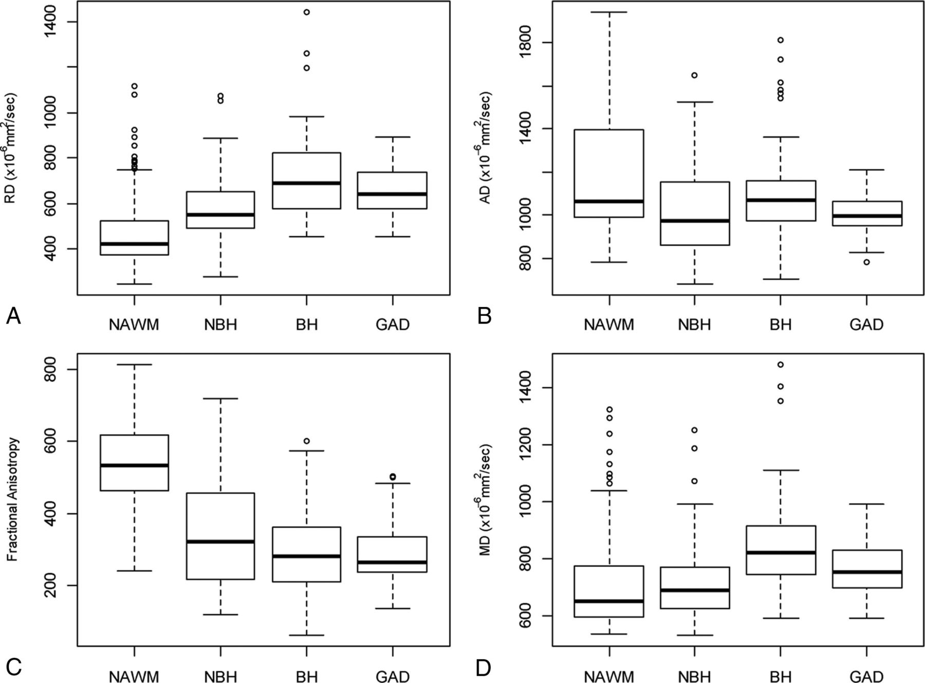
  • Fig 2.
    • Download figure
    • Open in new tab
    • Download powerpoint
    Fig 2.

    Baseline DTI characteristics of normal-appearing white matter, non-black holes, black holes, and gadolinium-enhancing lesions for radial diffusivity (A), axial diffusivity (B), fractional anisotropy (C), and mean diffusivity (D). Boxplots show median (dark line), upper and lower quartiles (box), maximum and minimum values excluding outliers (whiskers), and outliers (circles).

  • Fig 3.
    • Download figure
    • Open in new tab
    • Download powerpoint
    Fig 3.

    Longitudinal evolution of DTI metrics during 4 years for, non-black holes, black holes, and gadolinium-enhancing lesions for radial diffusivity (A), axial diffusivity (B), fractional anisotropy (C), and mean diffusivity (D). Mean values (symbol) and 95% confidence intervals (error bars) are presented. Statistical significance and effect from the mixed-model effect are presented in boxes.

Tables

  • Figures
    • View popup
    Table 1:

    Longitudinal DTI changes in NAWM and GAD

    Change/Month95% CI% Annualized ChangeP Value
    NAWM
        RD × 10−6 mm2/s0.2193−0.187 to 0.6250.57%.2898
        AD × 10−6 mm2/s−0.2109−0.614 to 0.192−0.21%.3052
        FA × 10−3−0.2572−0.551 to 0.037−0.57%.0865
        MD × 10−6 mm2/s0.03209−0.331 to 0.3960.05%.8626
    GAD
        RD × 10−6 mm2/s0.61070.039 to 1.2851.22%.0374
        AD × 10−6 mm2/s1.88311.135 to 2.6312.25%<.0001a
        FA × 10−30.50740.031 to 0.9842.10%.037
        MD × 10−6 mm2/s1.27880.695 to 1.8632.00%<.0001a
    • ↵a P < .05.

    • View popup
    Table 2:

    Longitudinal DTI changes in chronic lesional tissue

    Change/Month95% CI% Annualized ChangeP Value
    All T2 lesions
        RD × 10−6 mm2/s0.05683−0.181 to 0.2950.11%.6401
        AD × 10−6 mm2/s0.54110.251 to 0.8310.62%.0003a
        FA × 10−30.1630.001 to 0.3240.61%.0482a
        MD ×10−6 mm2/s0.26950.040 to 0.4990.41%.0211a
    BH
        RD × 10−6 mm2/s0.3392−0.028 to 0.7060.57%.0701
        AD × 10−6 mm2/s0.1967−0.200 to 0.5930.22%.331
        FA × 10−3−0.2137−0.422 to −0.005−0.91%.0444
        MD × 10−6 mm2/s0.2919−0.059 to 0.6430.42%.1026
    NBH
        RD ×10−6 mm2/s−0.4137−0.967 to 0.140−0.85%.1432
        AD × 10−6 mm2/s−0.2392−0.838 to 0.360−0.28%.434
        FA × 10−30.2907−0.025 to 0.6060.99%.0708
        MD × 10−6 mm2/s−0.355−0.174 to 0.884−0.58%.1883
    • ↵a P < .05.

    • View popup
    Table 3:

    Analysis of variance of fixed effects on longitudinal changes comparing evolution of DTI metrics from different tissue types during 4 years

    P Value
    NAWM vs GAD
        RD.11
        AD<.001a
        FA.004a
        MD<.001a
    NAWM vs T2 lesions
        RD.15
        AD.002a
        FA.09
        MD.037
    BH vs NBH
        RD.36
        AD.39
        FA.01a
        MD.07
    • ↵a P < .05.

PreviousNext
Back to top

In this issue

American Journal of Neuroradiology: 38 (1)
American Journal of Neuroradiology
Vol. 38, Issue 1
1 Jan 2017
  • Table of Contents
  • Index by author
  • Complete Issue (PDF)
Advertisement
Print
Download PDF
Email Article
Cite this article
0 Responses
Respond to this article
Share
Bookmark this article
Measuring Brain Tissue Integrity during 4 Years Using Diffusion Tensor Imaging
D. Ontaneda, K. Sakaie, J. Lin, X.-F. Wang, M.J. Lowe, M.D. Phillips, R.J. Fox
American Journal of Neuroradiology Jan 2017, 38 (1) 31-38; DOI: 10.3174/ajnr.A4946
del.icio.us logo Twitter logo Facebook logo Mendeley logo
  • Tweet Widget
Purchase

Jump to section

  • Article
    • Abstract
    • ABBREVIATIONS:
    • Materials and Methods
    • Results
    • Discussion
    • Conclusions
    • Acknowledgments
    • Footnotes
    • References
  • Figures & Data
  • Info & Metrics
  • Responses
  • References
  • PDF

Related Articles

  • No related articles found.
  • PubMed
  • Google Scholar

Cited By...

  • Lesion activity and chronic demyelination are the major determinants of brain atrophy in MS
  • Crossref
  • Google Scholar

This article has not yet been cited by articles in journals that are participating in Crossref Cited-by Linking.

More in this TOC Section

  • Diagnostic Neuroradiology of Monoclonal Antibodies
  • Clinical Outcomes After Chiari I Decompression
  • Segmentation of Brain Metastases with BLAST
Show more Adult Brain

Similar Articles

  • DTI Quantifies Shunt Surgery Outcomes in iNPH
  • Tissue Damage Characterization in MS Using DWI
  • Longitudinal Structural Network Analysis in MS
  • DMV relationship with SVD and DTI measures in RSSI
  • Beyond Diffusion Tensor MRI Methods for Improved Characterization of the Brain after Ischemic Stroke: A Review
See more
Advertisement

Indexed Content

  • Current Issue
  • Accepted Manuscripts
  • Article Preview
  • Past Issues
  • Editorials
  • Editor's Choice
  • Fellows' Journal Club
  • Letters to the Editor
  • Video Articles

Cases

  • Case Collection
  • Archive - Case of the Week
  • Archive - Case of the Month
  • Archive - Classic Case

More from AJNR

  • Trainee Corner
  • Imaging Protocols
  • MRI Safety Corner
  • Book Reviews

Multimedia

  • AJNR Podcasts
  • AJNR Scantastics

Resources

  • Turnaround Time
  • Submit a Manuscript
  • Submit a Video Article
  • Submit an eLetter to the Editor/Response
  • Manuscript Submission Guidelines
  • Statistical Tips
  • Fast Publishing of Accepted Manuscripts
  • Graphical Abstract Preparation
  • Imaging Protocol Submission
  • Evidence-Based Medicine Level Guide
  • Publishing Checklists
  • Author Policies
  • Become a Reviewer/Academy of Reviewers
  • News and Updates

About Us

  • About AJNR
  • Editorial Board
  • Editorial Board Alumni
  • Alerts
  • Permissions
  • Not an AJNR Subscriber? Join Now
  • Advertise with Us
  • Librarian Resources
  • Feedback
  • Terms and Conditions
  • AJNR Editorial Board Alumni

American Society of Neuroradiology

  • Not an ASNR Member? Join Now

© 2025 by the American Society of Neuroradiology All rights, including for text and data mining, AI training, and similar technologies, are reserved.
Print ISSN: 0195-6108 Online ISSN: 1936-959X

Powered by HighWire
Email this Article

Thank you for your interest in spreading the word on American Journal of Neuroradiology.

NOTE: We only request your email address so that the person you are recommending the page to knows that you wanted them to see it, and that it is not junk mail. We do not capture any email address.

Enter multiple addresses on separate lines or separate them with commas.
Measuring Brain Tissue Integrity during 4 Years Using Diffusion Tensor Imaging
(Your Name) has sent you a message from American Journal of Neuroradiology
(Your Name) thought you would like to see the American Journal of Neuroradiology web site.
CAPTCHA
This question is for testing whether or not you are a human visitor and to prevent automated spam submissions.
Cite this article
D. Ontaneda, K. Sakaie, J. Lin, X.-F. Wang, M.J. Lowe, M.D. Phillips, R.J. Fox
Measuring Brain Tissue Integrity during 4 Years Using Diffusion Tensor Imaging
American Journal of Neuroradiology Jan 2017, 38 (1) 31-38; DOI: 10.3174/ajnr.A4946

Citation Manager Formats

  • BibTeX
  • Bookends
  • EasyBib
  • EndNote (tagged)
  • EndNote 8 (xml)
  • Medlars
  • Mendeley
  • Papers
  • RefWorks Tagged
  • Ref Manager
  • RIS
  • Zotero

We use cookies on this site to enhance your user experience. By clicking any link on this page you are giving your consent for us to set cookies.