Advertisement

Main menu

  • Home
  • Content
    • Current Issue
    • Accepted Manuscripts
    • Article Preview
    • Past Issue Archive
    • Video Articles
    • AJNR Case Collection
    • Case of the Week Archive
    • Case of the Month Archive
    • Classic Case Archive
  • Special Collections
    • AJNR Awards
    • Low-Field MRI
    • Alzheimer Disease
    • ASNR Foundation Special Collection
    • Photon-Counting CT
    • View All
  • Multimedia
    • AJNR Podcasts
    • AJNR SCANtastic
    • Trainee Corner
    • MRI Safety Corner
    • Imaging Protocols
  • For Authors
    • Submit a Manuscript
    • Submit a Video Article
    • Submit an eLetter to the Editor/Response
    • Manuscript Submission Guidelines
    • Statistical Tips
    • Fast Publishing of Accepted Manuscripts
    • Graphical Abstract Preparation
    • Imaging Protocol Submission
    • Author Policies
  • About Us
    • About AJNR
    • Editorial Board
    • Editorial Board Alumni
  • More
    • Become a Reviewer/Academy of Reviewers
    • Subscribers
    • Permissions
    • Alerts
    • Feedback
    • Advertisers
    • ASNR Home

User menu

  • Alerts
  • Log in

Search

  • Advanced search
American Journal of Neuroradiology

American Journal of Neuroradiology

ASHNR American Society of Functional Neuroradiology ASHNR American Society of Pediatric Neuroradiology ASSR
  • Alerts
  • Log in

Advanced Search

  • Home
  • Content
    • Current Issue
    • Accepted Manuscripts
    • Article Preview
    • Past Issue Archive
    • Video Articles
    • AJNR Case Collection
    • Case of the Week Archive
    • Case of the Month Archive
    • Classic Case Archive
  • Special Collections
    • AJNR Awards
    • Low-Field MRI
    • Alzheimer Disease
    • ASNR Foundation Special Collection
    • Photon-Counting CT
    • View All
  • Multimedia
    • AJNR Podcasts
    • AJNR SCANtastic
    • Trainee Corner
    • MRI Safety Corner
    • Imaging Protocols
  • For Authors
    • Submit a Manuscript
    • Submit a Video Article
    • Submit an eLetter to the Editor/Response
    • Manuscript Submission Guidelines
    • Statistical Tips
    • Fast Publishing of Accepted Manuscripts
    • Graphical Abstract Preparation
    • Imaging Protocol Submission
    • Author Policies
  • About Us
    • About AJNR
    • Editorial Board
    • Editorial Board Alumni
  • More
    • Become a Reviewer/Academy of Reviewers
    • Subscribers
    • Permissions
    • Alerts
    • Feedback
    • Advertisers
    • ASNR Home
  • Follow AJNR on Twitter
  • Visit AJNR on Facebook
  • Follow AJNR on Instagram
  • Join AJNR on LinkedIn
  • RSS Feeds

AJNR Awards, New Junior Editors, and more. Read the latest AJNR updates

Review ArticleHead and Neck Imaging
Open Access

Spectrum of Third Window Abnormalities: Semicircular Canal Dehiscence and Beyond

M.-L. Ho, G. Moonis, C.F. Halpin and H.D. Curtin
American Journal of Neuroradiology January 2017, 38 (1) 2-9; DOI: https://doi.org/10.3174/ajnr.A4922
M.-L. Ho
aFrom the Department of Radiology (M.-L.H.), Mayo Clinic, Rochester, Minnesota
  • Find this author on Google Scholar
  • Find this author on PubMed
  • Search for this author on this site
  • ORCID record for M.-L. Ho
G. Moonis
bDepartment of Radiology (G.M.), Columbia University, New York, New York
  • Find this author on Google Scholar
  • Find this author on PubMed
  • Search for this author on this site
  • ORCID record for G. Moonis
C.F. Halpin
cDepartments of Otolaryngology (C.F.H.)
  • Find this author on Google Scholar
  • Find this author on PubMed
  • Search for this author on this site
  • ORCID record for C.F. Halpin
H.D. Curtin
dRadiology (H.D.C.), Massachusetts Eye and Ear Infirmary, Boston, Massachusetts.
  • Find this author on Google Scholar
  • Find this author on PubMed
  • Search for this author on this site
  • ORCID record for H.D. Curtin
  • Article
  • Figures & Data
  • Info & Metrics
  • Responses
  • References
  • PDF
Loading

Abstract

SUMMARY: Third window abnormalities are defects in the integrity of the bony structure of the inner ear, classically producing sound-/pressure-induced vertigo (Tullio and Hennebert signs) and/or a low-frequency air-bone gap by audiometry. Specific anatomic defects include semicircular canal dehiscence, perilabyrinthine fistula, enlarged vestibular aqueduct, dehiscence of the scala vestibuli side of the cochlea, X-linked stapes gusher, and bone dyscrasias. We discuss these various entities and provide key examples from our institutional teaching file with a discussion of symptomatology, temporal bone CT, audiometry, and vestibular-evoked myogenic potentials.

ABBREVIATIONS:

EVAS
enlarged vestibular aqueduct syndrome
SSCCD
superior semicircular canal dehiscence

Third window abnormalities are defects in the integrity of the bony structure of the inner ear, first described by Minor et al in 1998.1 In 2008, Merchant and Rosowski2 proposed a universal theory for the underlying mechanism of hearing loss accompanying these defects. Normal sound conduction is transmitted through the oval and round windows, which serve as fluid interfaces between air in the middle ear and perilymphatic fluid spaces of the inner ear. Various conditions can enlarge existing bony channels or create additional defects in the bony labyrinth, producing hydrodynamic third windows. Potential third windows include bony dehiscence of the semicircular canals, enlargement of the opening of the vestibular aqueduct, dehiscence of the scala vestibuli side of the cochlea, and abnormal bony thinning between the cochlea and vascular channels. At audiometry, there is a characteristic low-frequency air-bone gap due to decreased air conduction and increased bone conduction.2

Figure 1A illustrates the mechanism of air-conducted sound in normal ears, and Fig 1B demonstrates how third window shunting effects decrease air conduction. The 2 physiologic windows between the middle and inner ear are the oval window, which transmits vibrations from the auditory ossicles, and the round window of the cochlea. With air conduction, there is physiologic entrainment of the oval and round windows due to coupling by incompressible perilymph. Pressure differences between the cochlear perilymphatic spaces activate hair cells and create the perception of sound. In the presence of a third window, incoming acoustic energy from the oval window is shunted away, decreasing transmission to the round window. This result reduces sound perception because less acoustic energy is available to the hair cells.

Fig 1.
  • Download figure
  • Open in new tab
  • Download powerpoint
Fig 1.

Mechanisms of air- and bone-conducted sound in healthy and third window anatomy. A, Normal air conduction. Vibrations of the tympanic membrane are transmitted inward through the auditory ossicles and oval window. Energy is then conducted through the incompressible perilymph, producing equal and outward motion of the round window. The difference in vibration between the oval and round windows generates a pressure gradient across the basilar membrane, activating hair cells and creating the perception of sound. B, Decreased air conduction in third window anatomy. Due to shunting across third windows (semicircular canal dehiscence [SCCD], EVAS, cochlear dehiscence), there is decreased energy transmission from the oval window to the round window. The decrease in pressure gradient across the basilar membrane yields reduced sound perception. C, Normal bone conduction. Vibrations are transmitted throughout the otic capsule. This transmission results in differential outward motion of the oval and round windows due to unequal impedance of these 2 structures. The resulting pressure difference across the basilar membrane enables sound perception. D, Increased bone conduction in third window anatomy. Due to shunting across third windows, there is decreased motion of the oval window on the scala vestibuli side of the cochlea. However, the motion of the round window on the scala tympani side is unchanged. This phenomenon artifactually elevates the pressure difference across the basilar membrane, resulting in increased sound perception. TM indicates tympanic membrane; yellow, auditory ossicles; beige, otic capsule; red, oval window; green, round window; blue, perilymph; purple, basilar membrane. Adapted with permission from Merchant SN, Rosowski JJ. Conductive hearing loss caused by third window lesions of the inner ear. Otol Neurotol 2008;29:282–89.

In contrast, Fig 1C illustrates the mechanism of bone-conducted sound in normal ears, and Fig 1D demonstrates how third window shunting effects paradoxically increase bone conduction. With bone conduction, vibrations throughout the otic capsule produce differential outward motion of the oval and round windows, due to unequal impedance of these 2 structures. The pressure difference across the basilar membrane creates the perception of sound. The presence of a third window involving the semicircular canals, vestibular aqueduct, or scala vestibuli side of the cochlea lowers the apparent impedance. This feature has the opposite effect of increasing sound perception in proportion to the differential acoustic energy across the basilar membrane.2

In this article, we discuss the spectrum of third window abnormalities, including superior semicircular canal dehiscence (SSCCD), posterior semicircular canal dehiscence, perilabyrinthine fistula, enlarged vestibular aqueduct, X-linked stapes gusher, and bone dyscrasias. We review the literature for each disease entity and provide key examples from our institutional teaching file with discussion of symptomatology, temporal bone CT, audiometry, and vestibular-evoked myogenic potentials.

Superior Semicircular Canal Dehiscence

Superior semicircular canal dehiscence refers to focal loss of the bony wall of the superior semicircular canal. The prevalence is reported as 2.1%–10.7% on temporal bone CT and 0.5%–0.6% at postmortem studies. SSCCD is idiopathic, though the proposed risk factors include congenital underdevelopment of bone overlying the semicircular canal, shear stress from trauma, increased pressure due to Valsalva maneuvers, and gradual erosion by vascular pulsations.3⇓⇓⇓⇓⇓⇓–10 A special subset of SSCCD involves dehiscence of the superior semicircular canal into the superior petrosal sinus, which normally grooves the superior margin of the petrous portion of the temporal bone. Neurovascular foramina can serve as potential windows between the middle and inner ear but are normally not associated with abnormalities of sound transmission due to their small cross-sectional area. Vascular abnormalities that enlarge the foramina and/or communicate with the bony labyrinth can produce clinically significant acoustic dissipation. It is theorized that acoustic decompression through the dura mater acts as a third window equivalent, permitting shunting of acoustic energy into the subarachnoid space or into the vessel itself.2,11,12

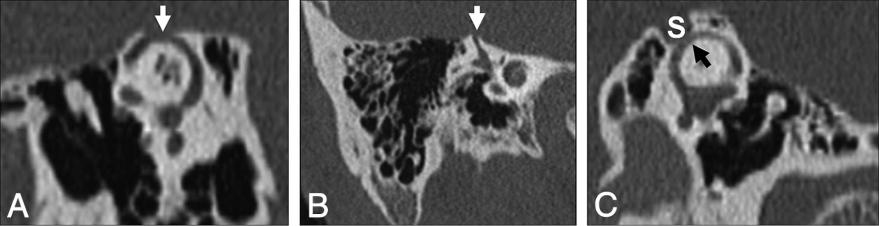
Patients with SSCCD typically present with vertigo and nystagmus induced by loud noises (Tullio phenomenon) or increases in external auditory canal pressure (Hennebert sign).5 High-resolution temporal bone CT images (maximum thickness, 0.5–0.625 mm) should be evaluated to avoid volume averaging, which can obscure a focal osseous defect. Multiplanar reconstructions and reformats parallel to (Pöschl plane) and perpendicular to (Stenvers plane) the superior semicircular canal are used to demonstrate the dehiscence to best advantage (Fig 2).3,8,9

Fig 2.
  • Download figure
  • Open in new tab
  • Download powerpoint
Fig 2.

Temporal bone CT in the Pöschl (A) and Stenvers (B) planes demonstrating a large defect (arrows) in the roof of the right superior semicircular canal. C, Temporal bone CT Pöschl reconstruction demonstrates dehiscence of the left superior semicircular canal (arrow) into the superior petrosal sinus (S).

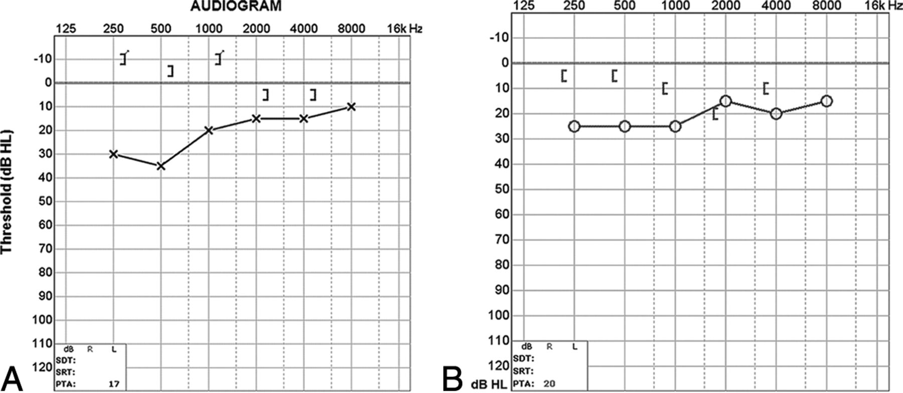
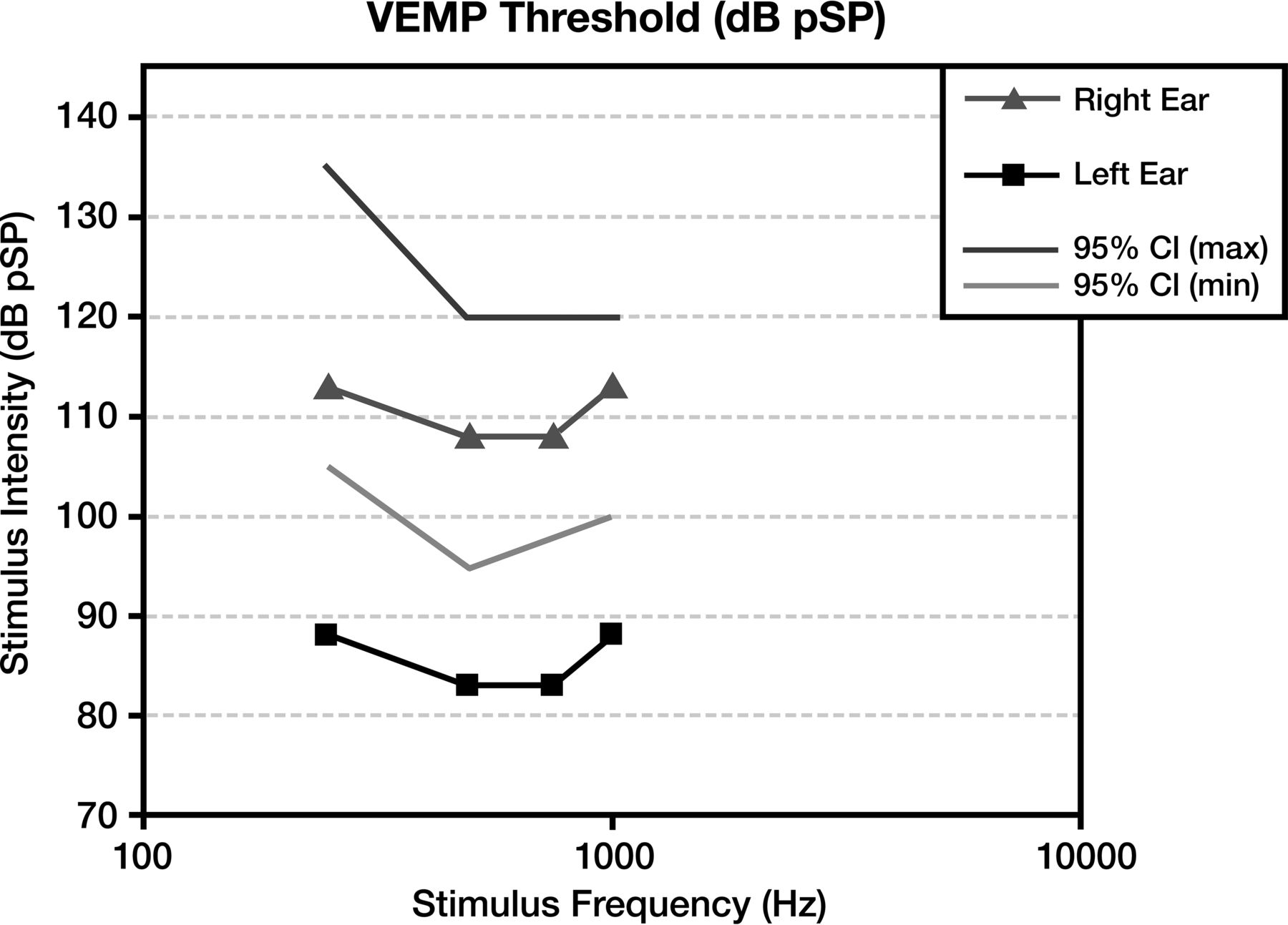
At audiometry, a characteristic air-bone gap results from increased bone and decreased air conduction. This phenomenon occurs most significantly at lower sound frequencies (below 1 kHz), a range at which acoustic energy is readily dissipated (Fig 3). At higher frequencies, there is a small or no gap because proportionally less acoustic energy is shunted by the third window. Vestibular-evoked myogenic potential testing may show abnormally low response thresholds on the side of pathology. The effective impedance is reduced; this reduction results in increased transmission of acoustic energy at the saccule (Fig 4). Because middle ear pathology can also produce an air-bone gap, tympanometry and acoustic reflexes may be tested to verify that the air-bone gap does not result from an inefficient middle ear.3,10

Fig 3.
  • Download figure
  • Open in new tab
  • Download powerpoint
Fig 3.

A, Audiogram of the right superior semicircular canal dehiscence with increased bone conduction (brackets) and decreased air conduction (crosses). The air-bone gap exceeds 10 dB at low sound frequencies (<1 kHz), the range at which acoustic energy is readily dissipated (described in the text). B, Audiogram of left superior semicircular canal dehiscence into the superior petrosal sinus. There is increased bone conduction (brackets) and decreased air conduction (circles), similar to typical SSCCD.

Fig 4.
  • Download figure
  • Open in new tab
  • Download powerpoint
Fig 4.

Vestibular-evoked myogenic potentials in superior semicircular canal dehiscence. Compared with the normal right ear (triangle) range (lines), the left ear demonstrates an abnormally sensitive response threshold (squares) across all tested stimulus frequencies. The lower thresholds indicate increased excitation of otolithic organs due to third window acoustic transmission.

At Massachusetts Eye and Ear Infirmary, 116 patients (161 ears) with SSCCD were included in the teaching file from 2000 to 2011. Of these, 5/161 ears demonstrated dehiscence into the superior petrosal sinus. Reported symptoms included hearing loss in 60/116 patients (52%), vertigo in 54/116 (46%), pulsatile tinnitus in 35/116 (30%), autophony in 31/116 (27%), and oscillopsia in 16/116 (14%). Audiometry was evaluable in 92 ears, with 71/92 ears (77%) supporting the diagnosis of semicircular canal dehiscence and 21/92 (23%) being normal. Vestibular-evoked myogenic potential testing was evaluable in 39 ears, with 22/39 ears (56%) supporting the diagnosis of SSCCD and 17/39 (44%) being normal.

Posterior Semicircular Canal Dehiscence

Posterior semicircular canal dehiscence is uncommon, with a reported frequency of 0.3%–4.5% on temporal bone CT and a postmortem prevalence of 0.2%. The condition can occur sporadically or in association with superior canal dehiscence. Clinically, patients may also demonstrate the Tullio and Hennebert signs. Thin-collimation CT demonstrates the focal bony defect in the posterior semicircular canal (Fig. 5). Audiometry reveals an air-bone gap at frequencies below 1 kHz (Fig 6).3,13⇓–15

Fig 5.
  • Download figure
  • Open in new tab
  • Download powerpoint
Fig 5.

Posterior semicircular canal dehiscence. Temporal bone CT in the coronal plane demonstrates a defect (arrow) in the roof of the posterior semicircular canal.

Fig 6.
  • Download figure
  • Open in new tab
  • Download powerpoint
Fig 6.

Audiogram of posterior semicircular canal dehiscence with increased bone conduction (brackets) and decreased air conduction (circles). Findings are compatible with posterior semicircular canal dehiscence (described in the text).

At Massachusetts Eye and Ear Infirmary, 5 patients (8 ears) with posterior semicircular canal dehiscence were included in the teaching file from 2000 to 2011. Reported symptoms included hearing loss in 3/5 (60%), pulsatile tinnitus in 4/5 (80%), autophony in 2/5 (40%), classic vertigo in 1/5 (20%), and nonclassic vertigo in 1/5 (20%). Audiometry was evaluable in 4 ears, with 3/4 ears (75%) supporting the diagnosis of third window abnormalities and 1/4 (25%) being normal.

Perilabyrinthine Fistula

Destructive middle ear processes that erode the attenuated otic capsule can produce inadvertent communication with the inner ear, known as a perilabyrinthine fistula. When this involves the semicircular canals, vestibule, and/or scala vestibuli side of the cochlea, third window mechanics can result. The most common etiology is chronic infection/inflammation, such as cholesteatoma or otitis media. The lateral semicircular canal is most frequently involved due to its location directly adjacent to the middle ear (Fig 7). On audiometry, cholesteatoma demonstrates a characteristic air-bone gap of middle ear origin, which is present at both low and high sound frequencies due to superimposed ossicular chain pathology (Fig 8).3,16,17 Rarely, cochlear-carotid dehiscence with the absence of the intervening bony partition can also occur (Fig 9). On audiometry, this condition demonstrates an air-bone gap that is greater at lower frequencies, similar to other third windows (Fig 10).18⇓–20

Fig 7.
  • Download figure
  • Open in new tab
  • Download powerpoint
Fig 7.

Inflammatory causes of perilabyrinthine fistula. A, Cholesteatoma. Axial CT shows an expansile, widely destructive soft-tissue mass centered in the middle ear with erosion into the cochlea, vestibule, and posterior semicircular canal (arrows). B, Recurrent otitis media. Axial CT identifies residual soft-tissue opacity in the middle ear, with a fistula (arrow) extending through the otic capsule into the lateral semicircular canal. C, Invasive fungal infection. Axial CT illustrates heterogeneous soft tissue centered in the middle ear, with invasion through the otic capsule into the lateral semicircular canal (arrow).

Fig 8.
  • Download figure
  • Open in new tab
  • Download powerpoint
Fig 8.

Audiogram of a cholesteatoma shows an air-bone gap of middle ear origin. There is decreased air conduction (crosses) relative to normal bone conduction (brackets). The resulting air-bone gap is present at both high and low sound frequencies. Given the background abnormality, it is not possible to detect the presence of superimposed third-window effects.

Fig 9.
  • Download figure
  • Open in new tab
  • Download powerpoint
Fig 9.

Carotid-cochlear dehiscence. Axial CT (A), Pöschl reformat (B), and Stenvers (A) reformat demonstrate communication between the apical turn of the cochlea and the carotid canal (arrows).

Fig 10.
  • Download figure
  • Open in new tab
  • Download powerpoint
Fig 10.

Audiogram of carotid-cochlear dehiscence. There is increased bone conduction (brackets) and decreased air conduction (circles), with a progressively larger air-bone gap at lower frequencies. In this case, the ipsilateral acoustic reflex was present, indicating that the ossicular chain was free to move with the action of the stapedius muscle. Therefore no middle ear pathology such as serous otitis, otosclerosis, or cholesteatoma was responsible for the air-bone gap.

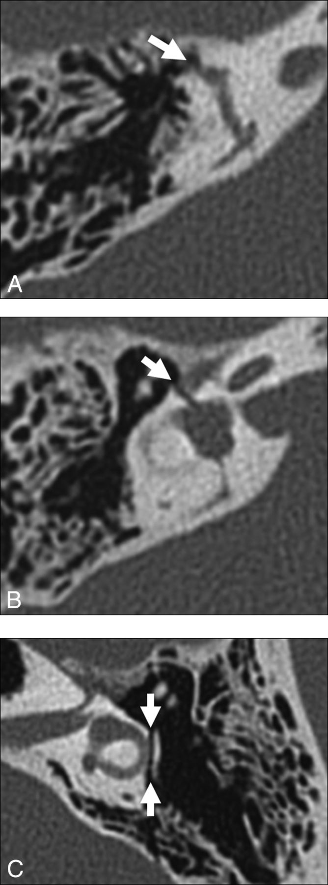
Other potential causes of perilabyrinthine fistula include trauma (Fig 11), an operation, and benign and neoplastic masses (Fig 12). Transverse temporal bone fractures are more likely to involve the petrous pyramid and violate the otic capsule. With this background abnormality, it is difficult or impossible to identify superimposed third window effects. In such cases, hearing is generally unrecoverable and audiometry is not performed, so a typical third window effect cannot be demonstrated.21,22

Fig 11.
  • Download figure
  • Open in new tab
  • Download powerpoint
Fig 11.

Traumatic causes of perilabyrinthine fistula. A, Transverse temporal bone fracture through the superior semicircular canal (arrow). B, Transverse temporal bone fracture across the bony vestibule (arrow). C, Transverse temporal bone fracture through the otic capsule (arrows) with unroofing of the lateral semicircular canal.

Fig 12.
  • Download figure
  • Open in new tab
  • Download powerpoint
Fig 12.

Neoplastic causes of perilabyrinthine fistula. A, Squamous cell carcinoma. Axial CT scan shows destructive soft tissue filling the middle ear and eroding through the otic capsule (arrow). B, Facial nerve venous malformation. Axial CT scan shows a lobulated soft-tissue density extending from the geniculate ganglion (white arrow) through the otic capsule and into the superior semicircular canal (black arrow).

At Massachusetts Eye and Ear Infirmary, 43 patients (60 ears) with perilabyrinthine fistula of the semicircular canals, vestibule, and/or scala vestibuli side of the cochlea were included in the teaching file from 2000 to 2011. Etiologies were inflammatory in 45/60 (75%), traumatic/iatrogenic in 9/60 (15%), and neoplastic in 6/60 (10%). Anatomic sites of involvement included the lateral semicircular canal in 31/60 (52%), the superior semicircular canal in 12/60 (20%), the posterior semicircular canal in 4/60 (7%), the cochlea in 13/60 (22%), and the vestibule in 11/60 (18%). Reported symptoms included hearing loss in 21/43 (49%), vertigo in 13/43 (31%), otorrhea in 13/43 (30%), and otalgia in 10/43 (23%).

Enlarged Vestibular Aqueduct

Enlarged vestibular aqueduct syndrome (EVAS) is a pathologic enlargement of the vestibular aqueduct at the level of the endolymphatic duct. In EVAS, the connection of the vestibular aqueduct to the vestibule is larger than normal, and this connection acts as a third window transmitting acoustic energy through the aqueduct to the dura. CT criteria are classically based on the transverse dimension of the vestibular aqueduct (Valvassori criterion: midpoint of ≥1.5 mm; Cincinnati criteria: midpoint of ≥1 mm or operculum of ≥2 mm), though the adjacent posterior semicircular canal often serves as a standard reference.23⇓⇓⇓–27 Normative values by using the 45° oblique (Pöschl) projection have recently been established as 0.3–0.9 mm (mean, 0.5 mm) at the midpoint.28 The finding is frequently bilateral and can be seen either in isolation or with various congenital conditions, including Pendred syndrome; coloboma, heart defect, atresia choanae (also known as choanal atresia), retarded growth and development, genital abnormality, and ear abnormality (CHARGE syndrome); and branchio-oto-renal syndrome. It has been reported that dehiscence of the vestibular aqueduct into the jugular bulb also exhibits third window mechanics (Fig 13).29⇓⇓⇓–33

Fig 13.
  • Download figure
  • Open in new tab
  • Download powerpoint
Fig 13.

A, Enlarged vestibular aqueduct syndrome. Axial CT scan shows a dilated vestibular aqueduct (arrow) at its junction with the vestibule. B, Axial CT scan shows dehiscence of the vestibular aqueduct (arrow) into the jugular foramen (J).

At audiometry, patients present with a complex and variable pattern of hearing loss. The sensorineural component of hearing loss is thought to result from potential associated cochleovestibular malformations and manifests at higher sound frequencies. The conductive component of hearing loss results from acoustic energy dissipation through an enlarged third window where the vestibular aqueduct joins the vestibule. This is evidenced by an audiometric air-bone gap at low frequencies but may be missed if bone conduction is not measured, particularly in young children who cannot tolerate a full audiologic examination.2,34⇓⇓–37 Vestibular-evoked myogenic potential studies may demonstrate increased vestibular organ responses on the side of pathology.38,39

At Massachusetts Eye and Ear Infirmary, 98 patients (165 ears) with EVAS were included in the teaching file from 2000 to 2011. Sixty of 165 ears (36%) demonstrated associated cochleovestibular malformations. Reported symptoms at the time of imaging included hearing loss in 67/98 patients (68%) and vertigo in 6/98 (6%). Audiogram findings for EVAS varied widely, demonstrating mixed sensorineural and conductive components. Six patients (7 ears) demonstrated vestibular aqueduct dehiscence into the jugular bulb. Audiometry was evaluable in 7 ears, with 1/7 (14%) showing the classic air-bone gap of the third window disorder.

X-Linked Stapes Gusher

X-linked stapes gusher is a congenital disorder resulting from a loss-of-function mutation in the POU3F4 gene at the DFN3 locus of the X chromosome. Patients are almost exclusively male and present with mixed hearing loss at birth, rapidly progressing to severe deafness in the first decade.40,41 Female carriers of the gene may be healthy or may have less severe hearing loss.42,43 The spectrum of abnormalities on CT includes cochlear hypoplasia with modiolar deficiency, absence of the lamina cribrosa, an enlarged internal auditory canal, and an enlarged labyrinthine facial nerve canal (Fig 14).44⇓⇓–47

Fig 14.
  • Download figure
  • Open in new tab
  • Download powerpoint
Fig 14.

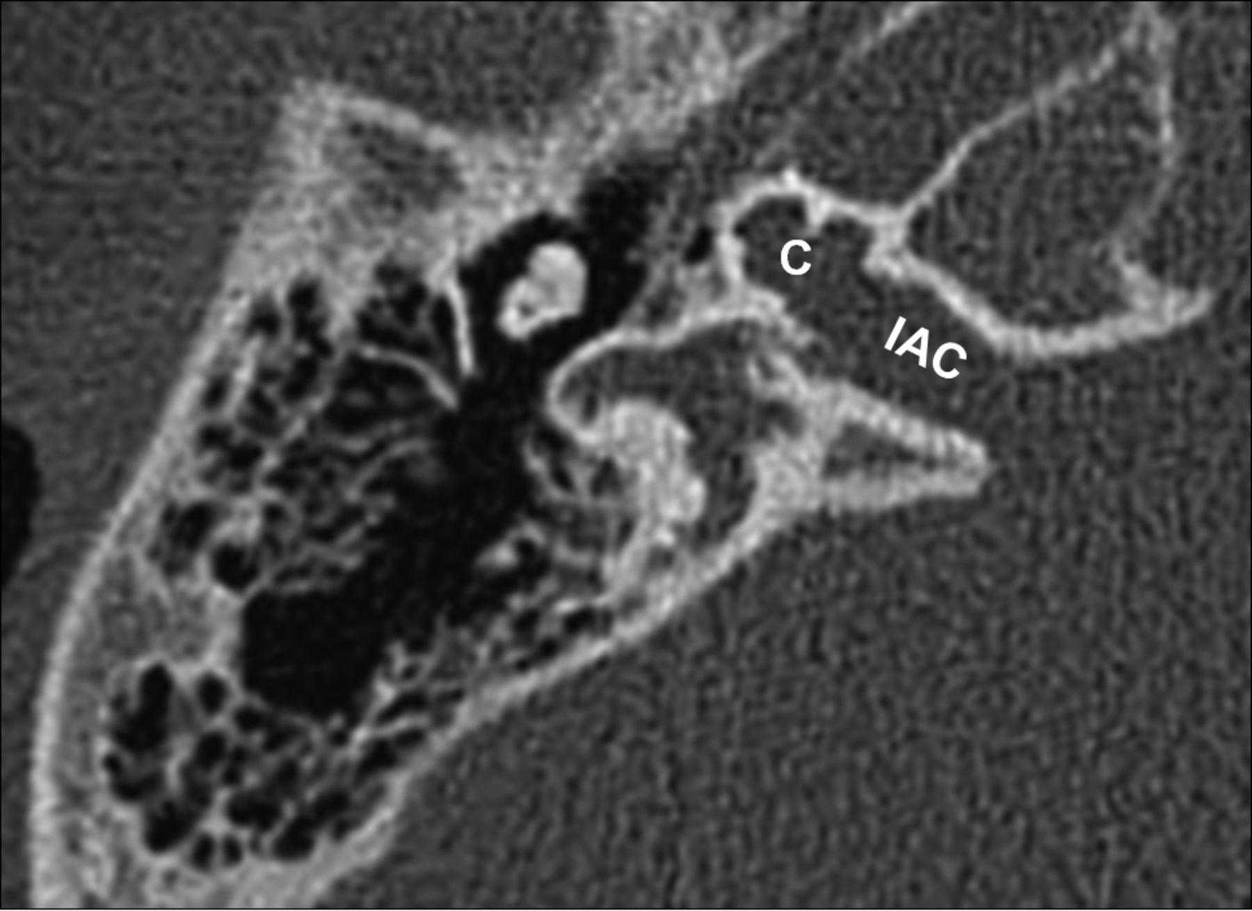
X-linked stapes gusher. Axial CT scan shows a dysplastic cochlea (C) with a modiolar deficiency and absence of the lamina cribrosa, resulting in a direct communication with an enlarged internal auditory canal (IAC).

On audiometry, patients with X-linked stapes gusher typically demonstrate a mixed hearing loss. The sensorineural component of hearing loss results from the cochleovestibular malformation. The conductive component results from absence of the lamina cribrosa, which normally separates the basal turn of the cochlea from the internal auditory canal. Therefore, there is direct communication of CSF between the subarachnoid and perilymphatic spaces, acting as a third window.2,48

At Massachusetts Eye and Ear Infirmary, 9 patients (18 ears) with X-linked stapes gusher were included in the teaching file from 2000 to 2011. On CT, all patients demonstrated bilateral temporal bone abnormalities. One hundred percent of patients had hearing loss, with mixed sensorineural and conductive components on audiometry.

Bone Dyscrasias

Bone dyscrasias such as Paget disease, osteogenesis imperfecta, and otospongiosis are rarely reported in conjunction with third window mechanics. In the adult, the bony labyrinth is composed of mature avascular enchondral bone, which undergoes virtually no remodeling after development. Metabolic bone diseases decrease the acoustic impedance both into and out of the bone, presumably acting as an anatomically diffuse or “distributed” third window (Figs 15 and 16). Postmortem micro-CT and temporal bone histology have demonstrated communications between the middle and inner ear that are too small to visualize on conventional CT. Air-bone gaps on audiometry have been reported in the literature but may be obscured by large conductive losses.2,49⇓⇓⇓⇓–54

Fig 15.
  • Download figure
  • Open in new tab
  • Download powerpoint
Fig 15.

Otospongiosis. A, Fenestral otospongiosis. Axial CT scan shows lucency at the fissula ante fenestram (arrow). B, Fenestral and retrofenestral otospongiosis. Axial CT scan shows lucency at the fissula ante fenestram (white arrow) and surrounding the cochlea (black arrows). C, Advanced otospongiosis. Axial CT scan late in the course of disease demonstrates a complete ring of pericochlear lucency.

Fig 16.
  • Download figure
  • Open in new tab
  • Download powerpoint
Fig 16.

Osteogenesis imperfecta. Axial CT scan shows otic capsule demineralization with multiple vague lucencies, including the fissula ante fenestram, pericochlear region, and area adjacent to the internal auditory canal. Radiologically, this is indistinguishable from otospongiosis.

Conclusions

Third window lesions classically present with auditory (hearing loss) and vestibular (Tullio and Hennebert syndromes) symptoms. On temporal bone CT, specific anatomic defects include superior semicircular canal dehiscence, posterior semicircular canal dehiscence, perilabyrinthine fistula, enlarged vestibular aqueduct, X-linked stapes gusher, and bone dyscrasias. Awareness of the various etiologies and correlation with an audiologic examination will help in diagnosing this multifaceted disorder.

Acknowledgment

We thank Garyfallia Pagonis for her expert assistance with graphics.

Footnotes

  • Disclosures: Gul Moonis—UNRELATED: Other: a blind reader for RadMD. Chris F. Halpin—UNRELATED: Payment for Lectures (including service on Speakers Bureaus): Auris Medical, Comments: travel to a conference in Dallas in 2015 on sudden sensory loss. Hugh D. Curtin—UNRELATED: Payment for Lectures (including service on Speakers Bureaus): International Institute for Continuing Medical Education, Comments: lectures in Continuing Medical Education courses; Royalties: Elsevier (book royalties).

  • Paper previously presented at: Morrison Research Day, May 23, 2011, Beth Israel Deaconess Radiology, Boston, Massachusetts; Resident Research Day, June 10, 2011, Beth Israel Deaconess Medical Center, Boston, Massachusetts; Annual Meeting of the American Society of Neuroradiology and the Foundation of the ASNR Symposium, June 4–9, 2011; Seattle, Washington; and Annual Meeting of the Eastern Neuroradiological Society, September 15–18, 2011, Chatham, Massachusetts.

Indicates open access to non-subscribers at www.ajnr.org

References

  1. 1.↵
    1. Minor LB,
    2. Solomon D,
    3. Zinreich JS, et al
    . Sound- and/or pressure-induced vertigo due to bone dehiscence of the superior semicircular canal. Arch Otolaryngol Head Neck Surg 1998;124:249–58 doi:10.1001/archotol.124.3.249 pmid:9525507
    CrossRefPubMed
  2. 2.↵
    1. Merchant SN,
    2. Rosowski JJ
    . Conductive hearing loss caused by third-window lesions of the inner ear. Otol Neurotol 2008;29:282–89 doi:10.1097/MAO.0b013e318161ab24 pmid:18223508
    CrossRefPubMed
  3. 3.↵
    1. Chien WW,
    2. Carey JP,
    3. Minor LB
    . Canal dehiscence. Curr Opin Neurology 2011;24:25–31 doi:10.1097/WCO.0b013e328341ef88 pmid:21124219
    CrossRefPubMed
  4. 4.↵
    1. Stimmer H,
    2. Hamann KF,
    3. Zeiter S, et al
    . Semicircular canal dehiscence in HR multislice computed tomography: distribution, frequency, and clinical relevance. Eur Arch Otorhinolaryngol 2012;269:475–80 doi:10.1007/s00405-011-1688-6 pmid:21739095
    CrossRefPubMed
  5. 5.↵
    1. Crovetto M,
    2. Whyte J,
    3. Rodriguez OM, et al
    . Anatomo-radiological study of the superior semicircular canal dehiscence: radiological considerations of superior and posterior semicircular canals. Eur J Radiol 2010;76:167–72 doi:10.1016/j.ejrad.2009.05.038 pmid:19540691
    CrossRefPubMed
  6. 6.↵
    1. Chen EY,
    2. Paladin A,
    3. Phillips G, et al
    . Semicircular canal dehiscence in the pediatric population. Int J Pediatr Otorhinolaryngol 2009;73:321–27 doi:10.1016/j.ijporl.2008.10.027 pmid:19091428
    CrossRefPubMed
  7. 7.↵
    1. Loke SC,
    2. Goh JP
    . Incidence of semicircular canal dehiscence in Singapore. Br J Radiol 2009;82:371–73 doi:10.1259/bjr/32471003 pmid:19098078
    Abstract/FREE Full Text
  8. 8.↵
    1. Cloutier JF,
    2. Bélair M,
    3. Saliba I
    . Superior semicircular canal dehiscence: positive predictive value of high-resolution CT scanning. Eur Arch Otorhinolaryngol 2008;265:1455–60 doi:10.1007/s00405-008-0672-2 pmid:18415114
    CrossRefPubMed
  9. 9.↵
    1. Sequeira SM,
    2. Whiting BR,
    3. Shimony JS, et al
    . Accuracy of computed tomography detection of superior canal dehiscence. Otol Neurotol 2011;32:1500–05 doi:10.1097/MAO.0b013e318238280c pmid:22072261
    CrossRefPubMed
  10. 10.↵
    1. Yuen HW,
    2. Boeddinghaus R,
    3. Eikelboom RH, et al
    . The relationship between the air-bone gap and the size of superior semicircular canal dehiscence. Otolaryngol Head Neck Surg 2009;141:689–94 doi:10.1016/j.otohns.2009.08.029 pmid:19932839
    Abstract/FREE Full Text
  11. 11.↵
    1. McCall AA,
    2. McKenna MJ,
    3. Merchant SN, et al
    . Superior canal dehiscence syndrome associated with the superior petrosal sinus in pediatric and adult patients. Otol Neurotol 2011;32:1312–19 doi:10.1097/MAO.0b013e31822e5b0a pmid:21918420
    CrossRefPubMed
  12. 12.↵
    1. Koo JW,
    2. Hong SK,
    3. Kim DK, et al
    . Superior semicircular canal dehiscence syndrome by the superior petrosal sinus. J Neurol Neurosurg Psychiatry 2010;81:465–67 doi:10.1136/jnnp.2008.155564 pmid:20176603
    Abstract/FREE Full Text
  13. 13.↵
    1. Krombach GA,
    2. DiMartino E,
    3. Schmitz-Rode T, et al
    . Posterior semicircular canal dehiscence: a morphologic cause of vertigo similar to superior semicircular canal dehiscence. Eur Radiol 2003;13:1444–50 pmid:12764665
    PubMed
  14. 14.↵
    1. Nomiya S,
    2. Cureoglu S,
    3. Kariya S, et al
    . Posterior semicircular canal dehiscence: a histopathologic human temporal bone study. Otol Neurotol 2010;31:1122–27 doi:10.1097/MAO.0b013e3181eb3309 pmid:20657329
    CrossRefPubMed
  15. 15.↵
    1. Gopen Q,
    2. Zhou G,
    3. Poe D, et al
    . Posterior semicircular canal dehiscence: first reported case series. Otol Neurotol 2010;31:339–44 doi:10.1097/MAO.0b013e3181be65a4 pmid:19841602
    CrossRefPubMed
  16. 16.↵
    1. Shinnabe A,
    2. Hara M,
    3. Hasegawa M, et al
    . Clinical characteristics and surgical benefits and problems of chronic otitis media and middle ear cholesteatoma in elderly patients older than 70 years. Otol Neurotol 2012;33:1213–17 doi:10.1097/MAO.0b013e31825f24ba pmid:22801042
    CrossRefPubMed
  17. 17.↵
    1. Ikeda R,
    2. Kobayashi T,
    3. Kawase T, et al
    . Risk factors for deterioration of bone conduction hearing in cases of labyrinthine fistula caused by middle ear cholesteatoma. Ann Otol Rhinol Laryngol 2012;121:162–67 doi:10.1177/000348941212100304 pmid:22530475
    Abstract/FREE Full Text
  18. 18.↵
    1. Lund AD,
    2. Palacios SD
    . Carotid artery-cochlear dehiscence: a review. Laryngoscope 2011;121:2658–60 doi:10.1002/lary.22391 pmid:22109767
    CrossRefPubMed
  19. 19.↵
    1. Young RJ,
    2. Shatzkes DR,
    3. Babb JS, et al
    . The cochlear-carotid interval: anatomic variation and potential clinical implications. AJNR Am J Neuroradiol 2006;27:1486–90 pmid:16908564
    Abstract/FREE Full Text
  20. 20.↵
    1. Penido Nde O,
    2. Borin A,
    3. Fukuda Y, et al
    . Microscopic anatomy of the carotid canal and its relations with cochlea and middle ear. Braz J Otorhinolaryngol 2005;71:410–14 doi:10.1016/S1808-8694(15)31191-5 pmid:16446952
    CrossRefPubMed
  21. 21.↵
    1. Kang HM,
    2. Kim MG,
    3. Boo SH, et al
    . Comparison of the clinical relevance of traditional and new classification systems of temporal bone fractures. Eur Arch Otorhinolaryngol 2012;269:1893–99 doi:10.1007/s00405-011-1849-7 pmid:22120750
    CrossRefPubMed
  22. 22.↵
    1. Dahiya R,
    2. Keller JD,
    3. Litofsky NS, et al
    . Temporal bone fractures: otic capsule sparing versus otic capsule violating clinical and radiographic considerations. J Trauma 1999;47:1079–83 doi:10.1097/00005373-199912000-00014 pmid:10608536
    CrossRefPubMed
  23. 23.↵
    1. Berrettini S,
    2. Forli F,
    3. Bogazzi F, et al
    . Large vestibular aqueduct syndrome: audiological, radiological, clinical, and genetic features. Am J Otolaryngol 2005;26:363–71 doi:10.1016/j.amjoto.2005.02.013 pmid:16275403
    CrossRefPubMed
  24. 24.↵
    1. Valvassori G,
    2. Clemis J
    . The large vestibular aqueduct syndrome. Laryngoscope 1978;88:273–78 pmid:306012
    PubMed
  25. 25.↵
    1. Boston M,
    2. Halsted M,
    3. Meinzen-Derr J, et al
    . The large vestibular aqueduct: a new definition based on audiologic and computed tomography correlation. Otolaryngol Head Neck Surg 2007;136:972–77 doi:10.1016/j.otohns.2006.12.011 pmid:17547990
    Abstract/FREE Full Text
  26. 26.↵
    1. Sennaroglu L,
    2. Saatci I
    . A new classification for cochleovestibular malformations. Laryngoscope 2002;112:2230–41 doi:10.1097/00005537-200212000-00019 pmid:12461346
    CrossRefPubMed
  27. 27.↵
    1. Saliba I,
    2. Gingras-Charland ME,
    3. St-Cyr K, et al
    . Coronal CT scan measurements and hearing evolution in enlarged vestibular aqueduct syndrome. Int J Pediatr Otorhinolaryngol 2012;76:492–99 doi:10.1016/j.ijporl.2012.01.004 pmid:22281371
    CrossRefPubMed
  28. 28.↵
    1. Lookabaugh S,
    2. Kelly HR,
    3. Carter MS, et al
    . Radiologic classification of superior canal dehiscence: implications for surgical repair. Otol Neurotol 2015;36:118–25 doi:10.1097/MAO.0000000000000523 pmid:25122602
    CrossRefPubMed
  29. 29.↵
    1. Hourani R,
    2. Carey J,
    3. Yousem DM
    . Dehiscence of the jugular bulb and vestibular aqueduct: findings on 200 consecutive temporal bone computed tomography scans. J Comput Assist Tomogr 2005;29:657–62 doi:10.1097/01.rct.0000175499.34213.5d pmid:16163038
    CrossRefPubMed
  30. 30.↵
    1. Friedmann DR,
    2. Eubig J,
    3. Winata LS, et al
    . Prevalence of jugular bulb abnormalities and resultant inner ear dehiscence: a histopathologic and radiologic study. Otolaryngol Head Neck Surg 2012;147:750–56 doi:10.1177/0194599812448615 pmid:22619257
    Abstract/FREE Full Text
  31. 31.↵
    1. Friedmann DR,
    2. Eubig J,
    3. Winata LS, et al
    . A clinical and histopathologic study of jugular bulb abnormalities. Arch Otolaryngol Head Neck Surg 2012;138:66–71 doi:10.1001/archoto.2011.231 pmid:22249632
    CrossRefPubMed
  32. 32.↵
    1. Kupfer RA,
    2. Hoesli RC,
    3. Green GE, et al
    . The relationship between jugular bulb-vestibular aqueduct dehiscence and hearing loss in pediatric patients. Otolaryngol Head Neck Surg 2012;146:473–77 doi:10.1177/0194599811430045 pmid:22114311
    Abstract/FREE Full Text
  33. 33.↵
    1. Friedmann DR,
    2. Le BT,
    3. Pramanik BK, et al
    . Clinical spectrum of patients with erosion of the inner ear by jugular bulb abnormalities. Laryngoscope 2010;120:365–72 doi:10.1002/lary.20699 pmid:19924772
    CrossRefPubMed
  34. 34.↵
    1. Mimura T,
    2. Sato E,
    3. Sugiura M, et al
    . Hearing loss in patients with enlarged vestibular aqueduct: air-bone gap and audiological Bing test. Int J Audiol 2005;44:466–69 doi:10.1080/14992020500057665 pmid:16149241
    CrossRefPubMed
  35. 35.↵
    1. Arjmand EM,
    2. Webber A
    . Audiometric findings in children with a large vestibular aqueduct. Arch Otolaryngol Head Neck Surg 2004;130:1169–74 doi:10.1001/archotol.130.10.1169 pmid:15492163
    CrossRefPubMed
  36. 36.↵
    1. Merchant SN,
    2. Nakajima HH,
    3. Halpin C, et al
    . Clinical investigation and mechanism of air-bone gaps in large vestibular aqueduct syndrome. Ann Otol Rhinol Laryngol 2007;116:532–41 doi:10.1177/000348940711600709 pmid:17727085
    Abstract/FREE Full Text
  37. 37.↵
    1. Zhou G,
    2. Gopen Q,
    3. Kenna MA
    . Delineating the hearing loss in children with enlarged vestibular aqueduct. Laryngoscope 2008;118:2062–66 doi:10.1097/MLG.0b013e31818208ad pmid:18665003
    CrossRefPubMed
  38. 38.↵
    1. Zhou G,
    2. Gopen Q
    . Characteristics of vestibular evoked myogenic potentials in children with enlarged vestibular aqueduct. Laryngoscope 2011;121:220–25 doi:10.1002/lary.21184 pmid:21132770
    CrossRefPubMed
  39. 39.↵
    1. Sheykholeslami K,
    2. Schmerber S,
    3. Habiby Kermany M, et al
    . Vestibular-evoked myogenic potentials in three patients with large vestibular aqueduct. Hear Res 2004;190:161–68 doi:10.1016/S0378-5955(04)00018-8 pmid:15051138
    CrossRefPubMed
  40. 40.↵
    1. Schild C,
    2. Prera E,
    3. Lüblinghoff N, et al
    . Novel mutation in the homeobox domain of transcription factor POU3F4 associated with profound sensorineural hearing loss. Otol Neurotol 2011;32:690–94 doi:10.1097/MAO.0b013e318210b749 pmid:21555964
    CrossRefPubMed
  41. 41.↵
    1. Friedman RA,
    2. Bykhovskaya Y,
    3. Tu G, et al
    . Molecular analysis of the POU3F4 gene in patients with clinical and radiographic evidence of X-linked mixed deafness with perilymphatic gusher. Ann Otol Rhinol Laryngol 1997;106:320–25 doi:10.1177/000348949710600411 pmid:9109724
    Abstract/FREE Full Text
  42. 42.↵
    1. Huang BY,
    2. Zdanski C,
    3. Castillo M
    . Pediatric sensorineural hearing loss, part 2: syndromic and acquired causes. AJNR Am J Neuroradiol 2012;33:399–406 doi:10.3174/ajnr.A2499 pmid:21596810
    Abstract/FREE Full Text
  43. 43.↵
    1. Marlin S,
    2. Moizard MP,
    3. David A, et al
    . Phenotype and genotype in females with POU3F4 mutations. Clin Genet 2009;76:558–63 doi:10.1111/j.1399-0004.2009.01215.x pmid:19930154
    CrossRefPubMed
  44. 44.↵
    1. Papadaki E,
    2. Prassopoulos P,
    3. Bizakis J, et al
    . X-linked deafness with stapes gusher in females. Eur J Radiol 1998;29:71–75 doi:10.1016/S0720-048X(98)00027-8 pmid:9934561
    CrossRefPubMed
  45. 45.↵
    1. Talbot JM,
    2. Wilson DF
    . Computed tomographic diagnosis of X-linked congenital mixed deafness, fixation of the stapedial footplate, and perilymphatic gusher. Am J Otol 1994;15:177–82 pmid:8172298
    PubMed
  46. 46.↵
    1. Kumar G,
    2. Castillo M,
    3. Buchman CA
    . X-linked stapes gusher: CT findings in one patient. AJNR Am J Neuroradiol 2003;24:1130–32 pmid:12812938
    Abstract/FREE Full Text
  47. 47.↵
    1. Cremers CW,
    2. Snik AF,
    3. Huygen PL, et al
    . X-linked mixed deafness syndrome with congenital fixation of the stapedial footplate and perilymphatic gusher (DFN3). Adv Otorhinolaryngol 2002;61:161–67 pmid:12408080
    PubMed
  48. 48.↵
    1. Snik AF,
    2. Hombergen GC,
    3. Mylanus EA, et al
    . Air-bone gap in patients with X-linked stapes gusher syndrome. Am J Otol 1995;16:241–46 pmid:8572127
    PubMed
  49. 49.↵
    1. Makarem AO,
    2. Linthicum FH
    . Cavitating otosclerosis. Otol Neurotol 2008;29:730–31 doi:10.1097/MAO.0b013e3181799763 pmid:18520624
    CrossRefPubMed
  50. 50.↵
    1. Makarem AO,
    2. Hoang TA,
    3. Lo WW, et al
    . Cavitating otosclerosis: clinical, radiologic, and histopathologic correlations. Otol Neurotol 2010;31:381–84 doi:10.1097/MAO.0b013e3181d275e8 pmid:20195188
    CrossRefPubMed
  51. 51.↵
    1. Richard C,
    2. Linthicum FH Jr.
    . An unexpected third window in a case of advanced cavitating otosclerosis. Otol Neurotol 2012;33:e47–48 doi:10.1097/MAO.0b013e318245cb3b pmid:22314921
    CrossRefPubMed
  52. 52.↵
    1. Merchant SN,
    2. Rosowski JJ,
    3. McKenna MJ
    . Superior semicircular canal dehiscence mimicking otosclerotic hearing loss. Adv Otorhinolaryngol 2007;65:137–45 pmid:17245035
    PubMed
  53. 53.↵
    1. Van Rompaey V,
    2. Potvin J,
    3. van den Hauwe L, et al
    . Third mobile window associated with suspected otosclerotic foci in two patients with an air-bone gap. J Laryngol Otol 2011;125:89–92 doi:10.1017/S0022215110001544 pmid:20727242
    CrossRefPubMed
  54. 54.↵
    1. Santos F,
    2. McCall AA,
    3. Chien W, et al
    . Otopathology in osteogenesis imperfecta. Otol Neurotol 2012;33:1562–66 doi:10.1097/MAO.0b013e31826bf19b pmid:22996160
    CrossRefPubMed
  • © 2017 by American Journal of Neuroradiology
View Abstract
PreviousNext
Back to top

In this issue

American Journal of Neuroradiology: 38 (1)
American Journal of Neuroradiology
Vol. 38, Issue 1
1 Jan 2017
  • Table of Contents
  • Index by author
  • Complete Issue (PDF)
Advertisement
Print
Download PDF
Email Article

Thank you for your interest in spreading the word on American Journal of Neuroradiology.

NOTE: We only request your email address so that the person you are recommending the page to knows that you wanted them to see it, and that it is not junk mail. We do not capture any email address.

Enter multiple addresses on separate lines or separate them with commas.
Spectrum of Third Window Abnormalities: Semicircular Canal Dehiscence and Beyond
(Your Name) has sent you a message from American Journal of Neuroradiology
(Your Name) thought you would like to see the American Journal of Neuroradiology web site.
CAPTCHA
This question is for testing whether or not you are a human visitor and to prevent automated spam submissions.
Cite this article
M.-L. Ho, G. Moonis, C.F. Halpin, H.D. Curtin
Spectrum of Third Window Abnormalities: Semicircular Canal Dehiscence and Beyond
American Journal of Neuroradiology Jan 2017, 38 (1) 2-9; DOI: 10.3174/ajnr.A4922

Citation Manager Formats

  • BibTeX
  • Bookends
  • EasyBib
  • EndNote (tagged)
  • EndNote 8 (xml)
  • Medlars
  • Mendeley
  • Papers
  • RefWorks Tagged
  • Ref Manager
  • RIS
  • Zotero
0 Responses
Respond to this article
Share
Bookmark this article
Spectrum of Third Window Abnormalities: Semicircular Canal Dehiscence and Beyond
M.-L. Ho, G. Moonis, C.F. Halpin, H.D. Curtin
American Journal of Neuroradiology Jan 2017, 38 (1) 2-9; DOI: 10.3174/ajnr.A4922
del.icio.us logo Twitter logo Facebook logo Mendeley logo
  • Tweet Widget
  • Facebook Like
  • Google Plus One
Purchase

Jump to section

  • Article
    • Abstract
    • ABBREVIATIONS:
    • Conclusions
    • Acknowledgment
    • Footnotes
    • References
  • Figures & Data
  • Info & Metrics
  • Responses
  • References
  • PDF

Related Articles

  • No related articles found.
  • PubMed
  • Google Scholar

Cited By...

  • Prevalence of Cochlear-Facial and Other Non-Superior Semicircular Canal Third Window Dehiscence on High-Resolution Temporal Bone CT
  • The Forgotten Second Window: A Pictorial Review of Round Window Pathologies
  • Carotid-cochlear dehiscence: a dangerous mimicker of inner ear pathologies
  • Cavitary Plaques in Otospongiosis: CT Findings and Clinical Implications
  • Crossref (69)
  • Google Scholar

This article has been cited by the following articles in journals that are participating in Crossref Cited-by Linking.

  • Third Window Syndrome: Surgical Management of Cochlea-Facial Nerve Dehiscence
    P. Ashley Wackym, Carey D. Balaban, Pengfei Zhang, David A. Siker, Jasdeep S. Hundal
    Frontiers in Neurology 2019 10
  • Vestibular Evoked Myogenic Potential (VEMP) Testing for Diagnosis of Superior Semicircular Canal Dehiscence
    Kimberley S. Noij, Steven D. Rauch
    Frontiers in Neurology 2020 11
  • Bone-conduction hyperacusis induced by superior canal dehiscence in human: the underlying mechanism
    Xiying Guan, Y. Song Cheng, Deepa J. Galaiya, John J. Rosowski, Daniel J. Lee, Hideko Heidi Nakajima
    Scientific Reports 2020 10 1
  • Cavitary Plaques in Otospongiosis: CT Findings and Clinical Implications
    P. Puac, A. Rodríguez, H.-C. Lin, V. Onofrj, F.-C. Lin, S.-C. Hung, C. Zamora, M. Castillo
    American Journal of Neuroradiology 2018 39 6
  • Third Window Lesions
    Mai-Lan Ho
    Neuroimaging Clinics of North America 2019 29 1
  • Audiometric and cVEMP Thresholds Show Little Correlation With Symptoms in Superior Semicircular Canal Dehiscence Syndrome
    Kimberley S. Noij, Kevin Wong, Maria J. Duarte, Salwa Masud, Nicholas A. Dewyer, Barbara S. Herrmann, John J. Guinan, Elliott D. Kozin, David H. Jung, Steven D. Rauch, Daniel J. Lee
    Otology & Neurotology 2018 39 9
  • Posterior Semicircular Canal Dehiscence: Case Series and Systematic Review
    Joshua A. Lee, Yuan F. Liu, Shaun A. Nguyen, Theodore R. McRackan, Ted A. Meyer, Habib G. Rizk
    Otology & Neurotology 2020 41 4
  • Proposal for a Unitary Anatomo-Clinical and Radiological Classification of Third Mobile Window Abnormalities
    Pierre Reynard, Samar Idriss, Aicha Ltaief-Boudrigua, Pierre Bertholon, Andreea Pirvan, Eric Truy, Hung Thai-Van, Eugen C. Ionescu
    Frontiers in Neurology 2022 12
  • The Forgotten Second Window: A Pictorial Review of Round Window Pathologies
    J.C. Benson, F. Diehn, T. Passe, J. Guerin, V.M. Silvera, M.L. Carlson, J. Lane
    American Journal of Neuroradiology 2020 41 2
  • Toward Optimizing cVEMP: 2,000-Hz Tone Bursts Improve the Detection of Superior Canal Dehiscence
    Kimberley S. Noij, Barbara S. Herrmann, John J. Guinan Jr., Steven D. Rauch
    Audiology and Neurotology 2018 23 6

More in this TOC Section

  • Contrast Enhanced Pituitary CISS/FIESTA
  • Temporal Evolution of Vestibular schwannoma
  • FDG Uptake and Thyroid Cancer Post-BRAF Therapy
Show more Head and Neck Imaging

Similar Articles

Advertisement

Indexed Content

  • Current Issue
  • Accepted Manuscripts
  • Article Preview
  • Past Issues
  • Editorials
  • Editor's Choice
  • Fellows' Journal Club
  • Letters to the Editor
  • Video Articles

Cases

  • Case Collection
  • Archive - Case of the Week
  • Archive - Case of the Month
  • Archive - Classic Case

More from AJNR

  • Trainee Corner
  • Imaging Protocols
  • MRI Safety Corner
  • Book Reviews

Multimedia

  • AJNR Podcasts
  • AJNR Scantastics

Resources

  • Turnaround Time
  • Submit a Manuscript
  • Submit a Video Article
  • Submit an eLetter to the Editor/Response
  • Manuscript Submission Guidelines
  • Statistical Tips
  • Fast Publishing of Accepted Manuscripts
  • Graphical Abstract Preparation
  • Imaging Protocol Submission
  • Evidence-Based Medicine Level Guide
  • Publishing Checklists
  • Author Policies
  • Become a Reviewer/Academy of Reviewers
  • News and Updates

About Us

  • About AJNR
  • Editorial Board
  • Editorial Board Alumni
  • Alerts
  • Permissions
  • Not an AJNR Subscriber? Join Now
  • Advertise with Us
  • Librarian Resources
  • Feedback
  • Terms and Conditions
  • AJNR Editorial Board Alumni

American Society of Neuroradiology

  • Not an ASNR Member? Join Now

© 2025 by the American Society of Neuroradiology All rights, including for text and data mining, AI training, and similar technologies, are reserved.
Print ISSN: 0195-6108 Online ISSN: 1936-959X

Powered by HighWire