
Article Figures & Data
Figures
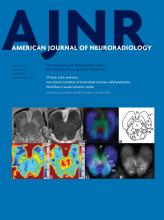
- Fig 1.
Improved workflow for the hydrocephalus imaging pathway. Imaging technologists are required to respond in a timely fashion. In case they could not respond on time, their phone numbers are outlined so that the ordering units can follow-up on their orders. HMED indicates HealthMatics Emergency Department (Allscripts, Chicago, Illinois); POE, Physician Order Entry; EPIC, Epic Systems (Madison, Wisconsin); ASCOM, tel.
- Fig 2.
Ultrafast brain MR imaging protocol: axial (A), coronal (B), and sagittal (C) T2-weighted HASTE of the brain. Note the clear visualization of the ventricular system and catheter tip.
- Fig 3.
Attribute capability analysis demonstrates that our control data are almost at the 95% confidence bounds.
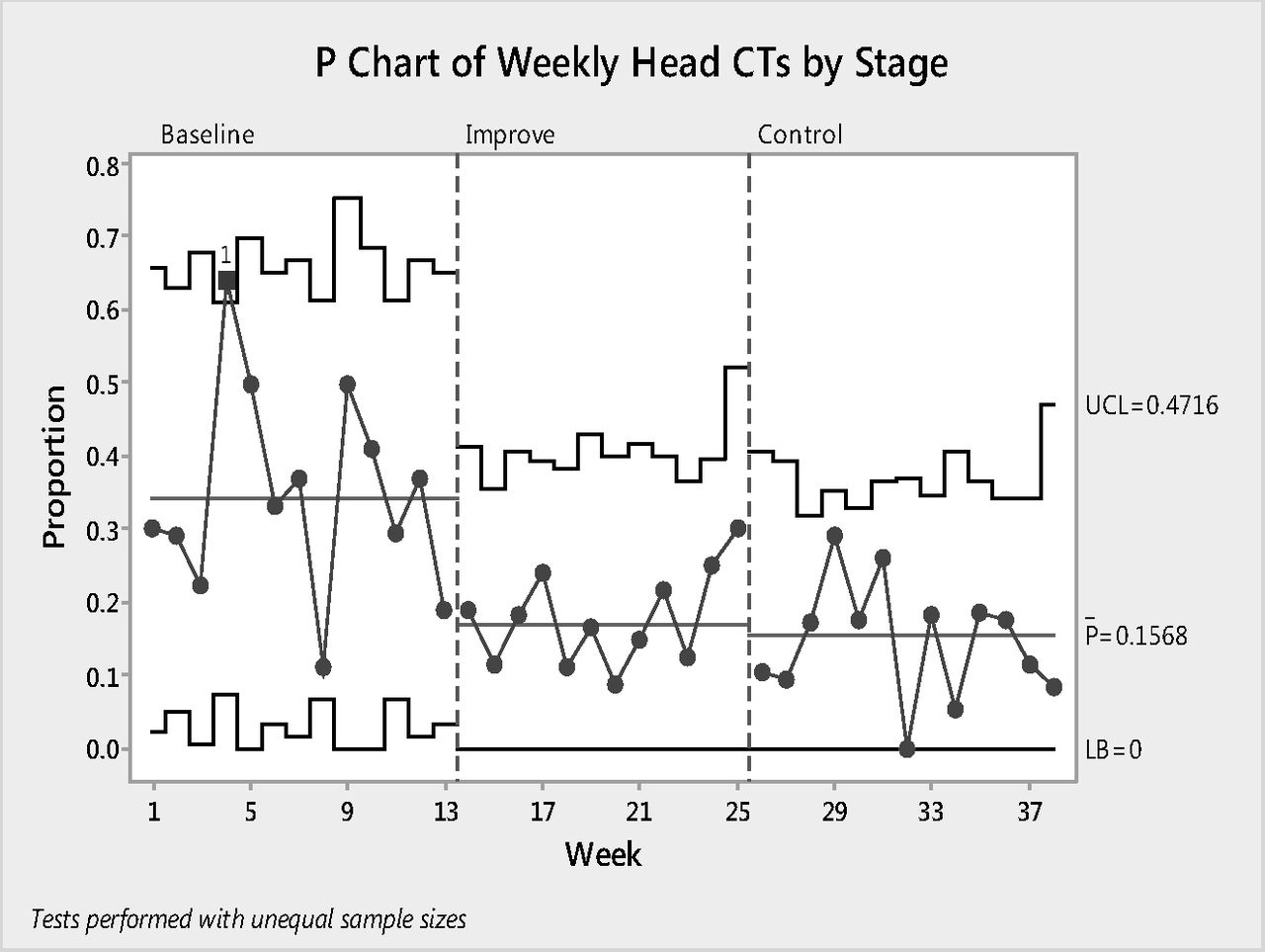
- Fig 4.
P-chart demonstrating weekly changes in head CT orders from baseline to control phases. Note the reduced fluctuations during improvement and control phases. UCL indicates upper control limit; LB, lower boundary.


Tables
Project Name: Reduce Head CT Studies in Children with Hydrocephalus Green Belt: Champion: Master Black Belt: Problem Statement: Radiation is dangerous especially in children. There is an increasing rate of head CT orders in children with hydrocephalus. Many children with hydrocephalus need repeat imaging, adding additional risk for cumulative radiation, which may lead to cancer. Project Goal: Reduce the percentage of head CT orders for hydrocephalus by 50 percent in 6 months (project start date: January 24, 2014) Project Y: Percentage of each modality (head CT, ultrafast brain MRI, head US) per ordering department, time of the day, ordering physician rank Scope: All children with known or suspected hydrocephalus, 0–18 years of age, presenting to emergency department, inpatient, and outpatient services Team Members: Project leader, pediatric radiology and pediatric neuroradiology Physician champion, pediatric radiology and pediatric neuroradiology Member, pediatric emergency department Member, pediatric neurosurgery Member, radiology administrator Member, radiology department, financial analyst Member, pediatric radiology manager Member, chief CT technologists Member, chief US technologists Member, radiology patient care coordinator Member, chief pediatric MRI technologists Benefits: 1) Eliminate radiation in evaluation of hydrocephalus 2) Reduce MRI time in the evaluation of hydrocephalus 3) Reduce cost with limited charge 4) Reduce shunt survey orders Timeline: Define/Measure: January 24–February 1, 2014 Analyze/Improve: March 10–May 30, 2014 Control: July 1–September 1, 2014 - Table 2:
Number and percentage of each modality per rank of ordering physician from baseline to control phasesa
Baseline I-Phase I I-Phase II I-Phase III C-Phase I C-Phase II C-Phase III No. % No. % No. % No. % No. % No. % No. % Attending RB-MRI 16 20.3 4 26.7 5 18.5 3 10.0 14 20.9 6 8.8 9 12.7 UF-MRI 18 22.8 4 26.7 16 59.3 10 33.3 31 46.3 34 50.0 38 53.5 Head CT 16 20.3 2 13.3 0.0 1 3.3 4 6.0 6 8.8 3 4.2 Head US 29 36.7 5 33.3 6 22.2 16 53.3 18 26.9 22 32.4 21 29.6 Attending total 79 38.5 15 24.2 27 31.4 30 32.6 67 70.5 68 58.1 71 63.4 Resident RB-MRI 22 17.5 5 10.6 5 8.5 5 8.1 4 14.3 5 10.2 7 17.1 UF-MRI 19 15.1 27 57.4 33 55.9 35 56.5 11 39.3 22 44.9 16 39.0 Head CT 46 36.5 3 6.4 6 10.2 6 9.7 6 21.4 8 16.3 6 14.6 Head US 39 31.0 12 25.5 15 25.4 16 25.8 7 25.0 14 28.6 12 29.3 Resident total 126 61.5 47 75.8 59 68.6 62 67.4 28 29.5 49 41.9 41 36.6 Grand total 205 100.0 62 100.0 86 100.0 92 100.0 95 100.0 117 100.0 112 100.0 Note:—I-Phase indicates improvement phase; C-phase, control phase; RB-MRI, routine brain MRI.
↵a Duration of baseline was 3 months, followed by 3 months of improvement (each phase for 1 month), and 3 months of control phases (each phase for 1 month).
- Table 3:
Number and percentage of each modality during different hours of the day from baseline to control phasesa
Baseline I-Phase I I-Phase II I-Phase III C-Phase I C-Phase II C-Phase III No. % No. % No. % No. % No. % No. % No. % Work hours RB-MRI 27 19.7 6 14.3 6 9.4 6 9.8 12 16.4 6 8.6 11 12.5 UF-MRI 34 24.8 21 50.0 40 62.5 26 42.6 34 46.6 38 54.3 44 50.0 Head CT 28 20.4 2 4.8 1 1.6 1 1.6 8 11.0 1 1.4 6 6.8 Head US 48 35.0 13 31.0 17 26.6 28 45.9 19 26.0 25 35.7 27 30.7 Work hour total 137 66.8 42 67.7 64 74.4 61 66.3 73 76.8 70 59.8 88 78.6 After hours RB-MRI 7 20.6 2 14.3 3 25.0 2 13.3 3 23.1 3 13.6 4 25.0 UF-MRI 2 5.9 8 57.1 5 41.7 9 60.0 6 46.2 9 40.9 8 50.0 Head CT 19 55.9 2 14.3 3 25.0 3 20.0 1 7.7 6 27.3 2 12.5 Head US 6 17.6 2 14.3 1 8.3 1 6.7 3 23.1 4 18.2 2 12.5 After hours total 34 16.6 14 22.6 12 14.0 15 16.3 13 13.7 22 18.8 16 14.3 Weekend RB-MRI 4 11.8 1 16.7 1 10.0 0.0 3 33.3 2 8.0 1 12.5 UF-MRI 1 2.9 2 33.3 4 40.0 10 62.5 2 22.2 9 36.0 2 25.0 Head CT 15 44.1 1 16.7 2 20.0 3 18.8 1 11.1 7 28.0 1 12.5 Head US 14 41.2 2 33.3 3 30.0 3 18.8 3 33.3 7 28.0 4 50.0 Weekend total 34 16.6 6 9.7 10 11.6 16 17.4 9 9.5 25 21.4 8 7.1 Grand total 205 100.0 62 100.0 86 100.0 92 100.0 95 100.0 117 100.0 112 100.0 Note:—I-Phase indicates improvement phase; C-phase, control phase; RB-MRI, routine brain MRI.
↵a Duration of baseline was 3 months, followed by 3 months of improvement (each phase for 1 month), and 3 months of control phases (each phase for 1 month).
Baseline I-Phase I I-Phase II I-Phase III C-Phase I C-Phase II C-Phase III No. % No. % No. % No. % No. % No. % No. % RB-MRI 38 18.5 9 14.5 10 11.6 8 8.7 18 18.9 11 9.4 16 14.3 UF-MRI 37 18.0 31 50.0 49 57.0 45 48.9 42 44.2 56 47.9 54 48.2 Head CT 62 30.2 5 8.1 6 7.0 7 7.6 10 10.5 14 12.0 9 8.0 Head US 68 33.2 17 27.4 21 24.4 32 34.8 25 26.3 36 30.8 33 29.5 Note:—I-Phase indicates improvement phase; C-phase, control phase; RB-MRI, routine brain MRI.
↵a Duration of baseline was 3 months, followed by 3 months of improvement (each phase for 1 month), and 3 months of control phases (each phase for 1 month).


