
Article Figures & Data
Figures
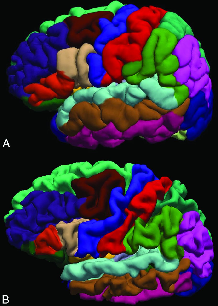
- Fig 1.
Surface representation of the FreeSurfer-based cortical parcellation for a control subject (A, 6.1 years of age) compared with that of an age-matched subject with CLN2 (B, 5.9 years of age; clinical CLN2 score = 1.5). Note the atrophy as evidenced by deeper sulci and less prominent gyri in the subject with more advanced disease.
- Fig 2.
The mean cerebral cortical gray matter thickness as a function of subject age (A) and clinical score (B) for CLN2 (solid diamond) and control (large filled square) groups. Subjects with CLN2 older than 72 months of age (open diamond) are not included in the linear fit.
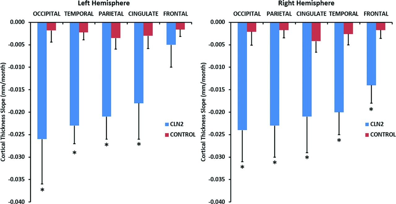
- Fig 3.
The rate of decline of cortical thickness with age for cerebral lobes in CLN2 (n = 42, blue) versus control (n = 52, red) groups. The 95% confidence interval of the slope is plotted as the error bar. Differences between CLN2 and control population of P < .05 are marked with an asterisk.
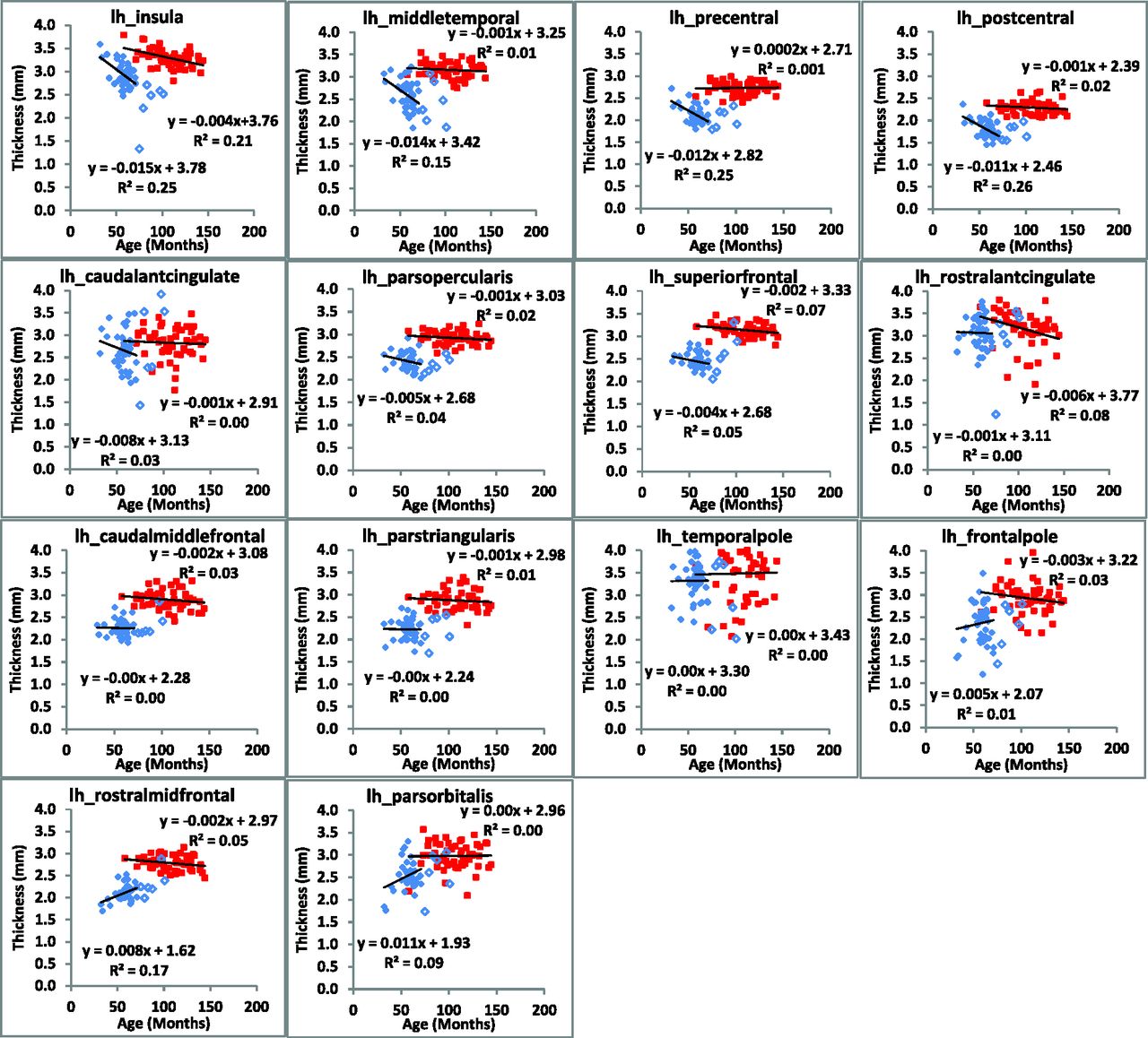
- Fig 4.
Regional changes in cortical thickness of the left hemisphere are plotted as a function of subject age for CLN2 (solid diamond) and control (large filled square) groups. Subjects with CLN2 older than 72 months of age (open diamond) are not included in the linear fit.
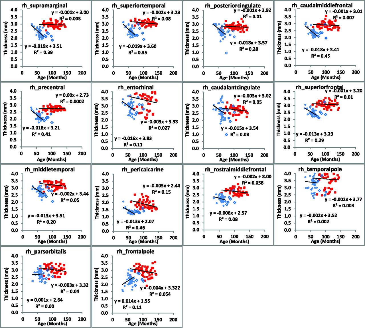
- Fig 5.
Regional changes in cortical thickness of the right hemisphere are plotted as a function of subject age for CLN2 (solid diamond) and control (large filled square) groups. Subjects with CLN2 older than 72 months of age (open diamond) are not included in the linear fit.


Tables
- Table 1:
Cortical regions grouped by lobe as defined by the Desikan-Killiany atlas in FreeSurfer
Temporal Lobe Parietal Lobe Occipital Lobe Cingulate Lobe Frontal Lobe Bankssts Inferior parietal Cuneus Caudal anterior cingulate Caudal middle frontal Entorhinal Postcentral Lateral occipital Isthmus cingulate Frontal pole Fusiform Precuneus Lingual Posterior cingulate Lateral orbitofrontal Inferior temporal Superior parietal Pericalcarine Rostral anterior cingulate Medial orbitofrontal Middle temporal Supramarginal Paracentral Parahippocampal Pars opercularis Superior temporal Pars orbitalis Temporal pole Pars triangularis Transverse temporal Precentral Rostral middle frontal Superior frontal Note:—Bankssts indicates banks of the superior temporal sulcus.
- Table 2:
The rate of decline (millimeters/month) and mean cortical thickness between CLN2 and healthy control populations in whole brain, hemispheres, and cerebral lobes
CLN2 Slope Control Slope P Value CLN2 Mean Control Mean P Value Whole brain −0.018(5) −0.002(1) <.001 2.26(23) 2.79(10) <.001 LH −0.016(5) −0.002(1) <.001 2.23(22) 2.78(10) <.001 RH −0.020(6) −0.002(1) <.001 2.29(24) 2.79(11) <.001 LH cingulate −0.018(8) −0.003(2) .002 2.69(32) 2.92(22) <.001 LH frontal −0.005(5) −0.002(2) .265 2.33(17) 2.87(12) <.001 LH occipital −0.026(10) −0.004(3) <.001 1.66(26) 2.16(19) <.001 LH parietal −0.021(5) −0.002(1) <.001 2.10(26) 2.71(13) <.001 LH temporal −0.023(4) −0.002(3) <.001 2.51(30) 2.99(19) <.001 RH cingulate −0.021(8) −0.003(3) <.001 2.69(40) 2.85(18) <.001 RH frontal −0.014(4) −0.002(2) <.001 2.41(21) 2.87(14) <.001 RH occipital −0.024(7) −0.004(2) <.001 1.68(24) 2.22(20) <.001 RH parietal −0.023(7) −0.002(2) <.001 2.16(27) 2.71(13) <.001 RH temporal −0.020(5) −0.002(3) <.001 2.61(28) 3.05(22) <.001 Note:—LH indicates left hemisphere; RH, right hemisphere.


Jump to section
Related Articles
Cited By...
- Enzyme Replacement Therapy for CLN2 Disease: MRI Volumetry Shows Significantly Slower Volume Loss Compared with a Natural History Cohort
- Slowing late infantile Batten disease by direct brain parenchymal administration of a rh.10 adeno-associated virus expressing CLN2
- Expanding the Neuroimaging Phenotype of Neuronal Ceroid Lipofuscinoses