Advertisement

Main menu

  • Home
  • Content
    • Current Issue
    • Accepted Manuscripts
    • Article Preview
    • Past Issue Archive
    • Video Articles
    • AJNR Case Collection
    • Case of the Week Archive
    • Case of the Month Archive
    • Classic Case Archive
  • Special Collections
    • AJNR Awards
    • Low-Field MRI
    • Alzheimer Disease
    • ASNR Foundation Special Collection
    • Photon-Counting CT
    • View All
  • Multimedia
    • AJNR Podcasts
    • AJNR SCANtastic
    • Trainee Corner
    • MRI Safety Corner
    • Imaging Protocols
  • For Authors
    • Submit a Manuscript
    • Submit a Video Article
    • Submit an eLetter to the Editor/Response
    • Manuscript Submission Guidelines
    • Statistical Tips
    • Fast Publishing of Accepted Manuscripts
    • Graphical Abstract Preparation
    • Imaging Protocol Submission
    • Author Policies
  • About Us
    • About AJNR
    • Editorial Board
    • Editorial Board Alumni
  • More
    • Become a Reviewer/Academy of Reviewers
    • Subscribers
    • Permissions
    • Alerts
    • Feedback
    • Advertisers
    • ASNR Home

User menu

  • Alerts
  • Log in

Search

  • Advanced search
American Journal of Neuroradiology

American Journal of Neuroradiology

ASHNR American Society of Functional Neuroradiology ASHNR American Society of Pediatric Neuroradiology ASSR
  • Alerts
  • Log in

Advanced Search

  • Home
  • Content
    • Current Issue
    • Accepted Manuscripts
    • Article Preview
    • Past Issue Archive
    • Video Articles
    • AJNR Case Collection
    • Case of the Week Archive
    • Case of the Month Archive
    • Classic Case Archive
  • Special Collections
    • AJNR Awards
    • Low-Field MRI
    • Alzheimer Disease
    • ASNR Foundation Special Collection
    • Photon-Counting CT
    • View All
  • Multimedia
    • AJNR Podcasts
    • AJNR SCANtastic
    • Trainee Corner
    • MRI Safety Corner
    • Imaging Protocols
  • For Authors
    • Submit a Manuscript
    • Submit a Video Article
    • Submit an eLetter to the Editor/Response
    • Manuscript Submission Guidelines
    • Statistical Tips
    • Fast Publishing of Accepted Manuscripts
    • Graphical Abstract Preparation
    • Imaging Protocol Submission
    • Author Policies
  • About Us
    • About AJNR
    • Editorial Board
    • Editorial Board Alumni
  • More
    • Become a Reviewer/Academy of Reviewers
    • Subscribers
    • Permissions
    • Alerts
    • Feedback
    • Advertisers
    • ASNR Home
  • Follow AJNR on Twitter
  • Visit AJNR on Facebook
  • Follow AJNR on Instagram
  • Join AJNR on LinkedIn
  • RSS Feeds

AJNR Awards, New Junior Editors, and more. Read the latest AJNR updates

Research ArticleADULT BRAIN

Imaging Findings Associated with Space-Occupying Edema in Patients with Large Middle Cerebral Artery Infarcts

A.D. Horsch, J.W. Dankbaar, T.A. Stemerdink, E. Bennink, T. van Seeters, L.J. Kappelle, J. Hofmeijer, H.W. de Jong, Y. van der Graaf and B.K. Velthuis on behalf of the DUST investigators
American Journal of Neuroradiology May 2016, 37 (5) 831-837; DOI: https://doi.org/10.3174/ajnr.A4637
A.D. Horsch
aFrom the Departments of Radiology (A.D.H., J.W.D., T.A.S., E.B., T.v.S., H.W.d.J., B.K.V.)
  • Find this author on Google Scholar
  • Find this author on PubMed
  • Search for this author on this site
  • ORCID record for A.D. Horsch
J.W. Dankbaar
aFrom the Departments of Radiology (A.D.H., J.W.D., T.A.S., E.B., T.v.S., H.W.d.J., B.K.V.)
  • Find this author on Google Scholar
  • Find this author on PubMed
  • Search for this author on this site
  • ORCID record for J.W. Dankbaar
T.A. Stemerdink
aFrom the Departments of Radiology (A.D.H., J.W.D., T.A.S., E.B., T.v.S., H.W.d.J., B.K.V.)
  • Find this author on Google Scholar
  • Find this author on PubMed
  • Search for this author on this site
  • ORCID record for T.A. Stemerdink
E. Bennink
aFrom the Departments of Radiology (A.D.H., J.W.D., T.A.S., E.B., T.v.S., H.W.d.J., B.K.V.)
  • Find this author on Google Scholar
  • Find this author on PubMed
  • Search for this author on this site
  • ORCID record for E. Bennink
T. van Seeters
aFrom the Departments of Radiology (A.D.H., J.W.D., T.A.S., E.B., T.v.S., H.W.d.J., B.K.V.)
  • Find this author on Google Scholar
  • Find this author on PubMed
  • Search for this author on this site
  • ORCID record for T. van Seeters
L.J. Kappelle
bNeurology (L.J.K.), Utrecht Stroke Center, University Medical Center Utrecht, Utrecht, the Netherlands
  • Find this author on Google Scholar
  • Find this author on PubMed
  • Search for this author on this site
  • ORCID record for L.J. Kappelle
J. Hofmeijer
cDepartment of Neurology (J.H.), Rijnstate Hospital, Arnhem, the Netherlands
  • Find this author on Google Scholar
  • Find this author on PubMed
  • Search for this author on this site
  • ORCID record for J. Hofmeijer
H.W. de Jong
aFrom the Departments of Radiology (A.D.H., J.W.D., T.A.S., E.B., T.v.S., H.W.d.J., B.K.V.)
  • Find this author on Google Scholar
  • Find this author on PubMed
  • Search for this author on this site
  • ORCID record for H.W. de Jong
Y. van der Graaf
dJulius Center for Health Sciences and Primary Care (Y.v.d.G.), Utrecht, the Netherlands.
  • Find this author on Google Scholar
  • Find this author on PubMed
  • Search for this author on this site
  • ORCID record for Y. van der Graaf
B.K. Velthuis
aFrom the Departments of Radiology (A.D.H., J.W.D., T.A.S., E.B., T.v.S., H.W.d.J., B.K.V.)
  • Find this author on Google Scholar
  • Find this author on PubMed
  • Search for this author on this site
  • ORCID record for B.K. Velthuis
  • Article
  • Figures & Data
  • Info & Metrics
  • Responses
  • References
  • PDF
Loading

Article Figures & Data

Figures

  • Tables
  • Fig 1.
    • Download figure
    • Open in new tab
    • Download powerpoint
    Fig 1.

    Inclusion flow chart. PH indicates parenchymal hemorrhage.
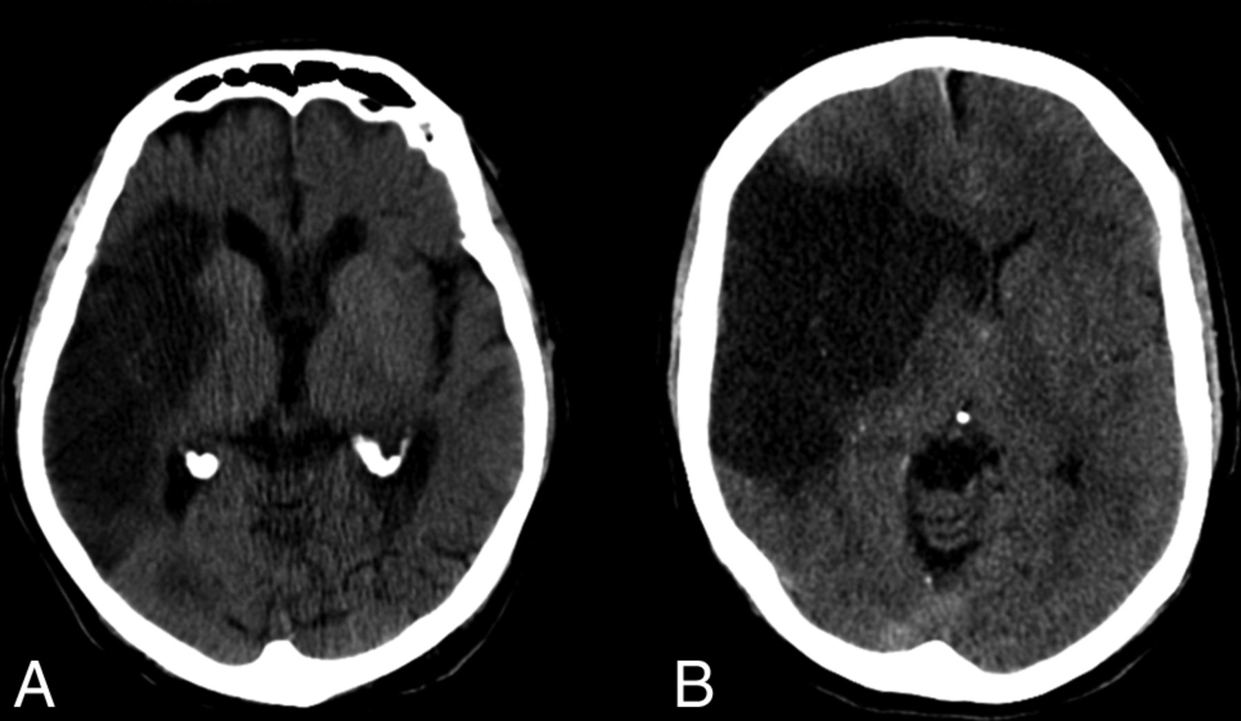
  • Fig 2.
    • Download figure
    • Open in new tab
    • Download powerpoint
    Fig 2.

    Large MCA infarct on follow-up, with and without prominent space-occupying edema. Patient A (87-year-old man, follow-up day 5) has a large MCA infarct and generalized atrophy but does not show a midline shift. Patient B (58-year-old man, follow-up day 3) has a large MCA infarct with a midline shift of ≥5 mm, representing prominent space-occupying edema.

Tables

  • Figures
    • View popup
    Table 1:

    Clinical and imaging characteristicsa

    CharacteristicsAll Patients (N = 137)Prominent Space-Occupying Edema (n = 51)No Prominent Space-Occupying Edema (n = 86)P Value
    Clinical parameters
        Age (yr) (median) (IQR)66 (53–73)63 (52–72)67 (54–73).31
        Female sex (No.) (%)48 (35)19 (37)29 (34).68
        Prior stroke (No.) (%)20 (15)10 (20)10 (12).18
        Atrial fibrillation (No.) (%)16 (12)8 (16)8 (9).27
        NIHSS (median) (IQR)15 (12–19)18 (13–21)14 (11–18).01b
        IV-rtPA (No.) (%)94 (69)32 (63)62 (72).25
        Intra-arterial treatment (No.) (%)29 (21)11 (22)18 (21).93
    Imaging parameters
        Time to admission scan (min) (median) (IQR)100 (64–152)106 (70–240)91 (63–135).11
        NCCT
            Early CT signs of infarction, ASPECTS, (median) (IQR)8 (6–10)7 (4–9)9 (7–10).0001b
        CTP
            CBV deficit, ASPECTS (mean) (SD)4.30 (2.57)3.31 (2.32)4.89 (2.54).001b
            MTT deficit, ASPECTS (median) (IQR)1 (0–3)1 (0–3)2 (0–3).11
            CBV deficit in lentiform nucleus (No.) (%)72 (53)32 (63)40 (47).15
            CBV deficit in caudate nucleus or ACA territory (No.) (%)54 (39)28 (55)26 (30).004b
            Permeability ratio (median) (IQR)c1.36 (1.13–1.88)1.66 (1.24–2.60)1.30 (1.09–1.54).002b
        CTA
            Clot burden score ≤6 (No.) (%)99 (73)44 (86)55 (65).006b
            Thrombus location ICA/M1 proximal (No.) (%)63 (48)33 (67)30 (36).001b
            Poor collateral score (No.) (%)62 (46)34 (68)28 (33).0001b
        Follow-up CTP and CTA
            No recanalizationc26 (33)6 (32)20 (33).89
            No reperfusionc45 (62)16 (76)29 (56).10
    • Note:—IQR indicates interquartile range.

    • ↵a χ2 was used to compare categoric variables, and a t test or Mann-Whitney U test, continuous variables.

    • ↵b All P < .05.

    • ↵c The analysis of permeability ratio, recanalization, and reperfusion is a subanalysis on 101, 79, and 73 cases, respectively.

    • View popup
    Table 2:

    Univariate and multivariate regression

    OR (95% CI)aOR (95% CI)
    Clinical parameters
        Age (per yr)0.99 (0.96–1.01)NA
        Female sex1.17 (0.57–2.40)0.88 (0.41–1.90)
        Prior stroke1.90 (0.73–4.95)2.08 (0.74–5.91)
        Atrial fibrillation1.79 (0.63–5.11)2.74 (0.86–8.70)
        NIHSS (per point)1.12 (1.04–1.21)aNA
        IV-rtPA0.65 (0.31–1.36)0.56 (0.25–1.22)
        Intra-arterial treatment1.04 (0.45–2.42)1.03 (0.42–2.49)
    Imaging parameters
        Time to admission scan (per min)1.002 (0.997–1.007)1.003 (0.999–1.006)
        NCCT
            Early CT signs of infarction, ASPECTS (0–10)1.32 (1.14–1.53)a1.32 (1.13–1.55)a
        CTP
            CBV deficit, ASPECTS (0–10)1.30 (1.12–1.52)a1.26 (1.07–1.49)a
            MTT deficit, ASPECTS (0–10)1.20 (0.98–1.46)1.14 (0.93–1.41)
            CBV deficit in lentiform nucleus1.90 (0.93–3.85)1.53 (0.72–3.22)
            CBV deficit in caudate nucleus or ACA territory2.81 (1.37–5.76)a2.01 (0.93–4.32)
            Permeability ratiob2.08 (1.21–3.60)a2.35 (1.30–4.24)a
        CTA
            Clot burden score ≤63.43 (1.38–8.55)a2.88 (1.11–7.45)a
            Thrombus location3.64 (1.73–7.69)a3.40 (1.57–7.37)a
            ICA/M1 proximal versus M1 distal, M2, or >M2
            Poor collateral score4.33 (2.05–9.13)a3.93 (1.78–8.69)a
        Follow-up CTP and CTA
            No recanalizationb0.92 (0.31–2.79)0.92 (0.30–2.81)
            No reperfusionb2.54 (0.81–7.96)2.18 (0.67–7.07)
    • Note:—aOR indicates adjusted odds ratio (for age and NIHSS); NA, not applicable.

    • ↵a All P < .05.

    • ↵b The analysis of permeability ratio, recanalization, and reperfusion is a subanalysis on 101, 79, and 73 cases respectively.

PreviousNext
Back to top

In this issue

American Journal of Neuroradiology: 37 (5)
American Journal of Neuroradiology
Vol. 37, Issue 5
1 May 2016
  • Table of Contents
  • Index by author
  • Complete Issue (PDF)
Advertisement
Print
Download PDF
Email Article

Thank you for your interest in spreading the word on American Journal of Neuroradiology.

NOTE: We only request your email address so that the person you are recommending the page to knows that you wanted them to see it, and that it is not junk mail. We do not capture any email address.

Enter multiple addresses on separate lines or separate them with commas.
Imaging Findings Associated with Space-Occupying Edema in Patients with Large Middle Cerebral Artery Infarcts
(Your Name) has sent you a message from American Journal of Neuroradiology
(Your Name) thought you would like to see the American Journal of Neuroradiology web site.
CAPTCHA
This question is for testing whether or not you are a human visitor and to prevent automated spam submissions.
Cite this article
A.D. Horsch, J.W. Dankbaar, T.A. Stemerdink, E. Bennink, T. van Seeters, L.J. Kappelle, J. Hofmeijer, H.W. de Jong, Y. van der Graaf, B.K. Velthuis
Imaging Findings Associated with Space-Occupying Edema in Patients with Large Middle Cerebral Artery Infarcts
American Journal of Neuroradiology May 2016, 37 (5) 831-837; DOI: 10.3174/ajnr.A4637

Citation Manager Formats

  • BibTeX
  • Bookends
  • EasyBib
  • EndNote (tagged)
  • EndNote 8 (xml)
  • Medlars
  • Mendeley
  • Papers
  • RefWorks Tagged
  • Ref Manager
  • RIS
  • Zotero
0 Responses
Respond to this article
Share
Bookmark this article
Imaging Findings Associated with Space-Occupying Edema in Patients with Large Middle Cerebral Artery Infarcts
A.D. Horsch, J.W. Dankbaar, T.A. Stemerdink, E. Bennink, T. van Seeters, L.J. Kappelle, J. Hofmeijer, H.W. de Jong, Y. van der Graaf, B.K. Velthuis
American Journal of Neuroradiology May 2016, 37 (5) 831-837; DOI: 10.3174/ajnr.A4637
del.icio.us logo Twitter logo Facebook logo Mendeley logo
  • Tweet Widget
  • Facebook Like
  • Google Plus One
Purchase

Jump to section

  • Article
    • Abstract
    • ABBREVIATIONS:
    • Materials and Methods
    • Results
    • Discussion
    • Conclusions
    • Acknowledgments
    • Footnotes
    • References
  • Figures & Data
  • Info & Metrics
  • Responses
  • References
  • PDF

Related Articles

  • No related articles found.
  • PubMed
  • Google Scholar

Cited By...

  • Absent Cortical Venous Filling Is Associated with Aggravated Brain Edema in Acute Ischemic Stroke
  • Predictors of malignant brain edema after mechanical thrombectomy for acute ischemic stroke
  • Thalamic Diaschisis in Acute Ischemic Stroke: Occurrence, Perfusion Characteristics, and Impact on Outcome
  • Early Imaging Prediction of Malignant Cerebellar Edema Development in Acute Ischemic Stroke
  • A Biophysical Model for Cytotoxic Cell Swelling
  • Crossref (22)
  • Google Scholar

This article has been cited by the following articles in journals that are participating in Crossref Cited-by Linking.

  • Cerebral Collateral Circulation in the Era of Reperfusion Therapies for Acute Ischemic Stroke
    Simone M. Uniken Venema, Jan Willem Dankbaar, Aad van der Lugt, Diederik W.J. Dippel, H. Bart van der Worp
    Stroke 2022 53 10
  • A Biophysical Model for Cytotoxic Cell Swelling
    Koen Dijkstra, Jeannette Hofmeijer, Stephan A. van Gils, Michel J.A.M. van Putten
    The Journal of Neuroscience 2016 36 47
  • Predictors of malignant brain edema after mechanical thrombectomy for acute ischemic stroke
    Xianjun Huang, Qian Yang, Xiaolei Shi, Xiangjun Xu, Liang Ge, Xianhui Ding, Zhiming Zhou
    Journal of NeuroInterventional Surgery 2019 11 10
  • Effect of Recanalization on Cerebral Edema in Ischemic Stroke Treated With Thrombolysis and/or Endovascular Therapy
    Magnus Thorén, Anand Dixit, Irene Escudero-Martínez, Zuzana Gdovinová, Lukas Klecka, Viiu-Marika Rand, Danilo Toni, Aleksandras Vilionskis, Nils Wahlgren, Niaz Ahmed
    Stroke 2020 51 1
  • Role of Decompressive Craniectomy in Ischemic Stroke
    Lars-Peder Pallesen, Kristian Barlinn, Volker Puetz
    Frontiers in Neurology 2019 9
  • Crossed cerebellar diaschisis in acute ischemic stroke: Impact on morphologic and functional outcome
    Wolfgang G Kunz, Wieland H Sommer, Christopher Höhne, Matthias P Fabritius, Felix Schuler, Franziska Dorn, Ahmed E Othman, Felix G Meinel, Louisa von Baumgarten, Maximilian F Reiser, Birgit Ertl-Wagner, Kolja M Thierfelder
    Journal of Cerebral Blood Flow & Metabolism 2017 37 11
  • Early Imaging Prediction of Malignant Cerebellar Edema Development in Acute Ischemic Stroke
    Matthias P. Fabritius, Kolja M. Thierfelder, Felix G. Meinel, Ahmed E. Othman, Franziska Dorn, Bastian O. Sabel, Pierre Scheffler, Birgit Ertl-Wagner, Wieland H. Sommer, Wolfgang G. Kunz
    Stroke 2017 48 9
  • Quantitative Serial CT Imaging-Derived Features Improve Prediction of Malignant Cerebral Edema after Ischemic Stroke
    Hossein Mohammadian Foroushani, Ali Hamzehloo, Atul Kumar, Yasheng Chen, Laura Heitsch, Agnieszka Slowik, Daniel Strbian, Jin-Moo Lee, Daniel S. Marcus, Rajat Dhar
    Neurocritical Care 2020 33 3
  • Absent Cortical Venous Filling Is Associated with Aggravated Brain Edema in Acute Ischemic Stroke
    H. Xia, H. Sun, S. He, M. Zhao, W. Huang, Z. Zhang, Y. Xue, P. Fu, W. Chen
    American Journal of Neuroradiology 2021 42 6
  • Thalamic Diaschisis in Acute Ischemic Stroke
    Paul Reidler, Kolja M. Thierfelder, Matthias P. Fabritius, Wieland H. Sommer, Felix G. Meinel, Franziska Dorn, Frank A. Wollenweber, Marco Duering, Wolfgang G. Kunz
    Stroke 2018 49 4

More in this TOC Section

ADULT BRAIN

  • Diagnostic Neuroradiology of Monoclonal Antibodies
  • Clinical Outcomes After Chiari I Decompression
  • Segmentation of Brain Metastases with BLAST
Show more ADULT BRAIN

Neurointervention

  • Effect of SARS-CoV2 on Endovascular Thrombectomy
  • Flow diversion for distal circulation aneurysms
  • Neuroform Atlas Stent for Intracranial Aneurysms
Show more Neurointervention

Similar Articles

Advertisement

Indexed Content

  • Current Issue
  • Accepted Manuscripts
  • Article Preview
  • Past Issues
  • Editorials
  • Editor's Choice
  • Fellows' Journal Club
  • Letters to the Editor
  • Video Articles

Cases

  • Case Collection
  • Archive - Case of the Week
  • Archive - Case of the Month
  • Archive - Classic Case

More from AJNR

  • Trainee Corner
  • Imaging Protocols
  • MRI Safety Corner
  • Book Reviews

Multimedia

  • AJNR Podcasts
  • AJNR Scantastics

Resources

  • Turnaround Time
  • Submit a Manuscript
  • Submit a Video Article
  • Submit an eLetter to the Editor/Response
  • Manuscript Submission Guidelines
  • Statistical Tips
  • Fast Publishing of Accepted Manuscripts
  • Graphical Abstract Preparation
  • Imaging Protocol Submission
  • Evidence-Based Medicine Level Guide
  • Publishing Checklists
  • Author Policies
  • Become a Reviewer/Academy of Reviewers
  • News and Updates

About Us

  • About AJNR
  • Editorial Board
  • Editorial Board Alumni
  • Alerts
  • Permissions
  • Not an AJNR Subscriber? Join Now
  • Advertise with Us
  • Librarian Resources
  • Feedback
  • Terms and Conditions
  • AJNR Editorial Board Alumni

American Society of Neuroradiology

  • Not an ASNR Member? Join Now

© 2025 by the American Society of Neuroradiology All rights, including for text and data mining, AI training, and similar technologies, are reserved.
Print ISSN: 0195-6108 Online ISSN: 1936-959X

Powered by HighWire