
Article Figures & Data
Figures
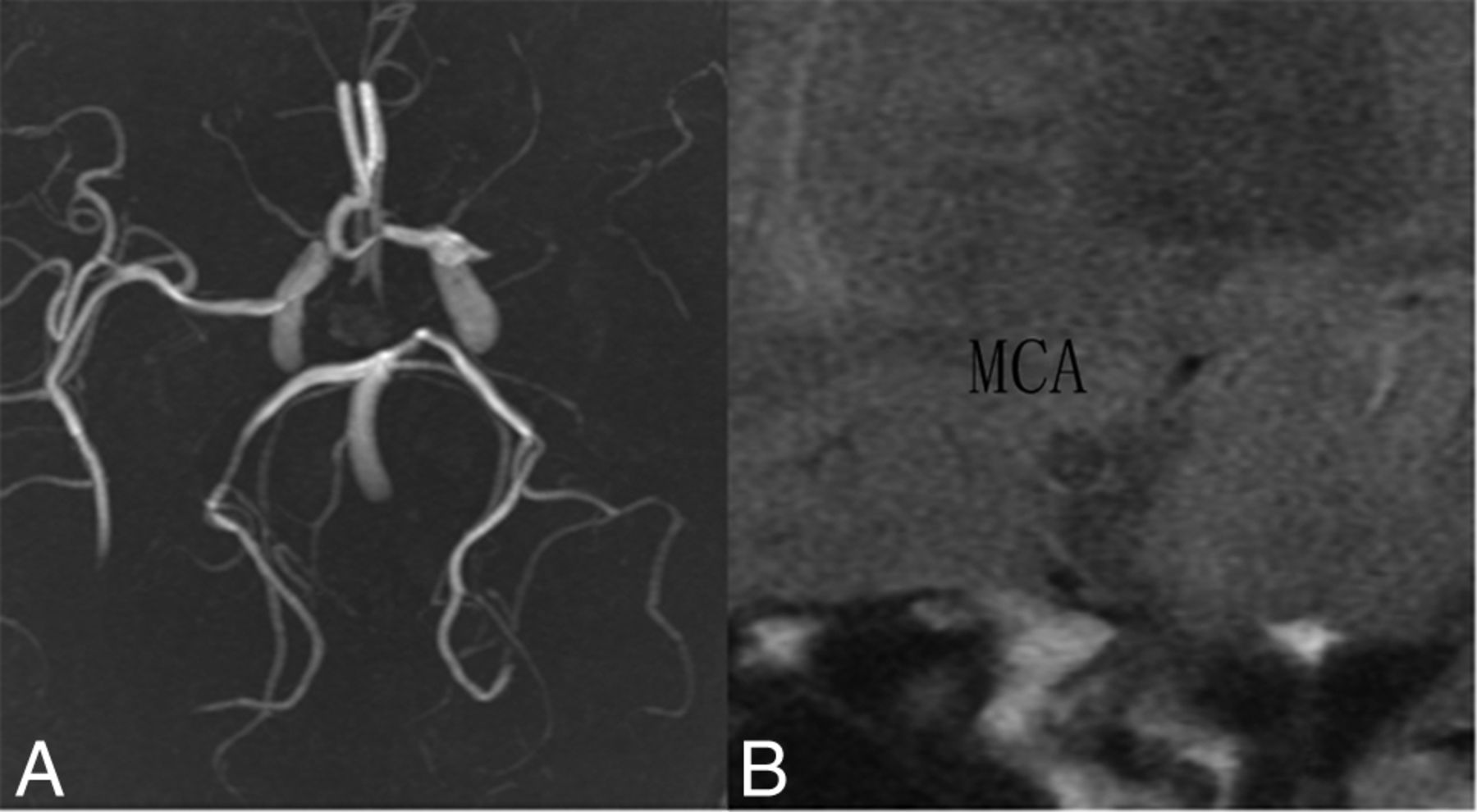
- Fig 1.
Representative MR angiography and high-resolution T2-weighted imaging findings. A, 3D time-of-flight MRA reveals an occlusive left middle cerebral artery. B, Plaque with heterogeneous signal intensity was present in the MCA lumen on HR-T2WI.
- Fig 2.
Illustration of MR susceptometry for determining Yv. A, A phase image obtained from susceptibility-weighted imaging. B, Zoomed view of the phase image for the gray matter, with the vein as the representative phase value within the vessel. The rectangle shows the ROI of the gray matter, including the vein.
- Fig 3.
Placement of the ROI for cerebral blood flow measurements on an arterial spin-labeling CBF map. The ROI was positioned from the top (A) to the level of the thalami (B) in a 50-year-old patient with an occlusive MCA. C and D, Anatomic images corresponding to A and B.


Tables
- Table 1:
Number, age, clinical characteristics, Hct, and Hb of the healthy subjects and patients
Groups No. Age Range (yr) Mean Age (yr) Males (No.) Females (No.) Hct (×10−2) Hb Healthy subjects 10 40–67 55.9 5 5 34.9 ± 12.6 13.1 ± 1.2 Mild stenotic MCA Group 1 13 37–65 50.4 6 7 40.2 ± 4.9 14.2 ± 1.4 Severe stenotic MCA Group 2 11 45–75 58.0 6 5 39.2 ± 5.1 13.1 ± 2.1 Occluded MCA Group 3 16 36–72 58.5 8 8 39.8 ± 4.4 13.8 ± 2.3 Patients with acute stroke Group 4 17 38–73 57.6 9 8 40.3 ± 6.6 13.6 ± 2.1 Note:—Hb indicates hemoglobin.
CBF (mL/100 g/min) OEF (%) CMRO2 (μmol/100 g/min) Left Right Left Right Left Right 53.4 ± 8.6 54.4 ± 9.6 36.8 ± 3.1 37.4 ± 3.3 146.8 ± 29.7 151.6 ± 32.6 T 1.028 1.269 1.366 P .331 .236 .205 - Table 3:
Statistical results of the paired t test for the CBF, OEF, and CMRO2 in the 4 patient subgroups
Contralateral Stenosis CBF (mL/100 g/min) OEF (%) CMRO2 (μmol/100 g/min) Group 1 51.9 ± 9.5 50.7 ± 11.2 38.5 ± 3.9 40.6 ± 4.2 153.2 ± 24.0 155.5 ± 22.9 T 0.796 4.027 0.377 P .442 .002 .713 Group 2 54.6 ± 5.9 47.9 ± 9.8 38.4 ± 1.9 42.6 ± 3.8 150.3 ± 28.5 149.4 ± 46.1 T 4.146 4.690 0.132 P .002 .001 .898 Group 3 54.0 ± 10.3 37.2 ± 9.7 37.1 ± 2.9 44.5 ± 3.9 154.0 ± 43.4 125.3 ± 35.3 T 6.295 6.128 2.926 P <.001 <.001 .010 Group 4 55.3 ± 9.5 40.8 ± 11.8 38.3 ± 4.1 43.9 ± 3.5 161.2 ± 47.4 135.0 ± 41.3 T 5.898 4.626 2.647 P <.001 <.001 .018 - Table 4:
Results of the analysis of variance test for the normal-to-lesion ratio of CBF, OEF, and CMRO2 in the 4 patient subgroups
Group 1 Group 2 Group 3 Group 4 F P rCBF 0.98 ± 0.11 0.87 ± 0.11 0.70 ± 0.17 0.74 ± 0.17 10.617 <.001 rOEF 1.06 ± 0.05 1.11 ± 0.08 1.21 ± 0.14 1.16 ± 0.14 4.316 .009 rCMRO2 1.03 ± 0.15 0.98 ± 0.15 0.84 ± 0.21 0.86 ± 0.23 3.041 .037 - Table 5:
Results of the Student-Newman-Keuls test for the normal-to-lesion ratio of CBF, OEF, and CMRO2 in the 4 patient subgroups with increasing MCA stenotic severity
rCBF rOEF rCMRO2 Group 1 Group 2 >0.05 >0.05 >0.05 Group 3 <0.05 <0.05 >0.05 Group 4 <0.05 >0.05 >0.05 Group 2 Group 3 <0.05 >0.05 >0.05 Group 4 <0.05 >0.05 >0.05 Group 3 Group 4 >0.05 >0.05 >0.05 - Table 6:
Comparison of OEF and CMRO2 with published literature values determined by using PET for healthy subjects
Study (Reference) OEF (Mean) (Range) CMRO2 (μmol/min/100 g) No. Mean Age (yr) (range) Ito et al22,a 0.44 ± 0.06 147.0 ± 22.3 70 53.1 (18–77) (0.36 ± 0.06–0.51 ± 0.04) Hattori et al23,a 0.39 ± 0.06 127.0 ± 17.4 16 35 (21–46) (0.30–0.51) Coles et al24,a 0.42 ± 0.04 124.8 ± 22.3 7 30 (18–60) Ibaraki et al25,b 0.35 ± 0.06 156.0 ± 22.3 8 NA (21–24) Bremmer et al26,a 0.43 ± 0.06 135.9 ± 8.9 7 69 (57–80) Kudomi et al27,b 0.39 ± 0.05 184.9 ± 20.1 7 25.3 (NA)


Jump to section
Related Articles
Cited By...
- Susceptibility Changes on Preoperative Acetazolamide-Loaded 7T MR Quantitative Susceptibility Mapping Predict Post-Carotid Endarterectomy Cerebral Hyperperfusion
- Lesion evolution and neurodegeneration in RVCL-S: A monogenic microvasculopathy
- Acetazolamide-Loaded Dynamic 7T MR Quantitative Susceptibility Mapping in Major Cerebral Artery Steno-Occlusive Disease: Comparison with PET
- Combining images and anatomical knowledge to improve automated vein segmentation in MRI
- Hematocrit Measurement with R2* and Quantitative Susceptibility Mapping in Postmortem Brain
- Preoperative Cerebral Oxygen Extraction Fraction Imaging Generated from 7T MR Quantitative Susceptibility Mapping Predicts Development of Cerebral Hyperperfusion following Carotid Endarterectomy
- Noninvasive Assessment of Oxygen Extraction Fraction in Chronic Ischemia Using Quantitative Susceptibility Mapping at 7 Tesla
- Association between large artery atherosclerosis and cerebral microbleeds: a systematic review and meta-analysis