
Article Figures & Data
Figures
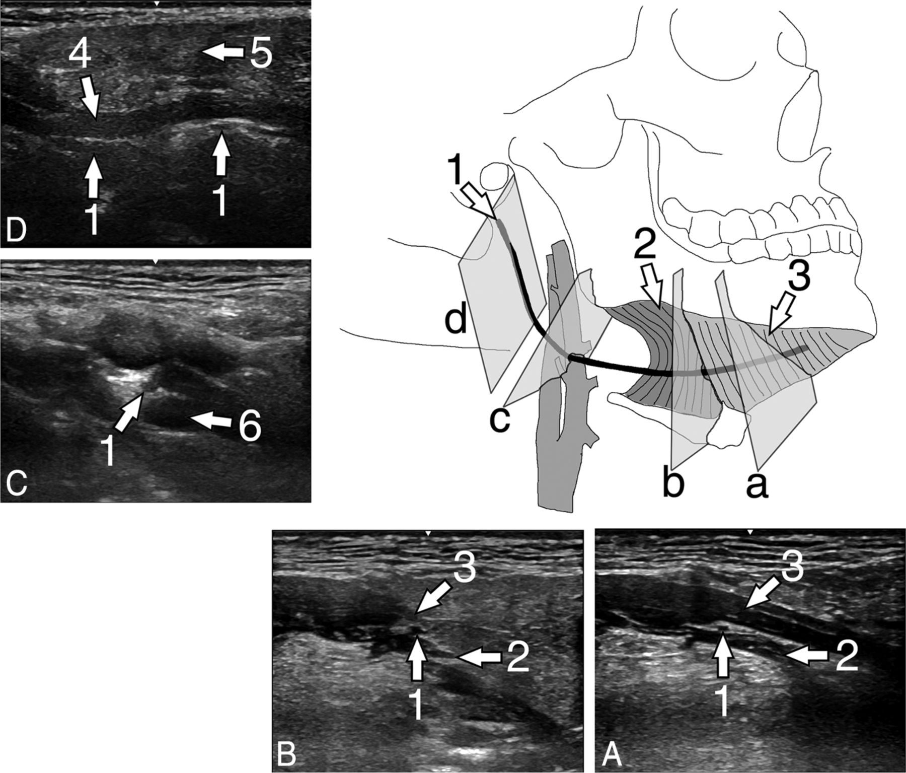
- Fig 1.
Illustration of the hypoglossal nerve with exemplary US scans of the hypoglossal nerve of a healthy volunteer. A, Transversal scan at the floor of the mouth in a coronal body plane. B, Transversal scan at the posterior rim of the hyoglossal muscle in a coronal body plane. C, Transversal scan in a paracoronal body plane at the crossing of the nerve with the external carotid artery. D, Longitudinal scan at the carotid space in a paracoronal body plane. 1 = hypoglossal nerve, 2 = hyoglossal muscle, 3 = mylohyoid muscle, 4, = stylohyoid and styloglossal muscles, 5 = parotid gland, 6 = external carotid artery.
- Fig 2.
Dissection situs of the right side of a neck. View from ventrolateral. Note the exposed hypoglossal nerve (arrowheads) with dark ink markings along its course. Cr indicates cranial; cd, caudal; r, right; l, left.
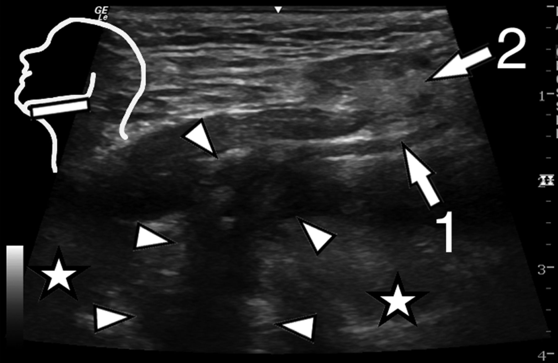
- Fig 3.
Parasagittal US scan at the floor of the mouth of patient 1 (Table 2). Note the hypoglossal nerve entering the region with extensive scar tissue after a neck dissection operation. Inset: position of the US transducer; 1 = hypoglossal nerve, asterisks = body of the tongue, arrowheads = scar tissue.
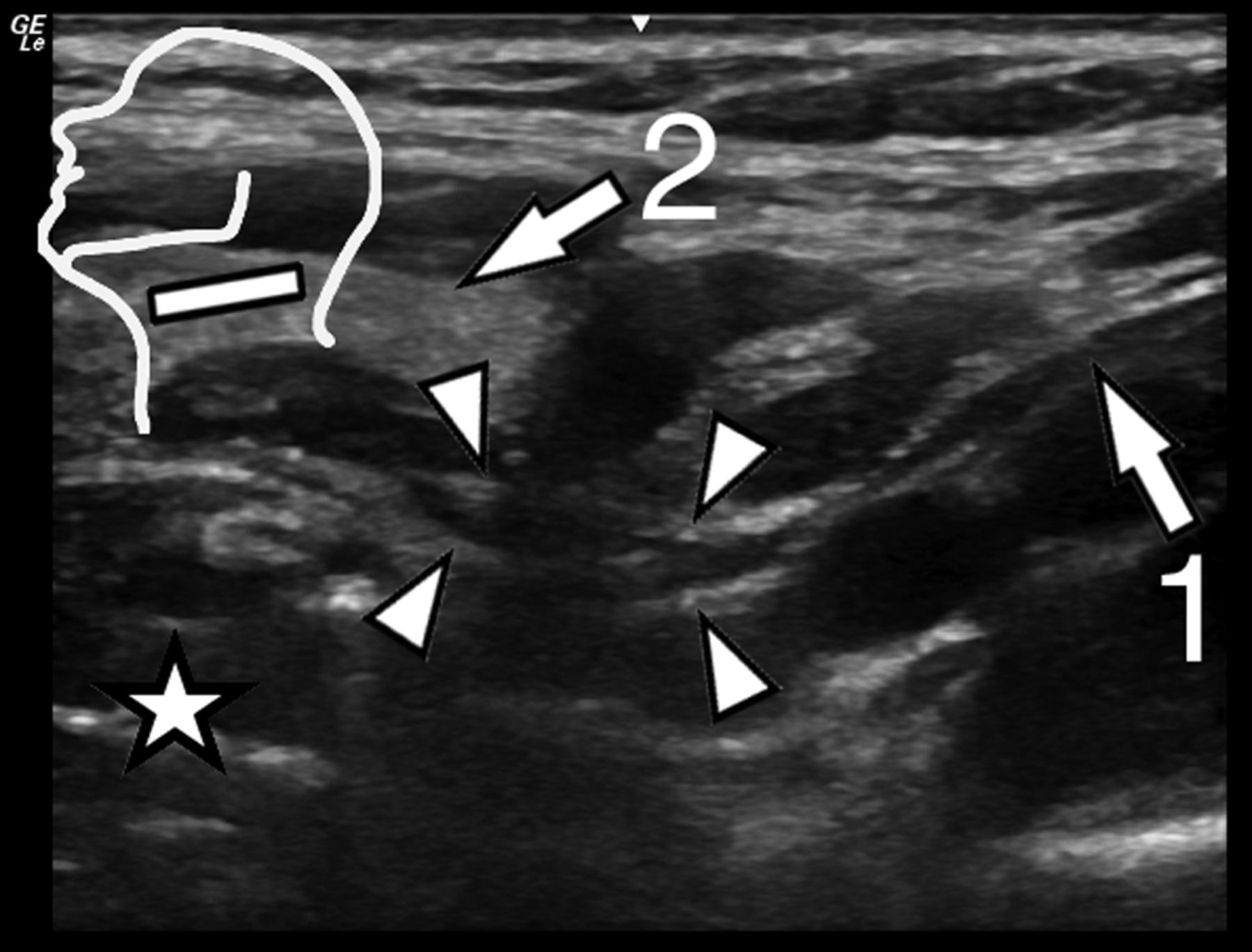
- Fig 4.
Parasagittal US scan at the floor of the mouth of patient 2 (Table 2). The hypoglossal nerve is directly infiltrated by a squamous cell carcinoma of the tongue base. Inset: position of the US transducer; 1 = hypoglossal nerve, 2 = submandibular gland, asterisks = body of the tongue, arrowheads = squamous cell carcinoma of the tongue base.
- Fig 5.
Parasagittal US scan at the carotid space of patient 3 (Table 2). Anterior to the carotid space the hypoglossal nerve is thickened focally after a facial nerve end-to-side coaptation. Inset: position of the US transducer; 1 = hypoglossal nerve, 2 = submandibular gland, asterisk = body of the tongue, arrowheads = coaptation site.
- Fig 6.
Parasagittal US scan at the carotid space of patient 6 (Table 2). The hypoglossal nerve is thickened markedly in the carotid space due to formation of a fibroma. Inset: position of the US transducer; 1 = hypoglossal nerve, 2 = submandibular gland, asterisk = body of the tongue, arrowheads = fibroma.


Tables
- Table 1:
Summary of US scan measurements of volunteer hypoglossal nerve cross-sectional areas
Right Side (Mean) (Median, SD, Maximum, Minimum) Left Side (Mean) (Median, SD, Maximum, Minimum) Location 1 At the posterolateral rim of the mylohyoid muscle (mm2) 1.9 (2, 0.6, 3, 0.8) 1.9 (2, 0.6, 4, 1) Location 2 At the crossing of the hypoglossal nerve with the external carotid artery (mm2) 2.1 (1.7, 0.6, 3, 0.8) 2.1 (2.1, 0.5, 3.4, 1.1) No. Age (yr) Sex History Clinical Details on Referral US findings 1 50 M Squamous cell carcinoma of the tonsil, neck dissection Hypoglossal nerve palsy Scar tissue around the nerve (Fig 3) 2 72 M Squamous cell carcinoma of the tongue base Hypoglossal nerve palsy Tumor infiltration of the nerve (Fig 4) 3 33 F Facial nerve end-to-side coaptation to the hypoglossal nerve after tongue palsy after cerebellopontine angle tumor resection Tongue paresis Nerve coaptation site with focal hypoglossal nerve thickening (Fig 5) 4 68 F Partial hypoglossal nerve transfer for facial nerve reconstruction after malignant parotid gland tumor resection Tongue weakness Nerve transfer site with focal thickening and fibrosis 5 48 M Squamous cell carcinoma of the tonsil, neck dissection, radiotherapy, chemotherapy Hypoglossal palsy Scar tissue entrapment of the hypoglossal nerve 6 55 M Neurofibromatosis Screening Neurofibroma (Fig 6)


Jump to section
Related Articles
Cited By...
- No citing articles found.