
Article Figures & Data
Figures
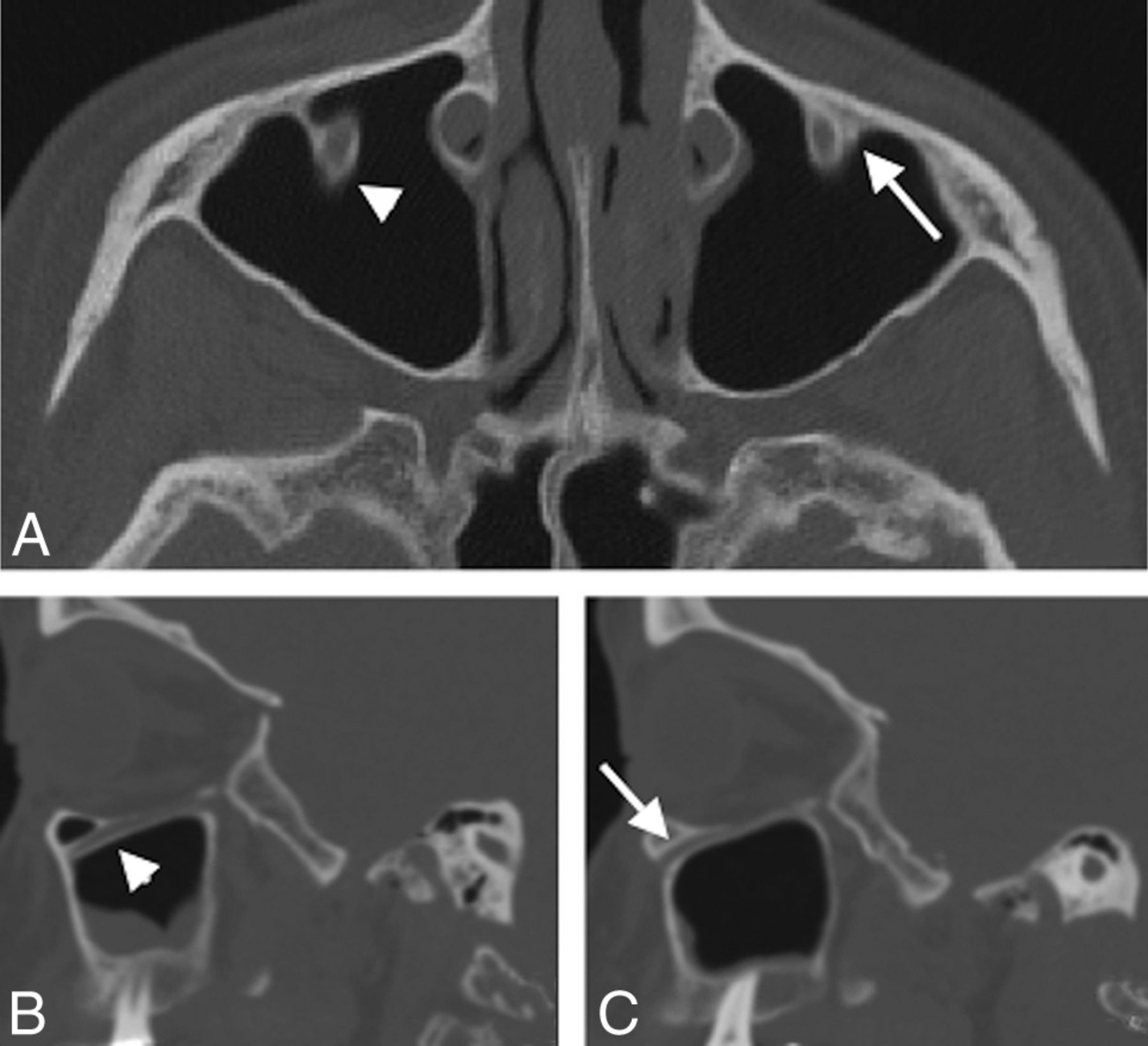
- Fig 1.
Axial (A), right parasagittal (B), and left parasagittal (C) sinus CT images in a 55-year-old woman show unilateral right-sided protrusion of the ION into the maxillary sinus (arrowhead in A and B). While part of the wall of the left IOC protrudes into the sinus, the entire circumference of the IOC is not distinct from the anterior maxillary sinus wall; this feature is confirmed on the sagittal image through the left maxillary sinus (arrows in A and C). Additionally, no measurable bony septum connects the IOC to the wall of the maxillary sinus. This distinction was chosen to define protrusion of the ION into the maxillary sinus.
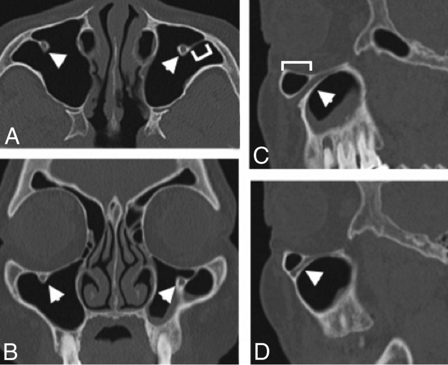
- Fig 2.
Axial (A), coronal (B), left parasagittal (C), and right parasagittal (D) CT images in a 72-year-old woman show bilateral protrusion of the ION (arrowheads) into the maxillary sinus. The septum attaching the IOC to the anterior wall of the sinus was measured (bracket in A) on the axial image. The distance at which protrusion begins posterior to the inferior orbital rim (bracket in C) was measured on the sagittal image.
- Fig 3.
Axial CT image in a 56-year-old man shows left-sided protrusion of the ION into the maxillary sinus (arrowhead) attached to a single posterior septum (arrow). This was the only patient with a septum attaching a protruding IOC to the posterior wall of the maxillary sinus without an additional septum attaching to the anterior wall.
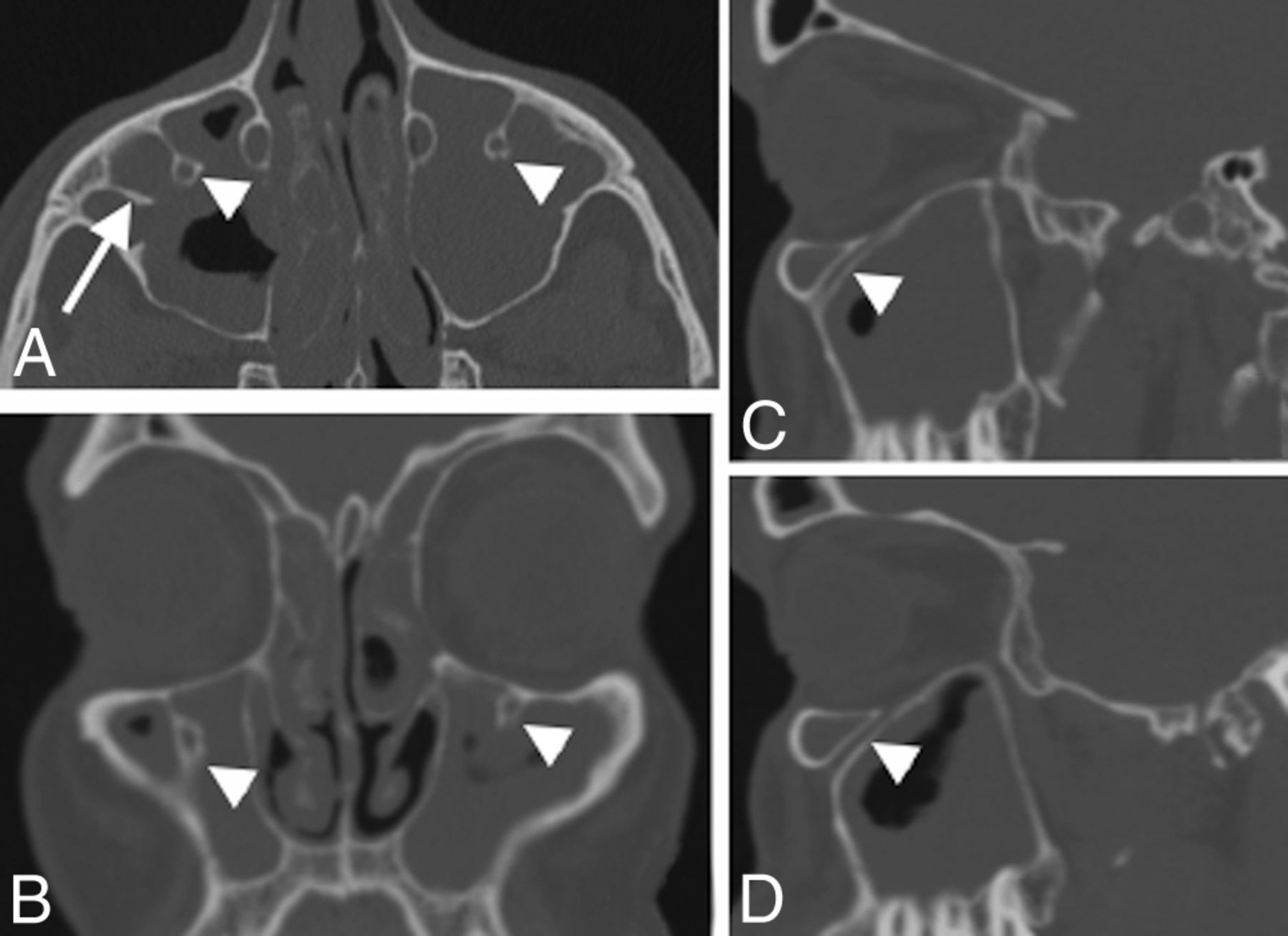
- Fig 4.
Coronal (A), axial (B), and right parasagittal (C) CT images in a 58-year-old man show bilateral protrusion of the IOC into the maxillary sinus. On the coronal image, an infraorbital ethmoid (Haller) cell is seen on the right (arrowhead) just medial and superior to the protruding IOC (arrow). Bilateral protrusion is seen on the axial image (arrows), and confirmation of protrusion is seen on the sagittal image through the right maxillary sinus. The presence of a protruding IOC near an infraorbital cell is important for the endoscopic surgeon to know preoperatively, in the event that the infraorbital cell is targeted during sinus surgery.
- Fig 5.
Axial (A), coronal (B), right (C), and left (D) parasagittal CT images of a 42-year-old man show extensive paranasal sinus inflammatory disease and also demonstrate bilateral protrusion of the IOC (arrowheads) into the maxillary sinus. Incidental note is made of an additional bony septum in the right maxillary sinus (arrow in A). For this surgical candidate, the IOC position should be mentioned in the report.


Tables
The 54 patients composing our group of patients with IOC protrusion broken down into 3 groupsa
Class Septum Length No. of Patients Median Age (yr) Female/Male Bilateral (%) 1 1–3 mm 26 62.5 15:11 12 (46%) 2 4–6 mm 18 48.5 9:9 8 (44%) 3 7–11 mm 10 43.5 5:5 8 (80%) ↵a The most medial septum was chosen in patients with multiple septa.


Jump to section
Related Articles
Cited By...
- No citing articles found.