Advertisement

Main menu

  • Home
  • Content
    • Current Issue
    • Accepted Manuscripts
    • Article Preview
    • Past Issue Archive
    • Video Articles
    • AJNR Case Collection
    • Case of the Week Archive
    • Case of the Month Archive
    • Classic Case Archive
  • Special Collections
    • AJNR Awards
    • Low-Field MRI
    • Alzheimer Disease
    • ASNR Foundation Special Collection
    • Photon-Counting CT
    • View All
  • Multimedia
    • AJNR Podcasts
    • AJNR SCANtastic
    • Trainee Corner
    • MRI Safety Corner
    • Imaging Protocols
  • For Authors
    • Submit a Manuscript
    • Submit a Video Article
    • Submit an eLetter to the Editor/Response
    • Manuscript Submission Guidelines
    • Statistical Tips
    • Fast Publishing of Accepted Manuscripts
    • Graphical Abstract Preparation
    • Imaging Protocol Submission
    • Author Policies
  • About Us
    • About AJNR
    • Editorial Board
    • Editorial Board Alumni
  • More
    • Become a Reviewer/Academy of Reviewers
    • Subscribers
    • Permissions
    • Alerts
    • Feedback
    • Advertisers
    • ASNR Home

User menu

  • Alerts
  • Log in

Search

  • Advanced search
American Journal of Neuroradiology

American Journal of Neuroradiology

ASHNR American Society of Functional Neuroradiology ASHNR American Society of Pediatric Neuroradiology ASSR
  • Alerts
  • Log in

Advanced Search

  • Home
  • Content
    • Current Issue
    • Accepted Manuscripts
    • Article Preview
    • Past Issue Archive
    • Video Articles
    • AJNR Case Collection
    • Case of the Week Archive
    • Case of the Month Archive
    • Classic Case Archive
  • Special Collections
    • AJNR Awards
    • Low-Field MRI
    • Alzheimer Disease
    • ASNR Foundation Special Collection
    • Photon-Counting CT
    • View All
  • Multimedia
    • AJNR Podcasts
    • AJNR SCANtastic
    • Trainee Corner
    • MRI Safety Corner
    • Imaging Protocols
  • For Authors
    • Submit a Manuscript
    • Submit a Video Article
    • Submit an eLetter to the Editor/Response
    • Manuscript Submission Guidelines
    • Statistical Tips
    • Fast Publishing of Accepted Manuscripts
    • Graphical Abstract Preparation
    • Imaging Protocol Submission
    • Author Policies
  • About Us
    • About AJNR
    • Editorial Board
    • Editorial Board Alumni
  • More
    • Become a Reviewer/Academy of Reviewers
    • Subscribers
    • Permissions
    • Alerts
    • Feedback
    • Advertisers
    • ASNR Home
  • Follow AJNR on Twitter
  • Visit AJNR on Facebook
  • Follow AJNR on Instagram
  • Join AJNR on LinkedIn
  • RSS Feeds

AJNR Awards, New Junior Editors, and more. Read the latest AJNR updates

Editorial

Point: Don't Call Me a DOC!

D.M. Yousem
American Journal of Neuroradiology December 2016, 37 (12) 2180; DOI: https://doi.org/10.3174/ajnr.A4927
D.M. Yousem
  • Find this author on Google Scholar
  • Find this author on PubMed
  • Search for this author on this site
  • ORCID record for D.M. Yousem
  • Article
  • Figures & Data
  • Info & Metrics
  • Responses
  • References
  • PDF
Loading

The words we use matter. That is why we build lexicons. I want to petition for the removal of the phrase “disc osteophyte complex” (DOC) from the neuroradiology lexicon. I am seeing this phrase used very frequently in descriptions of every level of the cervical spine by residents coming to neuroradiology—and it galls me. To prepare for this editorial, I consulted the 3 most active orthopedic and neurosurgery spine surgeons at Johns Hopkins, and they endorsed my call to eliminate the use of disc osteophyte complex.

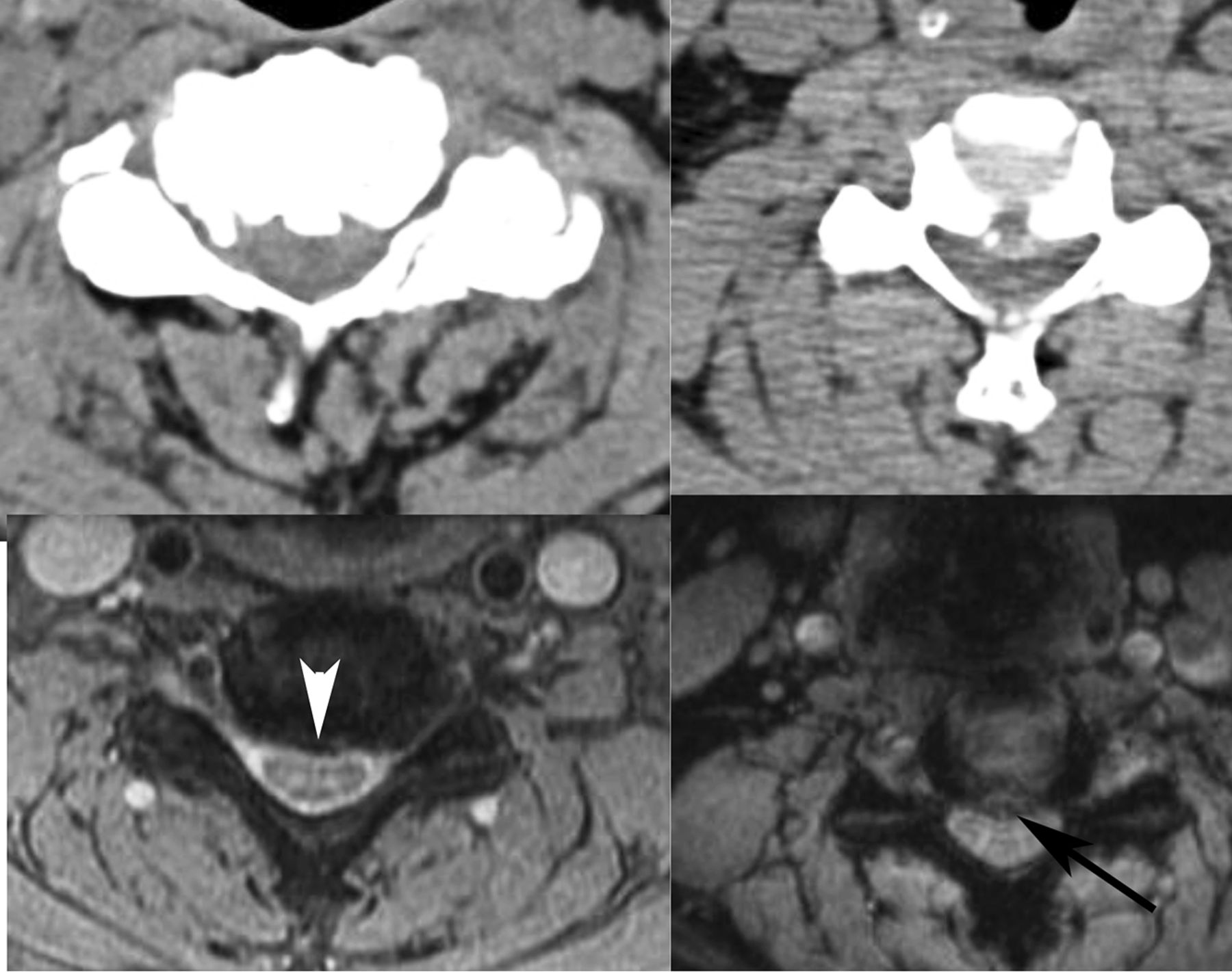
We can readily distinguish discs from osteophytes on the basis of our gradient-echo (GRE) low flip-angle thin-section cervical spine axial scans (Figure). On these sequences, the disc is bright and the osteophyte is dark. By contrast, on fast spin-echo T2-weighted images both may be dark, so the inclusion of this gradient-echo scan is critical to accurate interpretation. Rarely, we may see a severely desiccated disc that defies this GRE classification, but for most cases, we can tell a disc (bright) from an osteophyte (dark), and they do not necessarily coexist. Why should we care? Because the spine surgeons do! They told me!

Figure.
  • Download figure
  • Open in new tab
  • Download powerpoint
Figure.

Disc bright, osteophyte dark. Note the high-intensity disc herniation (black arrow) to be distinguished from the low-intensity osteophyte (white arrowhead), with confirmatory corresponding CT scans above.

If the disease is exclusively a disc, the surgeons may attempt a discectomy alone, or they may do watchful waiting for the disc to regress. Jay Khanna, orthopedic surgeon, said, “The larger, more hydrated discs tend to resorb. So if the patient is neuro-intact, we can try to treat them nonoperatively.” Osteophytes do not regress. If the disease is predominantly an osteophyte, it requires more drilling from an anterior approach, and according to Jean-Paul Wolinsky, neurosurgeon, it is a much more difficult procedure. He will warn the patient as such. Foraminotomies are more commonly required when osteophytes and uncovertebral joint spurs are present—the distraction of the intervertebral graft alone may not relieve the stenosis. From a posterior approach, Wolinsky says, a herniation is removed, whereas an “osteophyte is left alone, and instead the nerve is decompressed widely.” If there is coexistent ossification of the posterior longitudinal ligament, the approach is exclusively posterior, with indirect decompression. Neurosurgeons Ali Bydon and Dan Sciubba state, “lumping DOC generically doesn't help us determine the best approach” and “differentiating the two can change the management.”

Therefore, I implore you, my colleagues, to say that either there is a disc, there is an osteophyte, or there is a disc and osteophyte. Calling every level a disc osteophyte complex is a cop-out.

I used to say to my young children, “Use your words” (ie, do not hit). To my adult colleagues, I say, “Use your words wisely.”

  • © 2016 by American Journal of Neuroradiology
PreviousNext
Back to top

In this issue

American Journal of Neuroradiology: 37 (12)
American Journal of Neuroradiology
Vol. 37, Issue 12
1 Dec 2016
  • Table of Contents
  • Index by author
  • Complete Issue (PDF)
Advertisement
Print
Download PDF
Email Article

Thank you for your interest in spreading the word on American Journal of Neuroradiology.

NOTE: We only request your email address so that the person you are recommending the page to knows that you wanted them to see it, and that it is not junk mail. We do not capture any email address.

Enter multiple addresses on separate lines or separate them with commas.
Point: Don't Call Me a DOC!
(Your Name) has sent you a message from American Journal of Neuroradiology
(Your Name) thought you would like to see the American Journal of Neuroradiology web site.
CAPTCHA
This question is for testing whether or not you are a human visitor and to prevent automated spam submissions.
Cite this article
D.M. Yousem
Point: Don't Call Me a DOC!
American Journal of Neuroradiology Dec 2016, 37 (12) 2180; DOI: 10.3174/ajnr.A4927

Citation Manager Formats

  • BibTeX
  • Bookends
  • EasyBib
  • EndNote (tagged)
  • EndNote 8 (xml)
  • Medlars
  • Mendeley
  • Papers
  • RefWorks Tagged
  • Ref Manager
  • RIS
  • Zotero
0 Responses
Respond to this article
Share
Bookmark this article
Point: Don't Call Me a DOC!
D.M. Yousem
American Journal of Neuroradiology Dec 2016, 37 (12) 2180; DOI: 10.3174/ajnr.A4927
del.icio.us logo Twitter logo Facebook logo Mendeley logo
  • Tweet Widget
  • Facebook Like
  • Google Plus One
Purchase

Jump to section

  • Article
  • Figures & Data
  • Info & Metrics
  • Responses
  • References
  • PDF

Related Articles

  • No related articles found.
  • PubMed
  • Google Scholar

Cited By...

  • 3D T2-SPACE versus T2-FSE or T2 Gradient Recalled-Echo: Which Is the Best Sequence?
  • Crossref (3)
  • Google Scholar

This article has been cited by the following articles in journals that are participating in Crossref Cited-by Linking.

  • The imaging of cervical spondylotic myeloradiculopathy
    Susan Hesni, David Baxter, Asif Saifuddin
    Skeletal Radiology 2023 52 12
  • 3D T2-SPACE versus T2-FSE or T2 Gradient Recalled-Echo: Which Is the Best Sequence?
    M.I. Vargas, J.L. Dietemann
    American Journal of Neuroradiology 2017 38 7
  • Radiopaedia.org
    Frank Gaillard, Tariq Walizai, Hoe Guan
    2016

Similar Articles

Advertisement

Indexed Content

  • Current Issue
  • Accepted Manuscripts
  • Article Preview
  • Past Issues
  • Editorials
  • Editor's Choice
  • Fellows' Journal Club
  • Letters to the Editor
  • Video Articles

Cases

  • Case Collection
  • Archive - Case of the Week
  • Archive - Case of the Month
  • Archive - Classic Case

More from AJNR

  • Trainee Corner
  • Imaging Protocols
  • MRI Safety Corner

Multimedia

  • AJNR Podcasts
  • AJNR Scantastics

Resources

  • Turnaround Time
  • Submit a Manuscript
  • Submit a Video Article
  • Submit an eLetter to the Editor/Response
  • Manuscript Submission Guidelines
  • Statistical Tips
  • Fast Publishing of Accepted Manuscripts
  • Graphical Abstract Preparation
  • Imaging Protocol Submission
  • Evidence-Based Medicine Level Guide
  • Publishing Checklists
  • Author Policies
  • Become a Reviewer/Academy of Reviewers
  • News and Updates

About Us

  • About AJNR
  • Editorial Board
  • Editorial Board Alumni
  • Alerts
  • Permissions
  • Not an AJNR Subscriber? Join Now
  • Advertise with Us
  • Librarian Resources
  • Feedback
  • Terms and Conditions
  • AJNR Editorial Board Alumni

American Society of Neuroradiology

  • Not an ASNR Member? Join Now

© 2025 by the American Society of Neuroradiology All rights, including for text and data mining, AI training, and similar technologies, are reserved.
Print ISSN: 0195-6108 Online ISSN: 1936-959X

Powered by HighWire