Advertisement

Main menu

  • Home
  • Content
    • Current Issue
    • Accepted Manuscripts
    • Article Preview
    • Past Issue Archive
    • Video Articles
    • AJNR Case Collection
    • Case of the Week Archive
    • Case of the Month Archive
    • Classic Case Archive
  • Special Collections
    • Advancing NeuroMRI with High-Relaxivity Contrast Agents
    • AJNR Awards
    • Low-Field MRI
    • Alzheimer Disease
    • ASNR Foundation Special Collection
    • Photon-Counting CT
    • View All
  • Multimedia
    • AJNR Podcasts
    • AJNR SCANtastic
    • Trainee Corner
    • MRI Safety Corner
    • Imaging Protocols
  • For Authors
    • Submit a Manuscript
    • Submit a Video Article
    • Submit an eLetter to the Editor/Response
    • Manuscript Submission Guidelines
    • Statistical Tips
    • Fast Publishing of Accepted Manuscripts
    • Graphical Abstract Preparation
    • Imaging Protocol Submission
    • Author Policies
  • About Us
    • About AJNR
    • Editorial Board
    • Editorial Board Alumni
  • More
    • Become a Reviewer/Academy of Reviewers
    • Subscribers
    • Permissions
    • Alerts
    • Feedback
    • Advertisers
    • ASNR Home

User menu

  • Alerts
  • Log in

Search

  • Advanced search
American Journal of Neuroradiology

American Journal of Neuroradiology

ASHNR American Society of Functional Neuroradiology ASHNR American Society of Pediatric Neuroradiology ASSR
  • Alerts
  • Log in

Advanced Search

  • Home
  • Content
    • Current Issue
    • Accepted Manuscripts
    • Article Preview
    • Past Issue Archive
    • Video Articles
    • AJNR Case Collection
    • Case of the Week Archive
    • Case of the Month Archive
    • Classic Case Archive
  • Special Collections
    • Advancing NeuroMRI with High-Relaxivity Contrast Agents
    • AJNR Awards
    • Low-Field MRI
    • Alzheimer Disease
    • ASNR Foundation Special Collection
    • Photon-Counting CT
    • View All
  • Multimedia
    • AJNR Podcasts
    • AJNR SCANtastic
    • Trainee Corner
    • MRI Safety Corner
    • Imaging Protocols
  • For Authors
    • Submit a Manuscript
    • Submit a Video Article
    • Submit an eLetter to the Editor/Response
    • Manuscript Submission Guidelines
    • Statistical Tips
    • Fast Publishing of Accepted Manuscripts
    • Graphical Abstract Preparation
    • Imaging Protocol Submission
    • Author Policies
  • About Us
    • About AJNR
    • Editorial Board
    • Editorial Board Alumni
  • More
    • Become a Reviewer/Academy of Reviewers
    • Subscribers
    • Permissions
    • Alerts
    • Feedback
    • Advertisers
    • ASNR Home
  • Follow AJNR on Twitter
  • Visit AJNR on Facebook
  • Follow AJNR on Instagram
  • Join AJNR on LinkedIn
  • RSS Feeds

AJNR Awards, New Junior Editors, and more. Read the latest AJNR updates


Improved Turnaround Times | Median time to first decision: 12 days

Research ArticleAdult Brain
Open Access

Diffusion-Weighted Imaging for Predicting and Monitoring Primary Central Nervous System Lymphoma Treatment Response

W.-Y. Huang, J.-B. Wen, G. Wu, B. Yin, J.-J. Li and D.-Y. Geng
American Journal of Neuroradiology November 2016, 37 (11) 2010-2018; DOI: https://doi.org/10.3174/ajnr.A4867
W.-Y. Huang
aFrom the Departments of Radiology (W.-Y.H., J.-J.L.)
  • Find this author on Google Scholar
  • Find this author on PubMed
  • Search for this author on this site
  • ORCID record for W.-Y. Huang
J.-B. Wen
cDepartment of Radiology (J.-B.W., B.Y., D.-Y.G.), Huashan Hospital, Fudan University, Shanghai, China.
  • Find this author on Google Scholar
  • Find this author on PubMed
  • Search for this author on this site
  • ORCID record for J.-B. Wen
G. Wu
bRadiotherapy (G.W.), Hainan General Hospital, Haikou, Hainan, China
  • Find this author on Google Scholar
  • Find this author on PubMed
  • Search for this author on this site
  • ORCID record for G. Wu
B. Yin
cDepartment of Radiology (J.-B.W., B.Y., D.-Y.G.), Huashan Hospital, Fudan University, Shanghai, China.
  • Find this author on Google Scholar
  • Find this author on PubMed
  • Search for this author on this site
  • ORCID record for B. Yin
J.-J. Li
aFrom the Departments of Radiology (W.-Y.H., J.-J.L.)
  • Find this author on Google Scholar
  • Find this author on PubMed
  • Search for this author on this site
  • ORCID record for J.-J. Li
D.-Y. Geng
cDepartment of Radiology (J.-B.W., B.Y., D.-Y.G.), Huashan Hospital, Fudan University, Shanghai, China.
  • Find this author on Google Scholar
  • Find this author on PubMed
  • Search for this author on this site
  • ORCID record for D.-Y. Geng
  • Article
  • Figures & Data
  • Info & Metrics
  • Responses
  • References
  • PDF
Loading

Article Figures & Data

Figures

  • Tables
  • Fig 1.
    • Download figure
    • Open in new tab
    • Download powerpoint
    Fig 1.

    MR images in a 54-year-old man with diffuse large B-cell PCNSL, belonging to the CR group (A1, A2, A3, before therapy; B1, B2, B3, after 1 cycle of chemotherapy; C1, C2, C3, after 5 cycles of chemotherapy). Contrast-enhanced T1-weighted image shows an apparent enhanced tumor on the right temporal lobe (A1). The tumor shows hyperintense on the DWI (A2, B2). The pretherapeutic ADCmin of the tumor was 668 × 10−6 mm2/s (A3). After 1 cycle of chemotherapy, the size of tumor has decreased significantly (B1, B2) and the ADCmin of the tumor has increased to 1014 × 10−6 mm2/s (B3). After 5 cycles of chemotherapy, the tumor has almost disappeared (C1), and the ADCmin has increased to 1026 × 10−6 mm2/s (C2, C3).

  • Fig 2.
    • Download figure
    • Open in new tab
    • Download powerpoint
    Fig 2.

    MR images in a 53-year-old man with diffuse large B-cell PCNSL belonging to the PR group (A1, A2, A3, before therapy; B1, B2, B3, after 1 cycle of chemotherapy; C1, C2, C3, after 5 cycles of chemotherapy). An apparent enhanced tumor on the bilateral frontal lobe and corpus callosum (A1). On the DWI, the tumor is apparently hyperintense in relation to adjacent structures (A2, B2, C2). The baseline ADCmin of the tumor was 420 × 10−6 mm2/s (A3). After 1 cycle of chemotherapy, the size of the tumor has decreased slightly (B1, B2) and the ADCmin has increased to 764 × 10−6 mm2/s (B3). After 5 cycles of chemotherapy, the size of the tumor has continued to decrease (C1), while the ADCmin has decreased to 617 × 10−6 mm2/s (C2, C3).

  • Fig 3.
    • Download figure
    • Open in new tab
    • Download powerpoint
    Fig 3.

    MR images in a 68-year-old man with diffuse large B-cell PCNSL, belonging to the PD group (A1, A2, A3, before therapy; B1, B2, B3, after 1 cycle of chemotherapy; C1, C2, C3, after 5 cycles of chemotherapy). An apparent enhanced tumor on the left basal ganglia region (A1). On the DWI, the tumor is apparent hyperintense (A2, B2, C2). The baseline ADCmin of the tumor was 554 × 10−6 mm2/s (A3). After 1 cycle of chemotherapy, the size of tumor has decreased slightly (B1, B2). However, the ADCmin of the tumor has not increased but decreased to 536 × 10−6 mm2/s (B3). After 5 cycles of chemotherapy, the size of tumor (C1) has increased compared with A1 and B1 and the ADCmin of the tumor continued to decrease to 478 × 10−6 mm2/s (C2, C3).
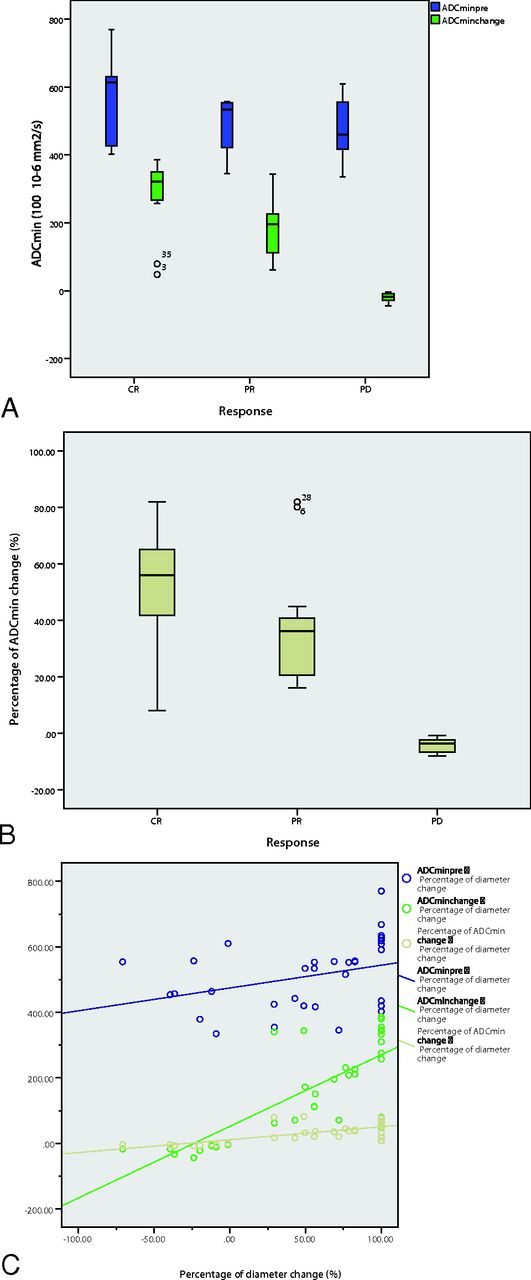
  • Fig 4.
    • Download figure
    • Open in new tab
    • Download powerpoint
    Fig 4.

    A boxplot of ADCminpre and ADCmin early change in the CR, PR, and PD groups (A). A boxplot of percentage ADCmin change in the CR, PR, and PD groups (B). ADCmin early change and percentage ADCmin change values can differentiate the 3 groups. The percentage ADCmin change performed better in differentiating the final treatment response, specifically differentiating the CR and PR groups from the PD group (A and B). C, Correlation between ADCminpre and percentage of the diameter (R2 = 0.046, blue), correlation between ADCmin early change and percentage of the diameter (R2 = 0.576, green), and the correlation between the percentage of ADCmin early change and percentage of the diameter (R2 = 0.717, yellow).

Tables

  • Figures
    • View popup
    Table 1:

    Response criteria for primary central nervous system lymphoma

    ResponseBrain ImagingCorticosteroid DoseEye Examination FindingsCSF Cytology Findings
    CRNo contrast enhancementNoneNormalNegative
    CRuNo contrast enhancementAnyNormalNegative
    Minimal abnormalityAnyMinor RPE abnormalityNegative
    PR50% Decrease in enhancing tumorIrrelevantMinor RPE abnormality or normalNegative
    No contrast enhancementIrrelevantDecrease in vitreous cells or retinal infiltratePersistent or suspicious
    PD25% Increase in lesionIrrelevantRecurrent or new ocular diseaseRecurrent or positive
    Any new site of disease: CNS or systemic
    • Note:—CRu indicates unconfirmed complete response; RPE, retinal pigment epithelium.

    • View popup
    Table 2:

    Comparison of ADCmin values among CR, PR, and PD groupsa

    No.ADCminpreADCminearlyADCminpostADCmin ChangebPercentage ADCmin Changec
    CR12566.56 ± 120.84849.09 ± 182.45858.06 ± 185.89282.54 ± 110.4051.17 ± 21.44
    PR15487.54 ± 78.00669.73 ± 130.28677.14 ± 131.47182.19 ± 88.2837.52 ± 20.02
    PD8476.13 ± 93.36456.65 ± 93.36432.55 ± 88.85−19.49 ± 13.46−4.17 ± 2.60
    Total35512.03 ± 103.15682.52 ± 203.52683.26 ± 213.34170.50 ± 142.0332.68 ± 27.67
    P value.072.000.000.000.000
        CR-PR.045.003.003.006.63
        CR-PD.051.000.000.000.000
        PR-PD.792.002.001.000.000
    • Note:—ADCminpost indicates ADCmin after 5 cycles.

    • ↵a All ADC values are reported as 100 × 10−6 mm2/s. Data are means.

    • ↵b ADCmin Change = ADCminearly − ADCminpre.

    • ↵c Percentage ADCmin Change = (ADCminearly − ADCminpre)/ADCminpre.

    • View popup
    Table 3:

    Comparison of tumor diameter among CR, PR, and PD groups

    No.Diameterpre (cm)Diameterearly (cm)Diameterpost (cm)Diameter Change (cm)aPercentage Diameter Changeb
    CR124.41 ± 1.462.45 ± 1.080.00 ± 0.004.41 ± 1.46100
    PR155.60 ± 2.354.71 ± 2.392.03 ± 1.193.57 ± 2.0760.75 ± 18.53
    PD85.06 ± 2.025.23 ± 2.426.21 ± 2.50−1.16 ± 0.8726.62 ± 22.16
    Total355.07 ± 2.024.03 ± 2.312.29 ± 2.712.78 ± 2.7354.24 ± 50.38
    P value.325.007.000.000.000
        CR-PR.137.008.001.201.000
        CR-PD.486.005.000.000.000
        PR-PD.543.570.000.000.000
    • ↵a Diameter Change = Diameterpre − Diameterpost.

    • ↵b Percentage Diameter Change = (Diameterpre − Diameterpost)/Diameterpre.

PreviousNext
Back to top

In this issue

American Journal of Neuroradiology: 37 (11)
American Journal of Neuroradiology
Vol. 37, Issue 11
1 Nov 2016
  • Table of Contents
  • Index by author
  • Complete Issue (PDF)
Advertisement
Print
Download PDF
Email Article

Thank you for your interest in spreading the word on American Journal of Neuroradiology.

NOTE: We only request your email address so that the person you are recommending the page to knows that you wanted them to see it, and that it is not junk mail. We do not capture any email address.

Enter multiple addresses on separate lines or separate them with commas.
Diffusion-Weighted Imaging for Predicting and Monitoring Primary Central Nervous System Lymphoma Treatment Response
(Your Name) has sent you a message from American Journal of Neuroradiology
(Your Name) thought you would like to see the American Journal of Neuroradiology web site.
CAPTCHA
This question is for testing whether or not you are a human visitor and to prevent automated spam submissions.
Cite this article
W.-Y. Huang, J.-B. Wen, G. Wu, B. Yin, J.-J. Li, D.-Y. Geng
Diffusion-Weighted Imaging for Predicting and Monitoring Primary Central Nervous System Lymphoma Treatment Response
American Journal of Neuroradiology Nov 2016, 37 (11) 2010-2018; DOI: 10.3174/ajnr.A4867

Citation Manager Formats

  • BibTeX
  • Bookends
  • EasyBib
  • EndNote (tagged)
  • EndNote 8 (xml)
  • Medlars
  • Mendeley
  • Papers
  • RefWorks Tagged
  • Ref Manager
  • RIS
  • Zotero
0 Responses
Respond to this article
Share
Bookmark this article
Diffusion-Weighted Imaging for Predicting and Monitoring Primary Central Nervous System Lymphoma Treatment Response
W.-Y. Huang, J.-B. Wen, G. Wu, B. Yin, J.-J. Li, D.-Y. Geng
American Journal of Neuroradiology Nov 2016, 37 (11) 2010-2018; DOI: 10.3174/ajnr.A4867
del.icio.us logo Twitter logo Facebook logo Mendeley logo
  • Tweet Widget
  • Facebook Like
  • Google Plus One
Purchase

Jump to section

  • Article
    • Abstract
    • ABBREVIATIONS:
    • Materials and Methods
    • Results
    • Discussion
    • Conclusions
    • Footnotes
    • References
  • Figures & Data
  • Info & Metrics
  • Responses
  • References
  • PDF

Related Articles

  • No related articles found.
  • PubMed
  • Google Scholar

Cited By...

  • Pretreatment ADC Histogram Analysis as a Prognostic Imaging Biomarker for Patients with Recurrent Glioblastoma Treated with Bevacizumab: A Systematic Review and Meta-analysis
  • Diffusion-Weighted Imaging of Brain Metastasis from Lung Cancer: Correlation of MRI Parameters with the Histologic Type and Gene Mutation Status
  • Crossref
  • Google Scholar

This article has not yet been cited by articles in journals that are participating in Crossref Cited-by Linking.

More in this TOC Section

  • Diagnostic Neuroradiology of Monoclonal Antibodies
  • Clinical Outcomes After Chiari I Decompression
  • Segmentation of Brain Metastases with BLAST
Show more Adult Brain

Similar Articles

Advertisement

Indexed Content

  • Current Issue
  • Accepted Manuscripts
  • Article Preview
  • Past Issues
  • Editorials
  • Editor's Choice
  • Fellows' Journal Club
  • Letters to the Editor
  • Video Articles

Cases

  • Case Collection
  • Archive - Case of the Week
  • Archive - Case of the Month
  • Archive - Classic Case

More from AJNR

  • Trainee Corner
  • Imaging Protocols
  • MRI Safety Corner
  • Book Reviews

Multimedia

  • AJNR Podcasts
  • AJNR Scantastics

Resources

  • Turnaround Time
  • Submit a Manuscript
  • Submit a Video Article
  • Submit an eLetter to the Editor/Response
  • Manuscript Submission Guidelines
  • Statistical Tips
  • Fast Publishing of Accepted Manuscripts
  • Graphical Abstract Preparation
  • Imaging Protocol Submission
  • Evidence-Based Medicine Level Guide
  • Publishing Checklists
  • Author Policies
  • Become a Reviewer/Academy of Reviewers
  • News and Updates

About Us

  • About AJNR
  • Editorial Board
  • Editorial Board Alumni
  • Alerts
  • Permissions
  • Not an AJNR Subscriber? Join Now
  • Advertise with Us
  • Librarian Resources
  • Feedback
  • Terms and Conditions
  • AJNR Editorial Board Alumni

American Society of Neuroradiology

  • Not an ASNR Member? Join Now

© 2025 by the American Society of Neuroradiology All rights, including for text and data mining, AI training, and similar technologies, are reserved.
Print ISSN: 0195-6108 Online ISSN: 1936-959X

Powered by HighWire