
Article Figures & Data
Figures
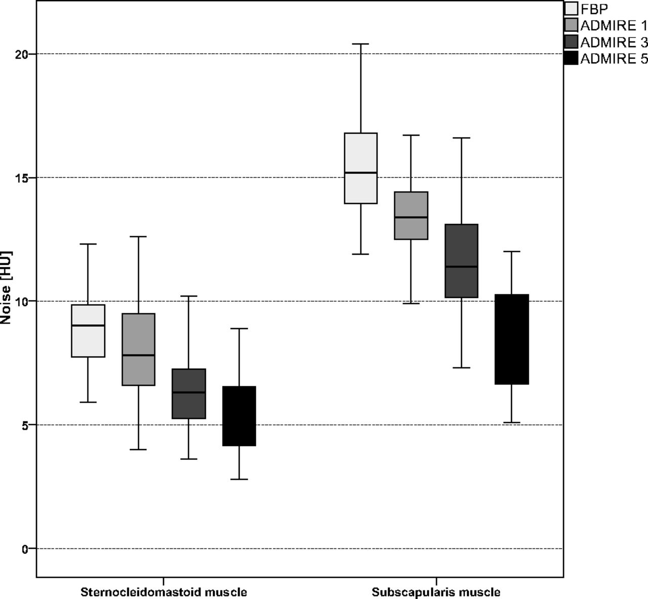
- Fig 1.
Boxplot graph shows comparison of image noise of the sternocleidomastoid and subscapularis muscles between the different image reconstruction settings. Image noise of the sternocleidomastoid muscle and subscapularis muscle was significantly lower with all ADMIRE levels, with the best results for ADMIRE 5 (all, P < .001). Noise in the lower part of the neck, represented by the subscapularis muscle, was significantly increased within each reconstruction mode compared with the upper part of the neck, represented by the sternocleidomastoid muscle (all, P < .001).
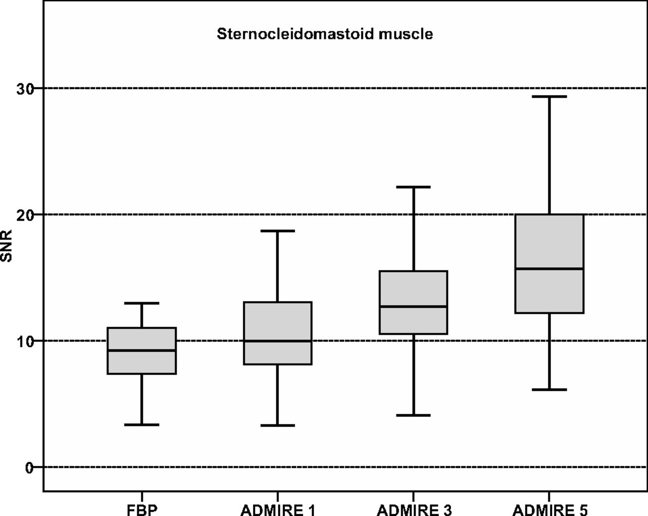
- Fig 2.
Boxplot graphs show comparison of signal-to-noise ratios of the sternocleidomastoid muscle. SNR was significantly higher for ADMIRE compared with FBP with the highest results observed for ADMIRE strength level 5 (all, P < .001). Significant differences were also shown within ADMIRE strength levels (all, P < .001).
- Fig 3.
Boxplot graphs show contrast-to-noise ratios of sternocleidomastoid muscle-to-fat (A) with significantly higher results in ADMIRE strength levels 3 and 5 compared with FBP and ADMIRE 1 (P < .001), while internal jugular vein–sternocleidomastoid muscle CNR (B) is significantly increased in all ADMIRE strength levels compared with FBP (all, P < .001).
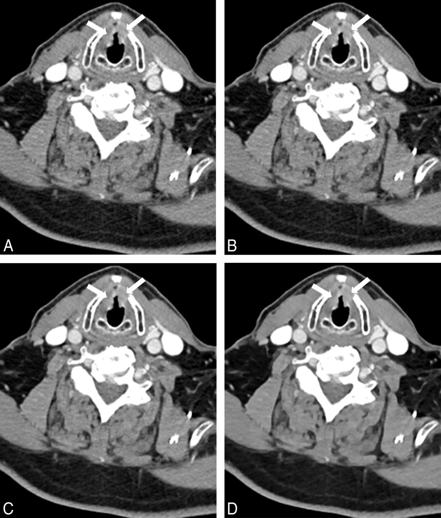
- Fig 4.
Images of a 46-year-old male patient examined with a low tube voltage of 90 kV on 192-section DSCT (window settings: width, 400 HU; level, 80 HU). Images were reconstructed by using filtered back-projection (A) and advanced modeled iterative reconstruction with strength levels 1 (B), 3 (C), and 5 (D). Axial images show histologically proved bilateral T2 squamous cell carcinoma of the glottic larynx (arrows). Image noise was highest by using FBP (A). The higher ADMIRE strength levels show consistently lower image noise (B–D). The internal jugular vein–sternocleidomastoid muscle CNR is highest by using ADMIRE 5 (D). Delineation of smaller structures was considered good by both observers in all images.
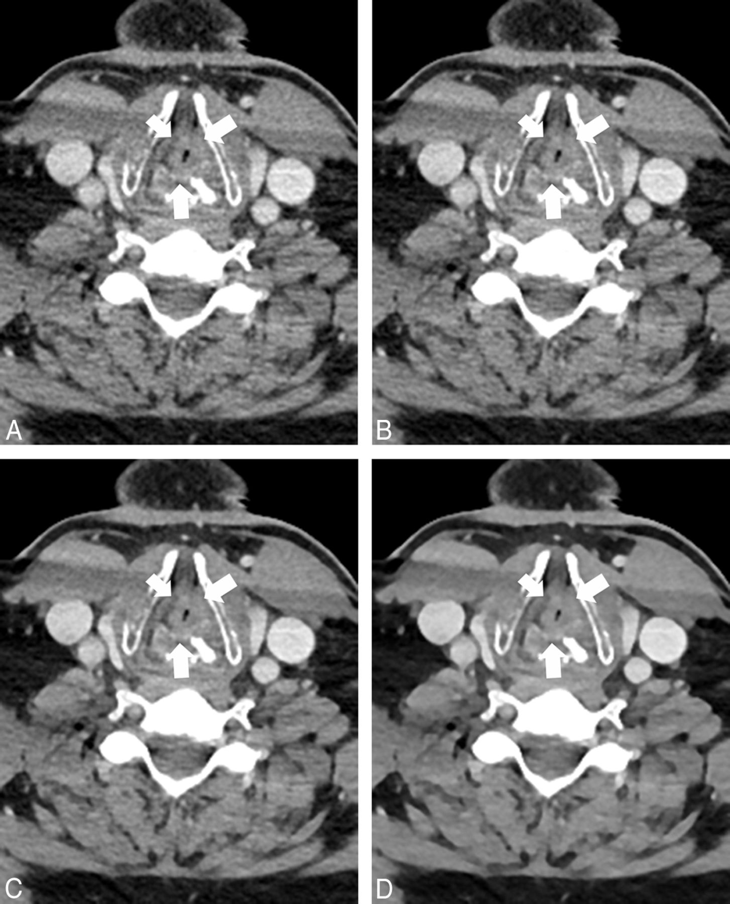
- Fig 5.
A 58-year-old male patient with sudden dyspnea. CT was performed with a tube voltage of 90 kV (window settings: width, 400 HU; level, 80 HU). Images were reconstructed by using filtered back-projection (A) and advanced modeled iterative reconstruction with strength levels 1 (B), 3 (C), and 5 (D). Images show histologically proved bilateral T4 laryngeal squamous cell carcinoma (arrows). The tumor reaches to the left thyroid cartilage but is separated from the right thyroid cartilage by a thin fat line. Image noise was lower in ADMIRE compared with FBP with the lowest image noise in ADMIRE 5 (D). Streak artifacts in the sternocleidomastoid muscle on both sides are visible in all images due to the shoulder region in the lower part of the neck.


Tables
FBP ADMIRE 1 ADMIRE 3 ADMIRE 5 Attenuation (HU) Sternocleidomastoid muscle 79.4 ± 13.2 80.1 ± 13.4 79.9 ± 13.6 79.2 ± 13.7 Internal jugular vein 288.5 ± 71.4 290.5 ± 72.6 289.0 ± 71.6 287.3 ± 72.2 Submandibular gland 131.4 ± 47.1 131.2 ± 48.2 131.6 ± 47.8 131.1 ± 47.2 Tongue 97.4 ± 14.1 97.5 ± 13.9 96.2 ± 13.5 95.7 ± 13.5 Subscapularis muscle 69.8 ± 12.9 69.9 ± 12.8 68.8 ± 12.6 69.0 ± 12.4 Fat −105.7 ± 9.3 −106.4 ± 11.3 −107.2 ± 10.9 −106.0 ± 11.3 Image noise (HU) Sternocleidomastoid muscle 9.4 ± 2.4 8.3 ± 2.8 6.7 ± 2.0 5.4 ± 1.7 Internal jugular vein 12.6 ± 6.3 11.2 ± 5.9 10.3 ± 7.1 8.5 ± 5.5 Submandibular gland 11.9 ± 2.6 11.2 ± 2.5 9.4 ± 2.9 7.5 ± 3.0 Tongue 10.3 ± 2.4 9.8 ± 2.6 8.6 ± 2.8 6.9 ± 3.2 Subscapularis muscle 15.3 ± 2.8 13.7 ± 2.7 11.8 ± 2.6 8.6 ± 2.8 Fat 15.1 ± 5.1 14.9 ± 4.5 12.6 ± 4.8 9.8 ± 4.0 ↵a Data are means.
FBP ADMIRE 1 ADMIRE 3 ADMIRE 5 Signal-to-noise ratio Sternocleidomastoid muscle 9.0 ± 2.5 10.8 ± 4.1 13.0 ± 4.4 16.4 ± 6.2 Submandibular gland 11.6 ± 4.6 12.3 ± 4.7 15.3 ± 6.8 19.6 ± 8.2 Contrast-to-noise ratio Sternocleidomastoid muscle–fat 14.5 ± 8.1 13.8 ± 4.9 17.3 ± 7.7 22.6 ± 11.4 Submandibular gland–fat 19.1 ± 11.6 17.9 ± 8.2 22.6 ± 11.7 28.6 ± 17.1 IJV–sternocleidomastoid muscle 24.1 ± 10.1 28.7 ± 13.5 34.8 ± 16.3 41.2 ± 22.1 Note:—IJV indicates internal jugular vein.
↵a Data are means.
FBP ADMIRE 1 ADMIRE 3 ADMIRE 5 Overall image quality 3.2 ± 0.5 (0.58) 3.3 ± 0.6 (0.57) 4.4 ± 0.9 (0.12) 4.7 ± 0.5 (0.67) Image noise 3.4 ± 0.5 (0.79) 3.8 ± 0.4 (0.83) 4.6 ± 0.5 (0.52) 4.9 ± 0.3 (0.32) Delineation of smaller structures 3.3 ± 0.4 (0.74) 3.7 ± 0.5 (0.78) 3.8 ± 0.4 (0.50) 3.8 ± 0.4 (0.74) Image contrast 3.5 ± 0.5 (0.80) 3.4 ± 0.5 (0.78) 4.8 ± 0.4 (0.34) 4.8 ± 0.4 (0.47) ↵a Data are means ± SD. Interobserver agreement (slight [κ < 0.3], moderate [κ = 0.3–0.7], good [κ > 0.7]).


