Advertisement

Main menu

  • Home
  • Content
    • Current Issue
    • Accepted Manuscripts
    • Article Preview
    • Past Issue Archive
    • Video Articles
    • AJNR Case Collection
    • Case of the Week Archive
    • Case of the Month Archive
    • Classic Case Archive
  • Special Collections
    • AJNR Awards
    • Low-Field MRI
    • Alzheimer Disease
    • ASNR Foundation Special Collection
    • Photon-Counting CT
    • View All
  • Multimedia
    • AJNR Podcasts
    • AJNR SCANtastic
    • Trainee Corner
    • MRI Safety Corner
    • Imaging Protocols
  • For Authors
    • Submit a Manuscript
    • Submit a Video Article
    • Submit an eLetter to the Editor/Response
    • Manuscript Submission Guidelines
    • Statistical Tips
    • Fast Publishing of Accepted Manuscripts
    • Graphical Abstract Preparation
    • Imaging Protocol Submission
    • Author Policies
  • About Us
    • About AJNR
    • Editorial Board
    • Editorial Board Alumni
  • More
    • Become a Reviewer/Academy of Reviewers
    • Subscribers
    • Permissions
    • Alerts
    • Feedback
    • Advertisers
    • ASNR Home

User menu

  • Alerts
  • Log in

Search

  • Advanced search
American Journal of Neuroradiology

American Journal of Neuroradiology

ASHNR American Society of Functional Neuroradiology ASHNR American Society of Pediatric Neuroradiology ASSR
  • Alerts
  • Log in

Advanced Search

  • Home
  • Content
    • Current Issue
    • Accepted Manuscripts
    • Article Preview
    • Past Issue Archive
    • Video Articles
    • AJNR Case Collection
    • Case of the Week Archive
    • Case of the Month Archive
    • Classic Case Archive
  • Special Collections
    • AJNR Awards
    • Low-Field MRI
    • Alzheimer Disease
    • ASNR Foundation Special Collection
    • Photon-Counting CT
    • View All
  • Multimedia
    • AJNR Podcasts
    • AJNR SCANtastic
    • Trainee Corner
    • MRI Safety Corner
    • Imaging Protocols
  • For Authors
    • Submit a Manuscript
    • Submit a Video Article
    • Submit an eLetter to the Editor/Response
    • Manuscript Submission Guidelines
    • Statistical Tips
    • Fast Publishing of Accepted Manuscripts
    • Graphical Abstract Preparation
    • Imaging Protocol Submission
    • Author Policies
  • About Us
    • About AJNR
    • Editorial Board
    • Editorial Board Alumni
  • More
    • Become a Reviewer/Academy of Reviewers
    • Subscribers
    • Permissions
    • Alerts
    • Feedback
    • Advertisers
    • ASNR Home
  • Follow AJNR on Twitter
  • Visit AJNR on Facebook
  • Follow AJNR on Instagram
  • Join AJNR on LinkedIn
  • RSS Feeds

AJNR Awards, New Junior Editors, and more. Read the latest AJNR updates

Research ArticleAdult Brain
Open Access

Mean Diffusional Kurtosis in Patients with Glioma: Initial Results with a Fast Imaging Method in a Clinical Setting

A. Tietze, M.B. Hansen, L. Østergaard, S.N. Jespersen, R. Sangill, T.E. Lund, M. Geneser, M. Hjelm and B. Hansen
American Journal of Neuroradiology August 2015, 36 (8) 1472-1478; DOI: https://doi.org/10.3174/ajnr.A4311
A. Tietze
aFrom the Department of Neuroradiology (A.T., L.Ø., M.G., M.H.), Aarhus University Hospital, Aarhus, Denmark
bCenter of Functionally Integrative Neuroscience and MINDLab, Institute of Clinical Medicine (A.T., M.B.H., L.Ø., S.N.J., R.S., T.E.L., M.G., B.H.)
  • Find this author on Google Scholar
  • Find this author on PubMed
  • Search for this author on this site
M.B. Hansen
bCenter of Functionally Integrative Neuroscience and MINDLab, Institute of Clinical Medicine (A.T., M.B.H., L.Ø., S.N.J., R.S., T.E.L., M.G., B.H.)
  • Find this author on Google Scholar
  • Find this author on PubMed
  • Search for this author on this site
L. Østergaard
aFrom the Department of Neuroradiology (A.T., L.Ø., M.G., M.H.), Aarhus University Hospital, Aarhus, Denmark
bCenter of Functionally Integrative Neuroscience and MINDLab, Institute of Clinical Medicine (A.T., M.B.H., L.Ø., S.N.J., R.S., T.E.L., M.G., B.H.)
  • Find this author on Google Scholar
  • Find this author on PubMed
  • Search for this author on this site
S.N. Jespersen
bCenter of Functionally Integrative Neuroscience and MINDLab, Institute of Clinical Medicine (A.T., M.B.H., L.Ø., S.N.J., R.S., T.E.L., M.G., B.H.)
cDepartment of Physics and Astronomy (S.N.J.), Aarhus University, Aarhus, Denmark.
  • Find this author on Google Scholar
  • Find this author on PubMed
  • Search for this author on this site
R. Sangill
bCenter of Functionally Integrative Neuroscience and MINDLab, Institute of Clinical Medicine (A.T., M.B.H., L.Ø., S.N.J., R.S., T.E.L., M.G., B.H.)
  • Find this author on Google Scholar
  • Find this author on PubMed
  • Search for this author on this site
T.E. Lund
bCenter of Functionally Integrative Neuroscience and MINDLab, Institute of Clinical Medicine (A.T., M.B.H., L.Ø., S.N.J., R.S., T.E.L., M.G., B.H.)
  • Find this author on Google Scholar
  • Find this author on PubMed
  • Search for this author on this site
M. Geneser
aFrom the Department of Neuroradiology (A.T., L.Ø., M.G., M.H.), Aarhus University Hospital, Aarhus, Denmark
bCenter of Functionally Integrative Neuroscience and MINDLab, Institute of Clinical Medicine (A.T., M.B.H., L.Ø., S.N.J., R.S., T.E.L., M.G., B.H.)
  • Find this author on Google Scholar
  • Find this author on PubMed
  • Search for this author on this site
M. Hjelm
aFrom the Department of Neuroradiology (A.T., L.Ø., M.G., M.H.), Aarhus University Hospital, Aarhus, Denmark
  • Find this author on Google Scholar
  • Find this author on PubMed
  • Search for this author on this site
B. Hansen
bCenter of Functionally Integrative Neuroscience and MINDLab, Institute of Clinical Medicine (A.T., M.B.H., L.Ø., S.N.J., R.S., T.E.L., M.G., B.H.)
  • Find this author on Google Scholar
  • Find this author on PubMed
  • Search for this author on this site
  • Article
  • Figures & Data
  • Supplemental
  • Info & Metrics
  • Responses
  • References
  • PDF
Loading

Article Figures & Data

Figures

  • Tables
  • Fig 1.
    • Download figure
    • Open in new tab
    • Download powerpoint
    Fig 1.

    The tumor core was defined as the contrast-enhancing part on postcontrast T1-weighted images, and the FLAIR masks were defined as hyperintense regions on T2 FLAIR images (minus the tumor core and necrosis, if present). In cases of nonenhancing tumors, hyperintensity areas on T2 FLAIR images were defined as the tumor core (the tumor core and edema were identical regions). The NAWM was outlined in the centrum semiovale of the contralateral hemisphere (on ≥4 consecutive sections). The NAGM was defined in the contralateral thalamus (on ≥3 sections).
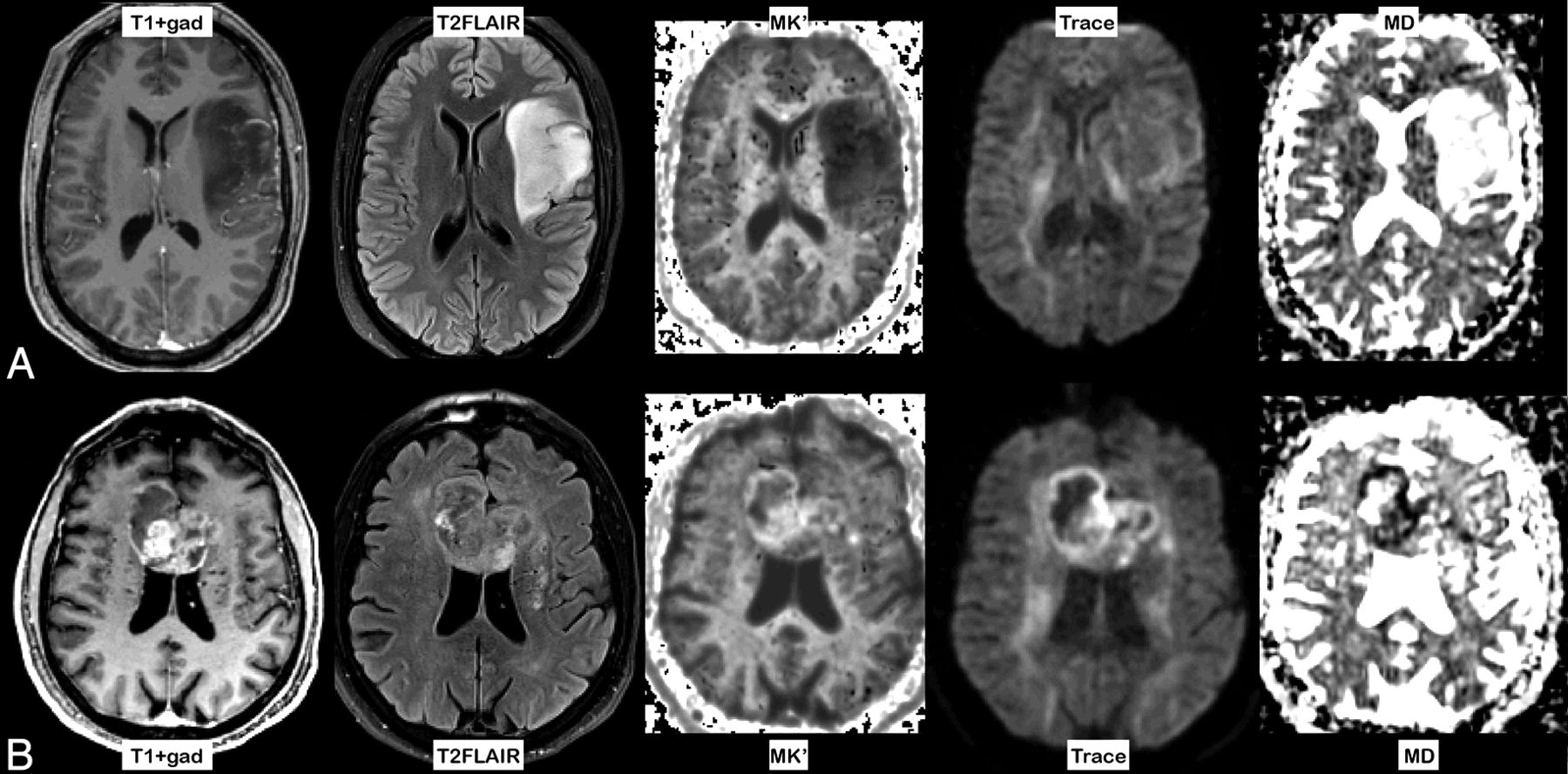
  • Fig 2.
    • Download figure
    • Open in new tab
    • Download powerpoint
    Fig 2.

    A, Contrast-enhanced T1-weighted and T2 FLAIR images of a grade II astrocytoma grade in the left hemisphere. The map of MK′ demonstrates low MK′, whereas the trace image and the MD map show high diffusivity (high signal changes on MD). B, Typical example of a glioblastoma, with contrast enhancement on postcontrast T1-weighted and complex signal changes on T2 FLAIR images. Increased MK′ is noted in most of the tumor. The trace image and MD show restricted diffusion, primarily in the periphery of the lesion.

  • Fig 3.
    • Download figure
    • Open in new tab
    • Download powerpoint
    Fig 3.

    Boxplots of average MK′ and normalized MK′ (in yellow) (A) and MD and normalized MD (in blue) (B) in the contrast-enhancing tumor core of all patients, grouped according to tumor types and grades, are shown. The horizontal lines in the boxes are the median values, the upper and lower box edges are the 25th and 75th percentiles, respectively, and the upper and lower whiskers represent the minimums and maximums, respectively. AC indicates grade III astrocytoma; ODG3, grade III oligodendroglioma; AC2, grade II astrocytoma; ODG2, grade II oligodendroglioma.

Tables

  • Figures
    • View popup
    Table 1:

    Significance of mean MK′, nMK′, MD, and nMD values in different tumor gradesa

    Tumor TypeMK′nMK′MDnMD
    HGG/LGG.028b.058.383.511
    GBM/all<.001b.002b.006b.006b
    GBM/HGG.003b.011b.004b.004b
    AC3/all.135.135.063.033b
    AC3/HGG.011b.031b.024b.019b
    • Note:—AC3 indicates grade III astrocytoma.

    • ↵a Mean MK′, nMK′, MD, and nMD values in the tumor core were compared between different tumor grades, and the resulting P values are listed.

    • ↵b A P value of <.05 indicates a significant difference between groups.

    • View popup
    Table 2:

    Results of regression analysisa

    Tumor TypeMK′PnMK′PMDPnMDPMK′ + MDPbnMK′ + nMDPb
    HGG/LGG0.731.045c0.701.0560.595.5120.572.4560.754.032c0.746.041c
    .220.213
    GBM/all0.842.004c0.811.004c0.775.017c0.772.014c0.842.045c0.807.047c
    .796.835
    GBM/HGG0.886.027c0.838.022c0.876.018c0.876.019c0.895.1270.905.181
    .204.184
    AC3/all0.717.1460.717.1700.766.0520.800.0600.793.7440.779.885
    .165.155
    AC3/HGG0.871.039c0.824.0550.835.038c0.847.042c0.882.1270.871.301
    .328.265
    • Note:—AC3 indicates grade III astrocytoma.

    • ↵a The areas under the receiver operating characteristic curves as a measure for diagnostic accuracy are reported.

    • ↵b Two P values occur when two values (MK′ and MD) are tested at the same time.

    • ↵c The variable contributes significantly to the regression model if the corresponding P value is <.05.

PreviousNext
Back to top

In this issue

American Journal of Neuroradiology: 36 (8)
American Journal of Neuroradiology
Vol. 36, Issue 8
1 Aug 2015
  • Table of Contents
  • Index by author
  • Complete Issue (PDF)
Advertisement
Print
Download PDF
Email Article

Thank you for your interest in spreading the word on American Journal of Neuroradiology.

NOTE: We only request your email address so that the person you are recommending the page to knows that you wanted them to see it, and that it is not junk mail. We do not capture any email address.

Enter multiple addresses on separate lines or separate them with commas.
Mean Diffusional Kurtosis in Patients with Glioma: Initial Results with a Fast Imaging Method in a Clinical Setting
(Your Name) has sent you a message from American Journal of Neuroradiology
(Your Name) thought you would like to see the American Journal of Neuroradiology web site.
CAPTCHA
This question is for testing whether or not you are a human visitor and to prevent automated spam submissions.
Cite this article
A. Tietze, M.B. Hansen, L. Østergaard, S.N. Jespersen, R. Sangill, T.E. Lund, M. Geneser, M. Hjelm, B. Hansen
Mean Diffusional Kurtosis in Patients with Glioma: Initial Results with a Fast Imaging Method in a Clinical Setting
American Journal of Neuroradiology Aug 2015, 36 (8) 1472-1478; DOI: 10.3174/ajnr.A4311

Citation Manager Formats

  • BibTeX
  • Bookends
  • EasyBib
  • EndNote (tagged)
  • EndNote 8 (xml)
  • Medlars
  • Mendeley
  • Papers
  • RefWorks Tagged
  • Ref Manager
  • RIS
  • Zotero
0 Responses
Respond to this article
Share
Bookmark this article
Mean Diffusional Kurtosis in Patients with Glioma: Initial Results with a Fast Imaging Method in a Clinical Setting
A. Tietze, M.B. Hansen, L. Østergaard, S.N. Jespersen, R. Sangill, T.E. Lund, M. Geneser, M. Hjelm, B. Hansen
American Journal of Neuroradiology Aug 2015, 36 (8) 1472-1478; DOI: 10.3174/ajnr.A4311
del.icio.us logo Twitter logo Facebook logo Mendeley logo
  • Tweet Widget
  • Facebook Like
  • Google Plus One
Purchase

Jump to section

  • Article
    • Abstract
    • ABBREVIATIONS:
    • Materials and Methods
    • Results
    • Discussion
    • Conclusions
    • Acknowledgments
    • Footnotes
    • References
  • Figures & Data
  • Supplemental
  • Info & Metrics
  • Responses
  • References
  • PDF

Related Articles

  • PubMed
  • Google Scholar

Cited By...

  • Pathogenic O-GlcNAc dyshomeostasis associated with cortical malformations and hyperactivity
  • CNS Embryonal Tumor with PLAGL Amplification, a New Tumor in Children and Adolescents: Insights from a Comprehensive MRI Analysis
  • Feasibility of generalised DKI approach for brain glioma grading
  • The effect of crack cocaine addiction and age on the microstructure and morphology of the human striatum and thalamus using shape analysis and fast diffusion kurtosis imaging
  • Role of diffusional kurtosis imaging in grading of brain gliomas: a protocol for systematic review and meta-analysis
  • Comparative Analysis of Diffusional Kurtosis Imaging, Diffusion Tensor Imaging, and Diffusion-Weighted Imaging in Grading and Assessing Cellular Proliferation of Meningiomas
  • Crossref (69)
  • Google Scholar

This article has been cited by the following articles in journals that are participating in Crossref Cited-by Linking.

  • The link between diffusion MRI and tumor heterogeneity: Mapping cell eccentricity and density by diffusional variance decomposition (DIVIDE)
    Filip Szczepankiewicz, Danielle van Westen, Elisabet Englund, Carl-Fredrik Westin, Freddy Ståhlberg, Jimmy Lätt, Pia C. Sundgren, Markus Nilsson
    NeuroImage 2016 142
  • Diffusion kurtosis imaging can efficiently assess the glioma grade and cellular proliferation
    Rifeng Jiang, Jingjing Jiang, Lingyun Zhao, Jiaxuan Zhang, Shun Zhang, Yihao Yao, Shiqi Yang, Jingjing Shi, Nanxi Shen, Changliang Su, Ju Zhang, Wenzhen Zhu
    Oncotarget 2015 6 39
  • Precision and accuracy of diffusion kurtosis estimation and the influence of b‐value selection
    Andrey Chuhutin, Brian Hansen, Sune Nørhøj Jespersen
    NMR in Biomedicine 2017 30 11
  • Imaging brain tumour microstructure
    Markus Nilsson, Elisabet Englund, Filip Szczepankiewicz, Danielle van Westen, Pia C. Sundgren
    NeuroImage 2018 182
  • Fast imaging of mean, axial and radial diffusion kurtosis
    Brian Hansen, Noam Shemesh, Sune Nørhøj Jespersen
    NeuroImage 2016 142
  • Glioma Grade Discrimination with MR Diffusion Kurtosis Imaging: A Meta-Analysis of Diagnostic Accuracy
    Anna Falk Delgado, Markus Nilsson, Danielle van Westen, Alberto Falk Delgado
    Radiology 2018 287 1
  • Kurtosis fractional anisotropy, its contrast and estimation by proxy
    Brian Hansen, Sune Nørhøj Jespersen
    Scientific Reports 2016 6 1
  • Noninvasive assessment of isocitrate dehydrogenase mutation status in cerebral gliomas by magnetic resonance spectroscopy in a clinical setting
    Anna Tietze, Changho Choi, Bruce Mickey, Elizabeth A. Maher, Benedicte Parm Ulhøi, Ryan Sangill, Yasmin Lassen-Ramshad, Slavka Lukacova, Leif Østergaard, Gorm von Oettingen
    Journal of Neurosurgery 2018 128 2
  • Adult Glioma WHO Classification Update, Genomics, and Imaging
    James Bai, Jerrin Varghese, Rajan Jain
    Topics in Magnetic Resonance Imaging 2020 29 2
  • Experimental considerations for fast kurtosis imaging
    Brian Hansen, Torben E. Lund, Ryan Sangill, Ebbe Stubbe, Jürgen Finsterbusch, Sune Nørhøj Jespersen
    Magnetic Resonance in Medicine 2016 76 5

More in this TOC Section

  • Diagnostic Neuroradiology of Monoclonal Antibodies
  • NCCT vs. MRI for Brain Atrophy in Acute Stroke
  • Clinical Outcomes After Chiari I Decompression
Show more ADULT BRAIN

Similar Articles

Advertisement

Indexed Content

  • Current Issue
  • Accepted Manuscripts
  • Article Preview
  • Past Issues
  • Editorials
  • Editor's Choice
  • Fellows' Journal Club
  • Letters to the Editor
  • Video Articles

Cases

  • Case Collection
  • Archive - Case of the Week
  • Archive - Case of the Month
  • Archive - Classic Case

More from AJNR

  • Trainee Corner
  • Imaging Protocols
  • MRI Safety Corner
  • Book Reviews

Multimedia

  • AJNR Podcasts
  • AJNR Scantastics

Resources

  • Turnaround Time
  • Submit a Manuscript
  • Submit a Video Article
  • Submit an eLetter to the Editor/Response
  • Manuscript Submission Guidelines
  • Statistical Tips
  • Fast Publishing of Accepted Manuscripts
  • Graphical Abstract Preparation
  • Imaging Protocol Submission
  • Evidence-Based Medicine Level Guide
  • Publishing Checklists
  • Author Policies
  • Become a Reviewer/Academy of Reviewers
  • News and Updates

About Us

  • About AJNR
  • Editorial Board
  • Editorial Board Alumni
  • Alerts
  • Permissions
  • Not an AJNR Subscriber? Join Now
  • Advertise with Us
  • Librarian Resources
  • Feedback
  • Terms and Conditions
  • AJNR Editorial Board Alumni

American Society of Neuroradiology

  • Not an ASNR Member? Join Now

© 2025 by the American Society of Neuroradiology All rights, including for text and data mining, AI training, and similar technologies, are reserved.
Print ISSN: 0195-6108 Online ISSN: 1936-959X

Powered by HighWire